Jump to content
In this section we have spot diagnoses posted on a daily basis since June 2010, now over 4000! You can review the archived cases and read the suggested diagnoses by users and the final comment by the contributors.
Case are uploaded each week day by 10 am UK time with the correct diagnosis will generally be posted at 8 pm UK time. Why not view the most recent spot diagnosis and proffer a diagnosis?

Case Number : Case 2194 - 6 November 2018 Posted By: Uma Sundram

Please read the clinical history and view the images by clicking on them before you proffer your diagnosis.
Submitted Date :
   (0 reviews)

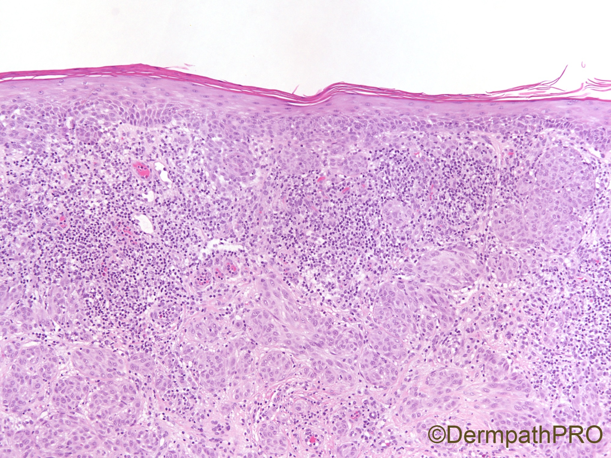
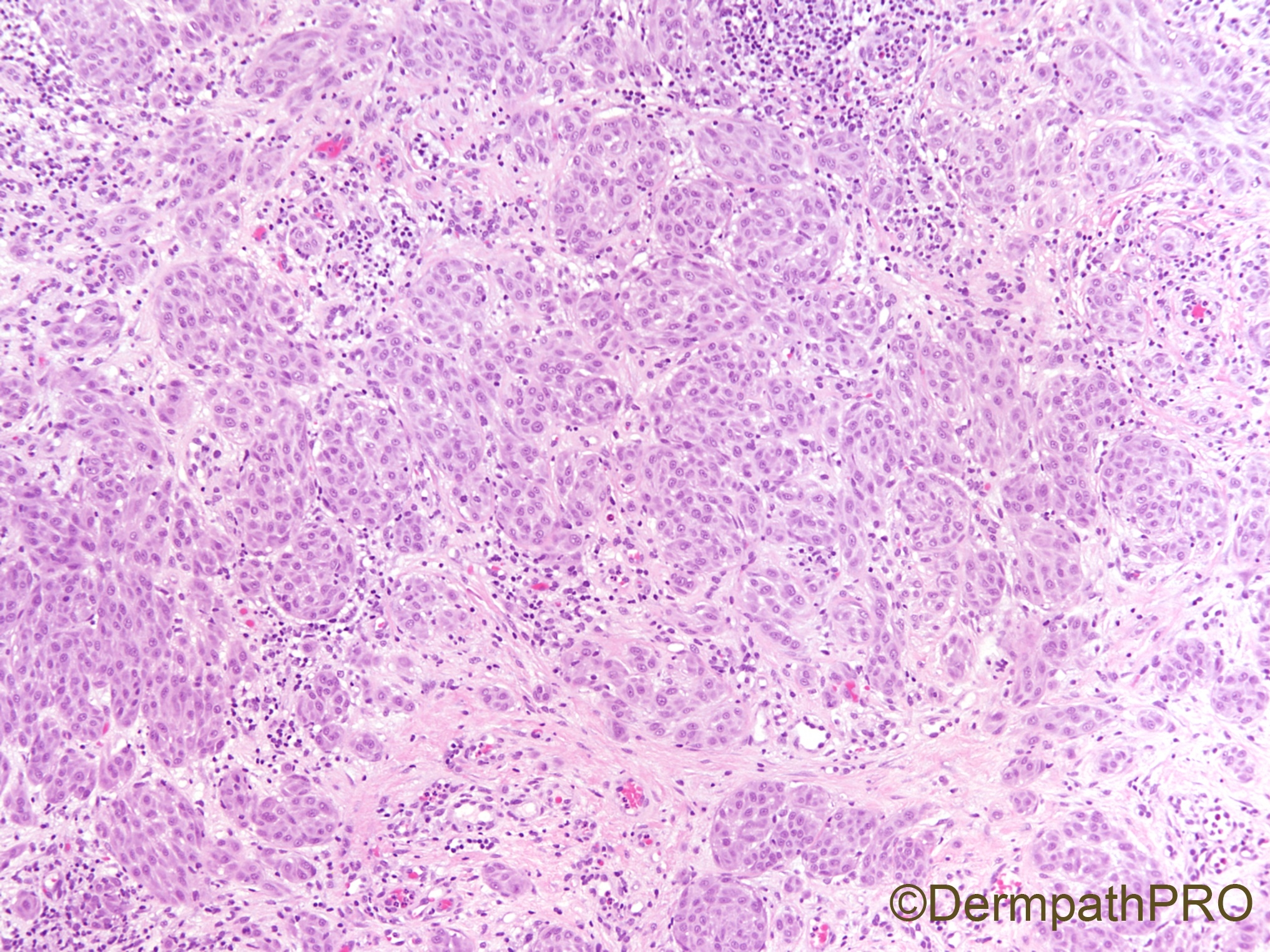
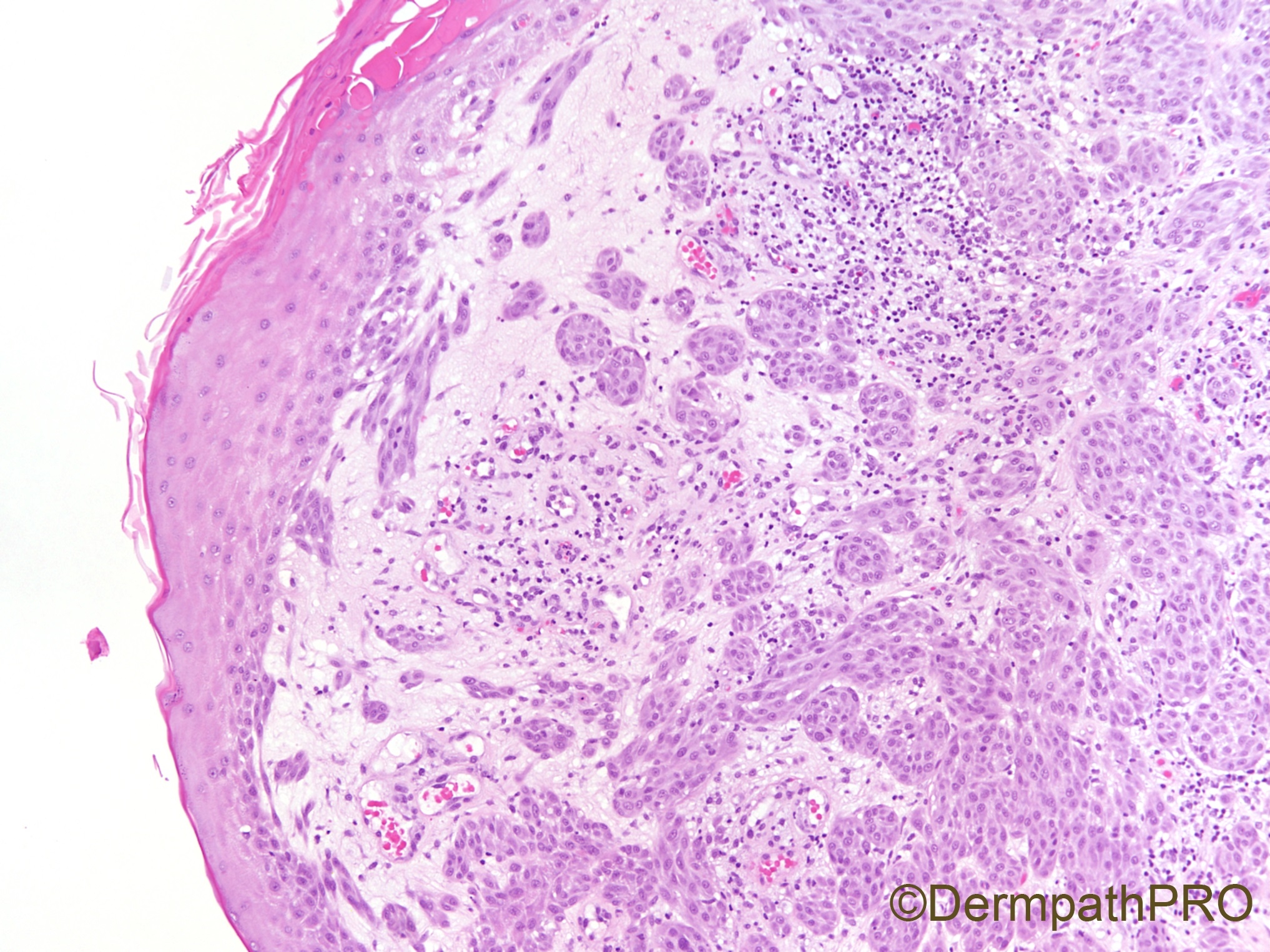
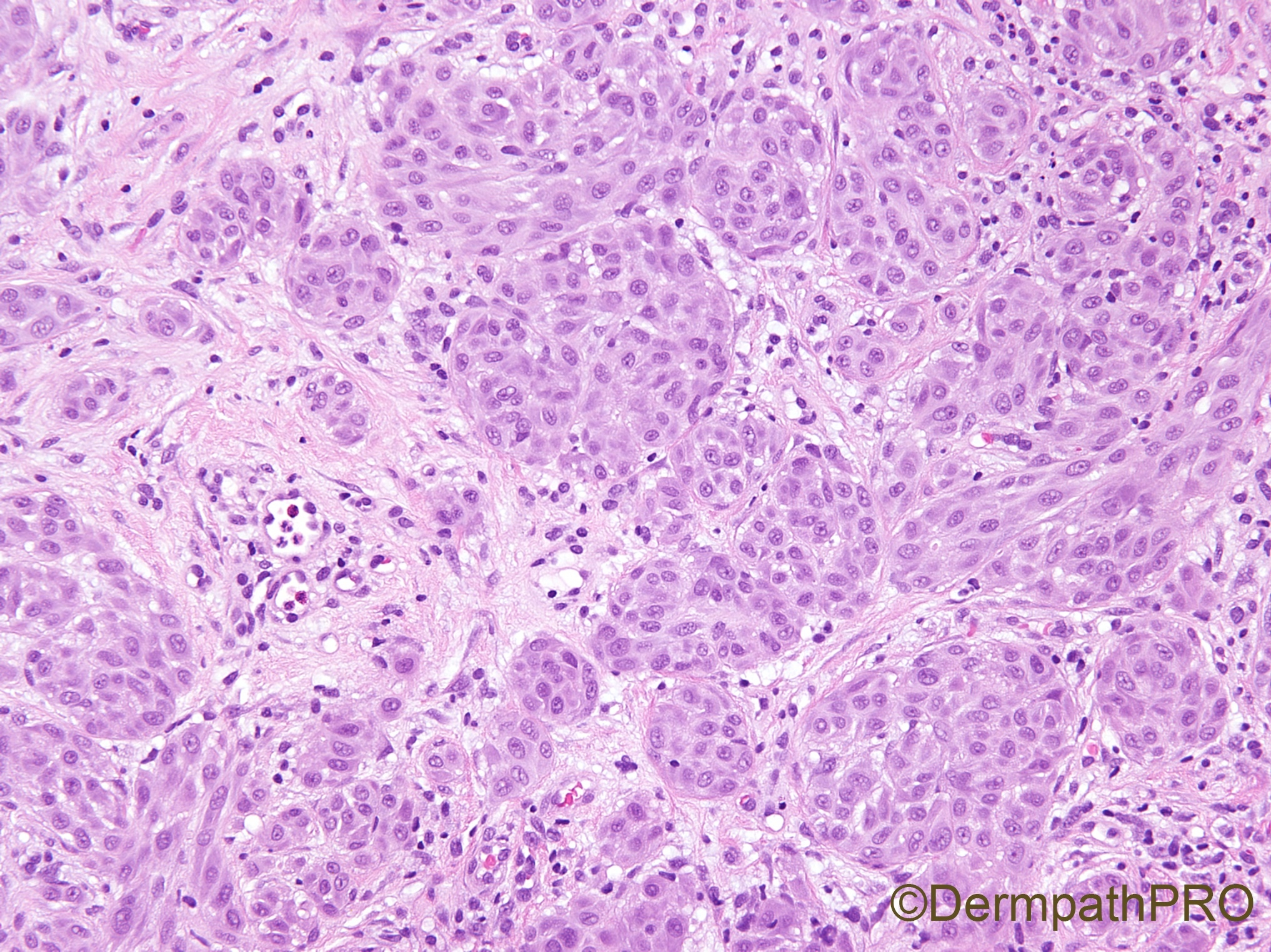
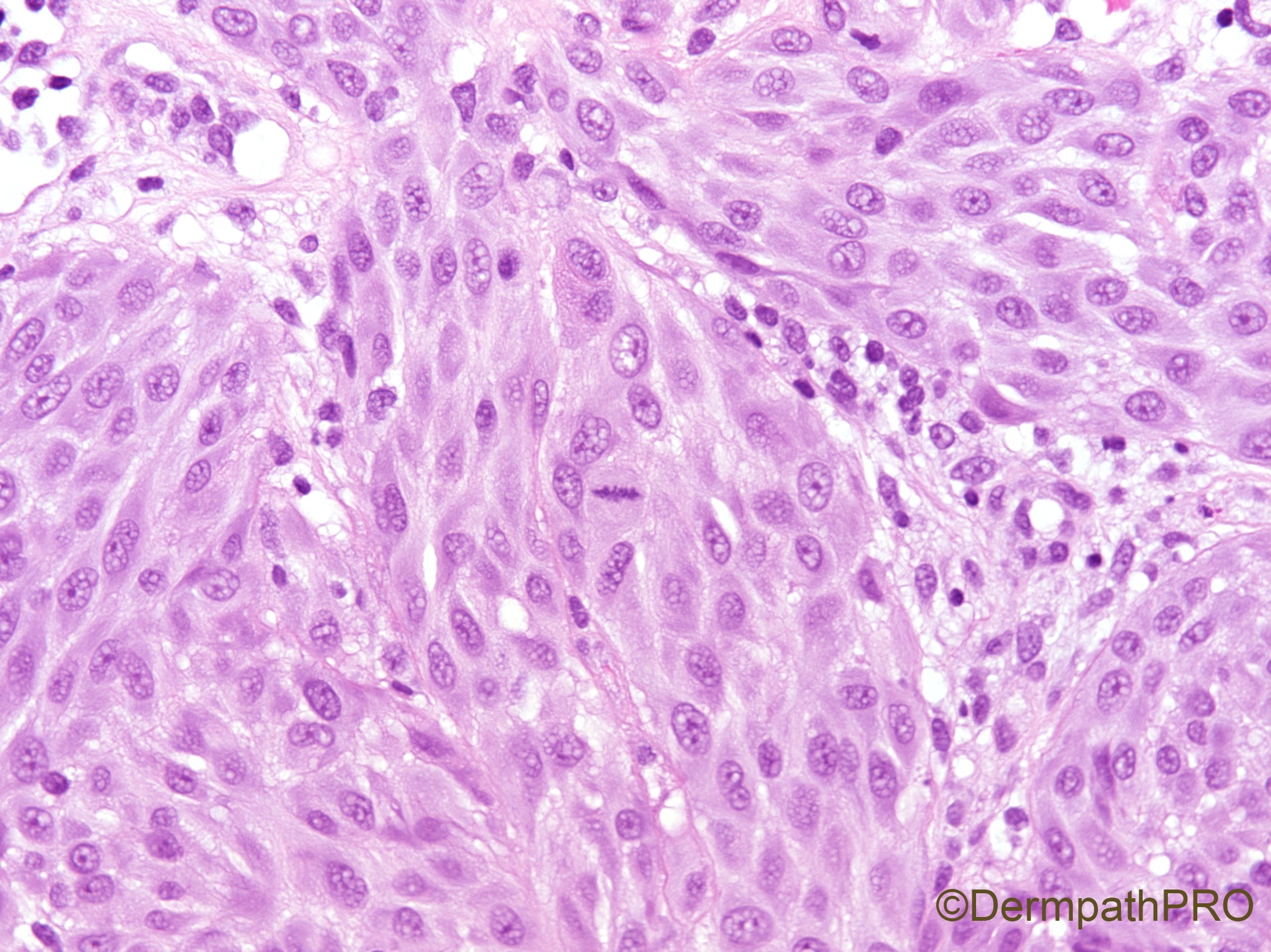
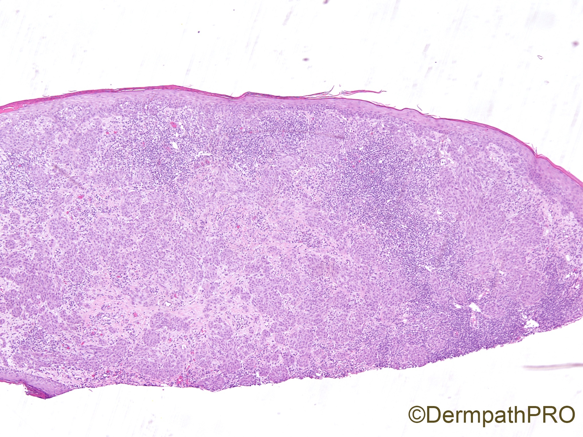
21 year old female with scalp lesion.

  Report Record

User Feedback


Lymphoepithelioma like carcinoma v/s inflamed SCC. Metastasis of LELC from other sites is also a possibility. 

Share this comment


Link to comment
share_externally

Alex-Ventura-Leon

Posted

Young patient. Difficult lesion (at lest for me). 

It seems epithelial whit connection to Epidermis and some vacuoles in the citoplasm. 

Favor SCC. I wonder if the patient have some underlying genetic alterations. 

Share this comment


Link to comment
share_externally

Spitz Nevus ( oriented fascicular pattern and vesicular monomorphic nuclei ).

Share this comment


Link to comment
share_externally

Don't really appreciate an epidermal component. So would do S-100 protein to make sure it is indeed a melanocytic lesion. Neurothekeoma sometimes can be mistaken for a melanocytic lesion.

Share this comment


Link to comment
share_externally

Krishnakumar subramanian

Posted

difficult for me, nevoid lesions with squamous morphology, but loss of polarity waiting for inputs

Share this comment


Link to comment
share_externally

Raul Perret

Posted

I would be very careful with this lesion. As Vincenzo suggested this looks like a spitzoid lesion not overtly malignant, would suggest complete excision and express the uncertainty on the report. Maybe some ancillary techniques could be useful.

Share this comment


Link to comment
share_externally

Alex-Ventura-Leon

Posted

After read your comments I think you all right. Melanocityc lesion it would fit more.

Cellular Neurothekeoma it's a great differential. 

Share this comment


Link to comment
share_externally

Uma Sundram

Posted

Love your comments. These are always challenging cases. This was S100 and Melan A positive, and cytokeratin negative. We opted for a Spitzoid lesion with atypical features and recommended re excision. Long term outcome is uncertain and the patient will be watched closely.

Share this comment


Link to comment
share_externally

Dr. Richard Carr

Posted

Agree with atypical Spitz tumour, uncertain for malignancy. Complete excision with clear margins is recommended (5mm). We'd usually run Ki67, HMB45, MelanA & p16 for STUMP/MELTUMPs. 

Share this comment


Link to comment
share_externally


Join the conversation

You can post now and register later. If you have an account, sign in now to post with your account.

Guest
Add a comment...

×   Pasted as rich text.   Paste as plain text instead

  Only 75 emoji are allowed.

×   Your link has been automatically embedded.   Display as a link instead

×   Your previous content has been restored.   Clear editor

×   You cannot paste images directly. Upload or insert images from URL.

Loading...

×
×
  • Create New...