Jump to content
In this section we have spot diagnoses posted on a daily basis since June 2010, now over 4000! You can review the archived cases and read the suggested diagnoses by users and the final comment by the contributors.
Case are uploaded each week day by 10 am UK time with the correct diagnosis will generally be posted at 8 pm UK time. Why not view the most recent spot diagnosis and proffer a diagnosis?

Case Number : Case 2196 - 8 November 2018 Posted By: Raul Perret

Please read the clinical history and view the images by clicking on them before you proffer your diagnosis.
Submitted Date :
   (0 reviews)

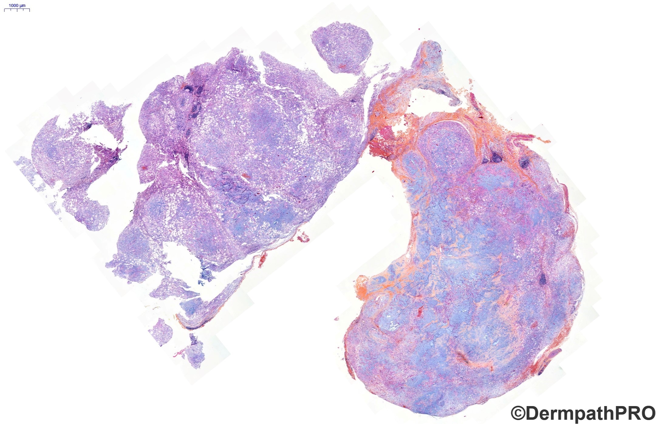
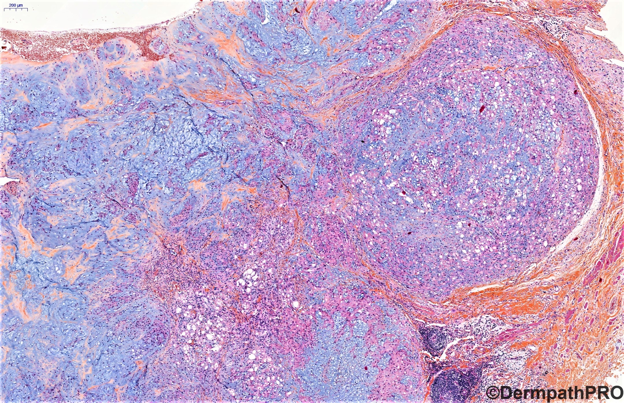
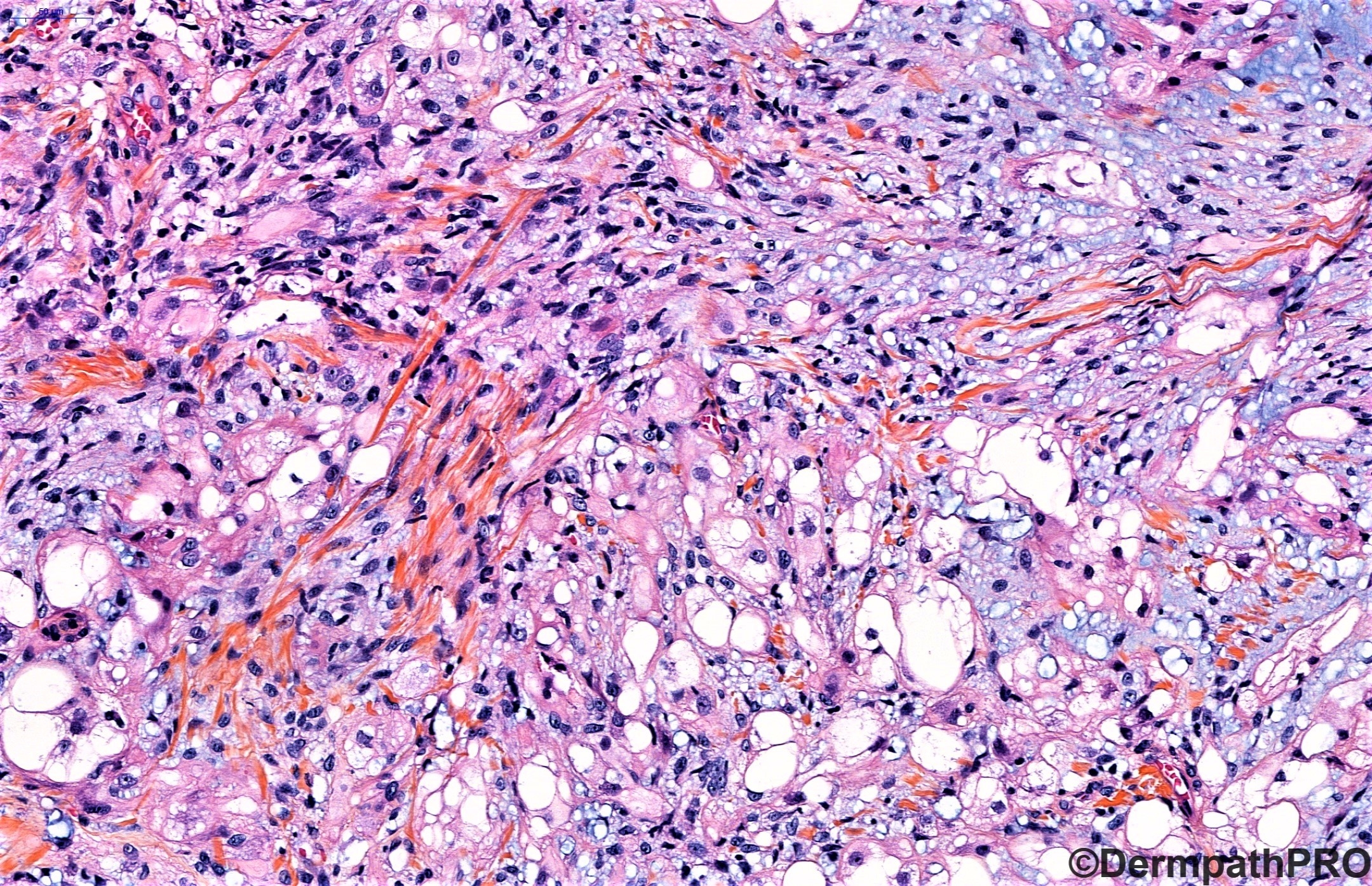
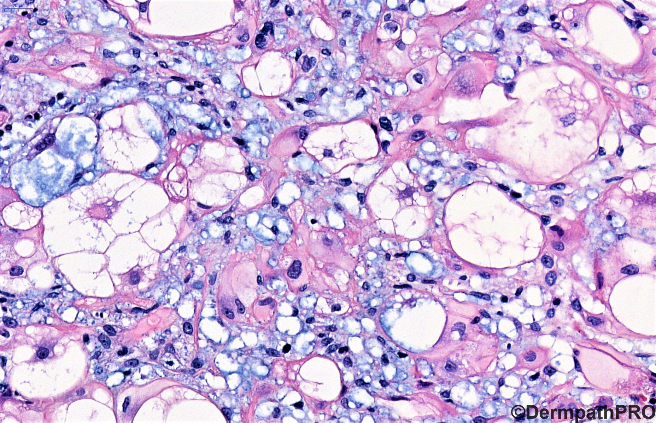
26y old male presenting with tumor in the masseter muscle evolving for 2 years.


  Report Record

User Feedback


Alex-Ventura-Leon

Posted

Odd location but agree whit anh. 

Chordoma is my first choice. 

Share this comment


Link to comment
share_externally

Chordoma periphericum or parachordoma? Would be interesting to know Raul's opinion on this controversy. He must have done IHC for brachyury. 

Share this comment


Link to comment
share_externally

My Colleagues are really skill in soft tissue tumors. I long thought of a Myoepithelioma/mixed tumor, but have to suck it up and agree with them! 

12 hours ago, anh said:

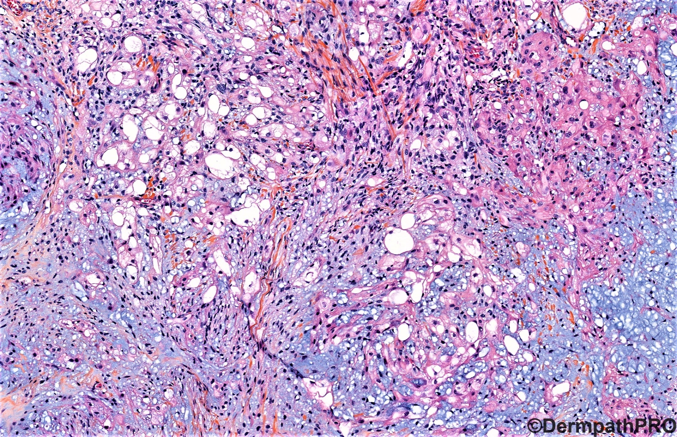
Definitely beautiful physaliphorous cells.

Yes! definitely phisaliphorous cells. As Anil said, Chordoma periphericum should be a better definition. 

Share this comment


Link to comment
share_externally

Vincenzo,

you might actually be right about myoepithelioma. Assuming this is not a metastatic chordoma (brachyury would help as Anil had mentioned), this tumor is probably a parachordoma / myoepithelioma. Some authors believe it's the same tumor (myoepithelioma) . I guess Raul will have some kind of molecular finding and explains the concept of parachordoma / myoepithelioma.

Share this comment


Link to comment
share_externally

Very interesting. Either metastatic chordoma or primary soft tissue chordoma. Clinical history is required in this case, I guess.

Share this comment


Link to comment
share_externally

Raul Perret

Posted (edited)

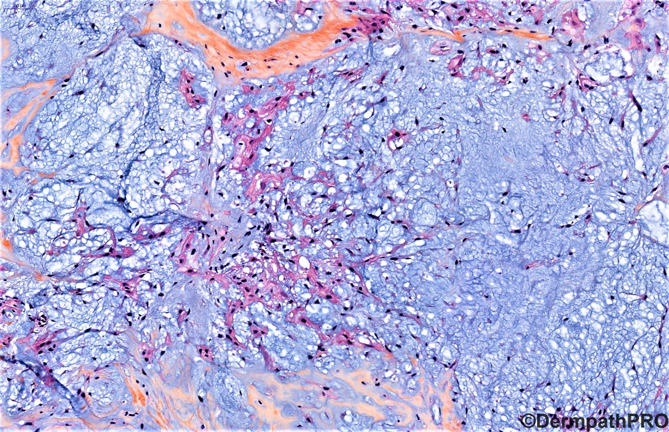
Yes, this is a case of extra-axial chordoma. In the original diagnosis I suggested to rule out the possibility of a metastasis from a primary axial chordoma. A CT-scan was performed which showed no tumor in the axial skeleton, proving that this was a true extra axial chordoma.

The term parachordoma has been used in the past for myoepithelial tumors showing massive vacuolation (hence simulating a chordoma). Some of these cases probably represented extra axial chordomas that were missed due to the absence of more specific markers like brachyury. Personally, I avoid using this terminology.

Interestingly, the diagnosis of the referring pathologist was a myoepithelioma, this is a comprehensible mistake due to the morphological and immunophenotypical overlap seen between this tumor and chordoma. The main clue is the physaliphorous cells and the expression of brachyury.

These tumors have overall a good prognosis with only rare cases developing metastasis (mainly lung). I suggest you to read this article for more information on this pathology.

Hope you guys liked the case and thanks for the comments.

Edited by Raul Perret

Share this comment


Link to comment
share_externally

Raul Perret

Posted

Here a little table I did with the "pearls" of this entity

cordoma (2).jpg

Share this comment


Link to comment
share_externally


Join the conversation

You can post now and register later. If you have an account, sign in now to post with your account.

Guest
Add a comment...

×   Pasted as rich text.   Paste as plain text instead

  Only 75 emoji are allowed.

×   Your link has been automatically embedded.   Display as a link instead

×   Your previous content has been restored.   Clear editor

×   You cannot paste images directly. Upload or insert images from URL.

Loading...

×
×
  • Create New...