Jump to content
In this section we have spot diagnoses posted on a daily basis since June 2010, now over 4000! You can review the archived cases and read the suggested diagnoses by users and the final comment by the contributors.
Case are uploaded each week day by 10 am UK time with the correct diagnosis will generally be posted at 8 pm UK time. Why not view the most recent spot diagnosis and proffer a diagnosis?

Case Number : Case 2199 - 13 November 2018 Posted By: Iskander H. Chaudhry

Please read the clinical history and view the images by clicking on them before you proffer your diagnosis.
Submitted Date :
   (0 reviews)

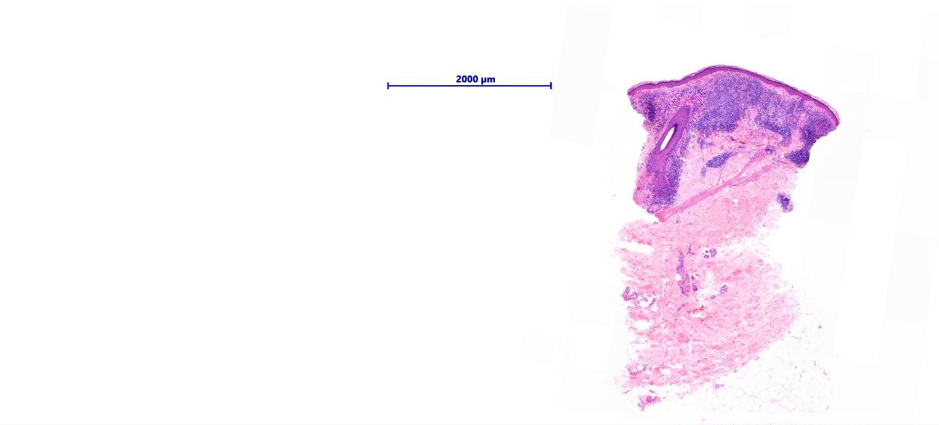
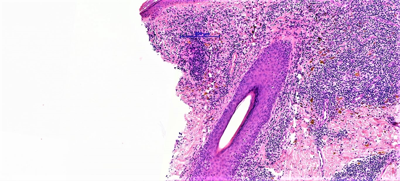
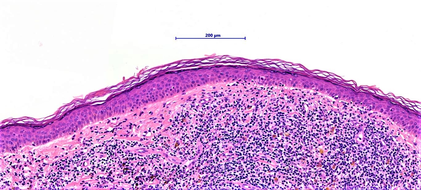
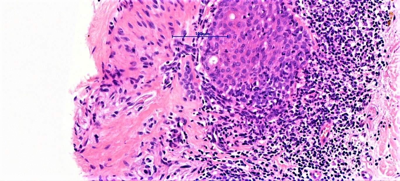
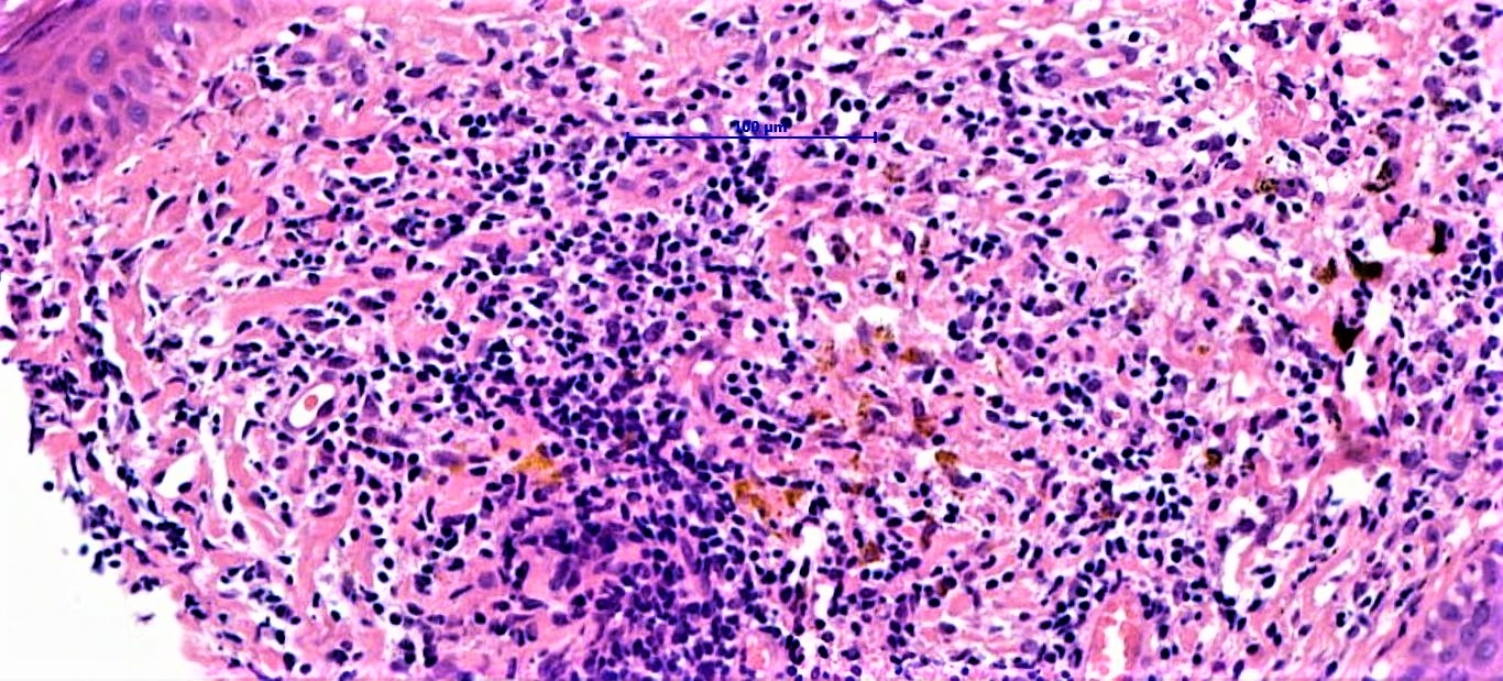
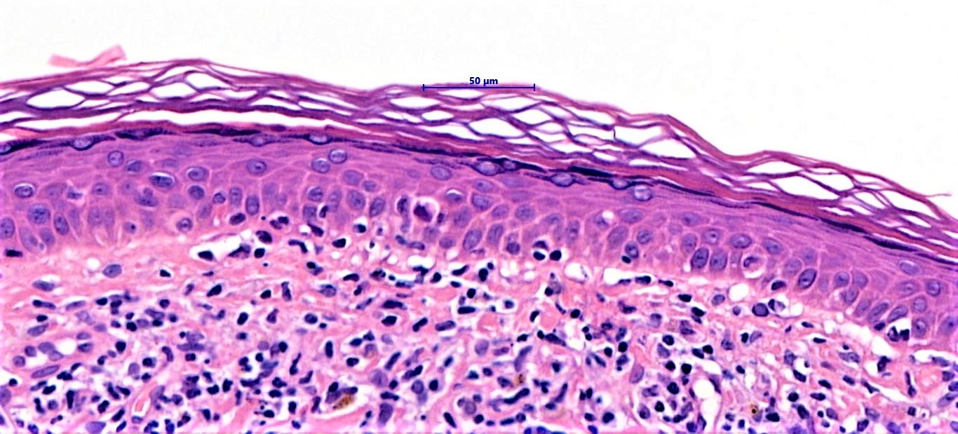
M48. Lesion right upper arm


  • Like 1
  Report Record

User Feedback


Alex-Ventura-Leon

Posted

Yes. It's my first possibility too. Folliculotropic MF. Agree. 

Share this comment


Link to comment
share_externally

Agree with Colleagues. LED as dd, but no dermal mucin and last pic is more in keeping with FMF. 

Share this comment


Link to comment
share_externally

Admin_Dermpath

Posted

Response from Dr Chaudhry:


This was a solitary lesion - any other suggestions ? 

Share this comment


Link to comment
share_externally

Yes Glauco. May be this is a tattoo ( fig 6 fit well ). Congrats for your skill.

Share this comment


Link to comment
share_externally

Arti Bakshi

Posted

?Lichenoid keratosis ?pseudo lymphomatous folliculitis

Share this comment


Link to comment
share_externally

Saman Fatah

Posted (edited)

A myraid of tissue reactions in a tattoo can occur and sometimes a combination of more than one pattern is seen.Clinically they look very similar with apperance of a papule/nodule or thickening within the tattoo.

This looks like a dense reactive lymphoid infiltrate with focal lichenoid/interface changes. “Pseudolymphoma” is commonly used in various case reports or small series to describe this reaction.

Usually to red colour tattoos (but certainly can be seen with other colours) as one of the hypothesis is a reaction to the mercury compounds in a red dye.

Was this a red tattoo or another colour? 


Edited by Saman Fatah

Share this comment


Link to comment
share_externally

Dr. Richard Carr

Posted

I thought it could be a pseudolymphomatous reaction to tattoo pigment. 

Share this comment


Link to comment
share_externally

Iskander H. Chaudhry

Posted

Dear all - no history of tatoo pigment and the pigment is positive for Perl's stain. The immuno' marks and clinical did not support folliculotropic MF - after discussion with clinicians the final diagnosis of lichen aureus was rendered. 

Share this comment


Link to comment
share_externally

Dr. Richard Carr

Posted

Oh dear we forgot haemosiderin! Nice reminder of lichen aureus.

Share this comment


Link to comment
share_externally


Join the conversation

You can post now and register later. If you have an account, sign in now to post with your account.

Guest
Add a comment...

×   Pasted as rich text.   Paste as plain text instead

  Only 75 emoji are allowed.

×   Your link has been automatically embedded.   Display as a link instead

×   Your previous content has been restored.   Clear editor

×   You cannot paste images directly. Upload or insert images from URL.

Loading...

×
×
  • Create New...