Jump to content
In this section we have spot diagnoses posted on a daily basis since June 2010, now over 4000! You can review the archived cases and read the suggested diagnoses by users and the final comment by the contributors.
Case are uploaded each week day by 10 am UK time with the correct diagnosis will generally be posted at 8 pm UK time. Why not view the most recent spot diagnosis and proffer a diagnosis?

Case Number : Case 2206 - 22 November 2018 Posted By: Raul Perret

Please read the clinical history and view the images by clicking on them before you proffer your diagnosis.
Submitted Date :
   (0 reviews)

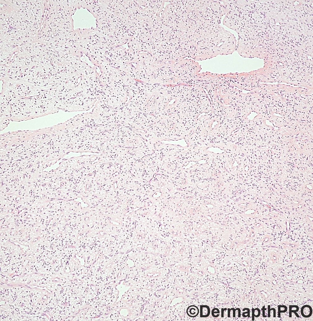
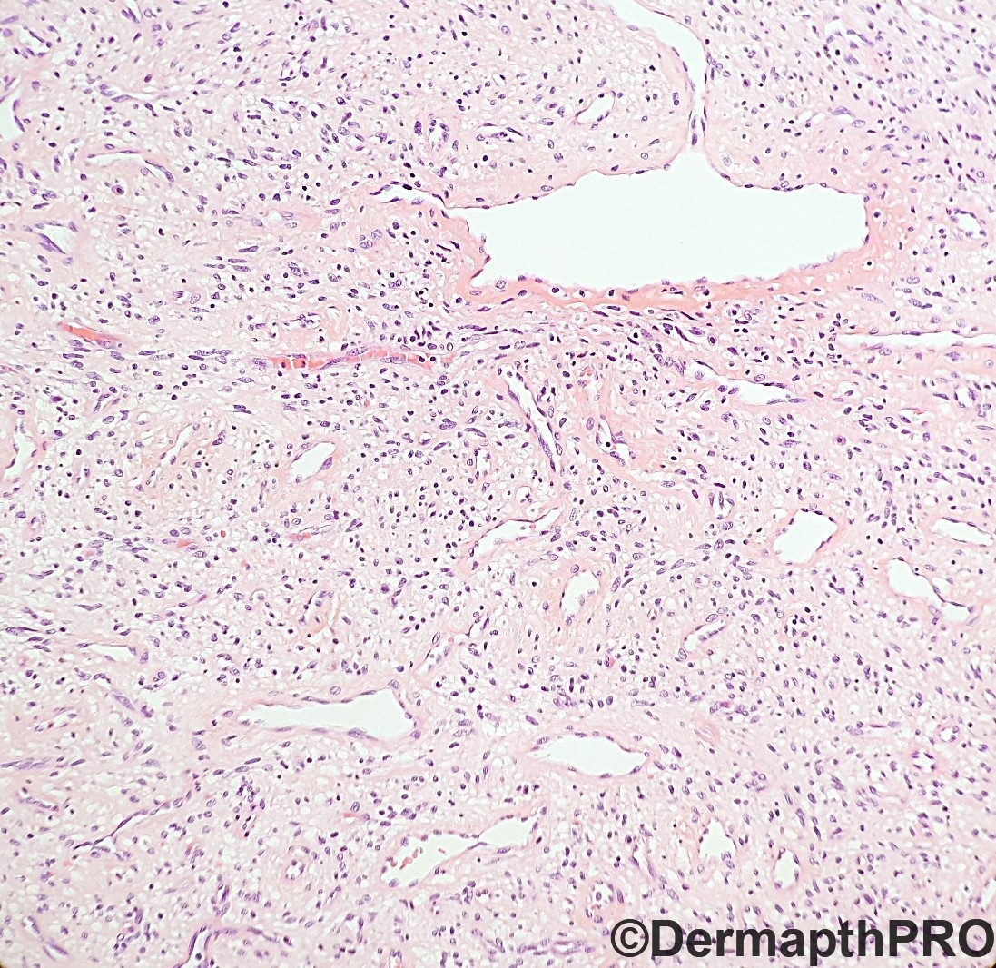
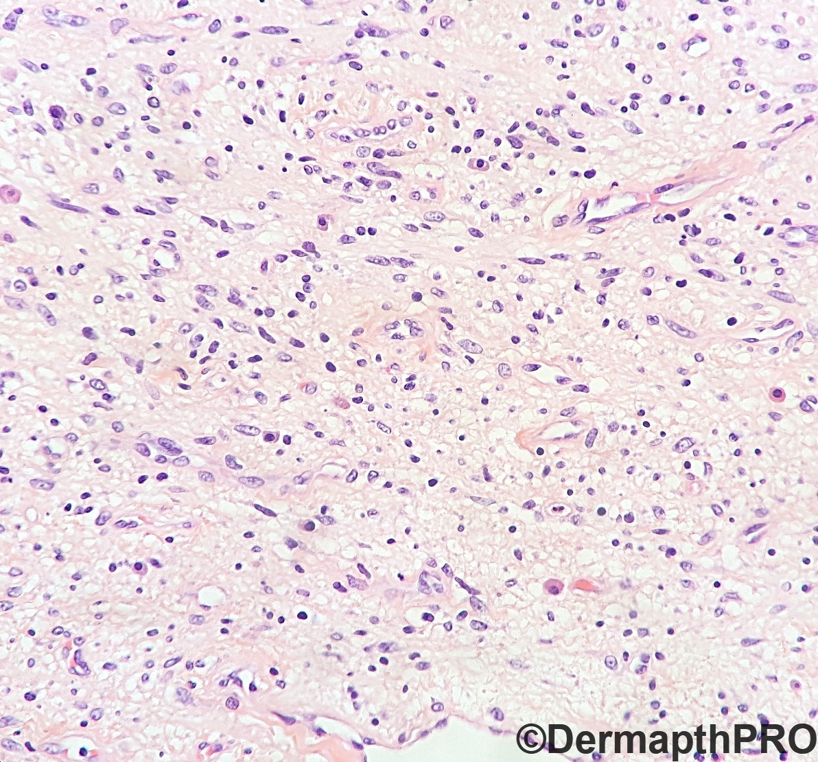
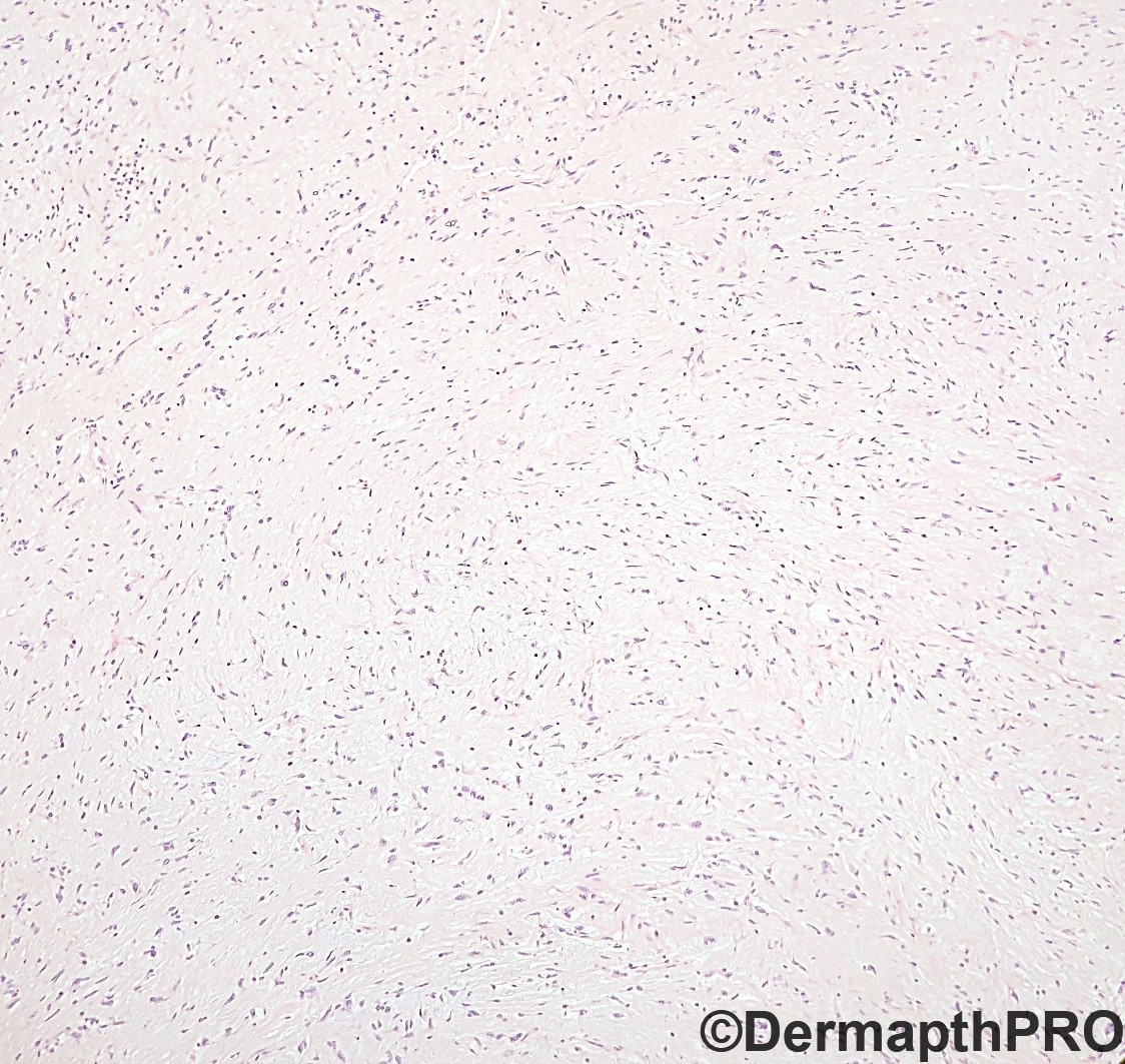
Male, 72 y deep thigh mass.


  • Like 1
  Report Record

User Feedback


vincenzo

Posted (edited)

There three histologic subtypes of angioleiomyoma(vascular  leiomyoma): solid, venous and cavernous. Thick vessels in venous but thin in solid variant. Odd age but my first spot is angioleiomyoma.  

Edited by vincenzo

Share this comment


Link to comment
share_externally

Alex-Ventura-Leon

Posted

I dont have a spot here. Difficult case.

I would add to the differential: Low grade fibromyxoid sarcoma and least likely Myxoid neurofibroma.

Share this comment


Link to comment
share_externally

The vascular pattern is very important in soft tissue tumor in addition to the architecture and cellular cytology. My experience is many soft tissue lesions are just spindle cell tumor so the cytology sometimes does not help. I can see some hemangiopericytoma-like vessels in the tumor so SFT is definitely in the differential diagnosis. So I would do STAT6.

On the other hand, there are too many small capillary-size vessels in the background in addition to the hemangiopericytoma-like vasculature, particularly in Fig 1 and Fig 2. SFT usually does not have so many small vessels like that. So I would like to raise the possibility of a soft tissue angiofibroma in the differential diagnosis. But for sure we need some immunos to guide us.

Share this comment


Link to comment
share_externally

Raul Perret

Posted

FiSH NCOA2: absence of rearrangement. 

Rest of IHC panel

EMA : neg
CD34 : neg
SMA : neg
Desmin : neg
Caldesmone : neg
MYOD1 : neg
MDM2 : neg
MUC4 : neg
STAT6 : neg
RP : neg
Ki67 : 2 %

Share this comment


Link to comment
share_externally

Too cellular for aggressive angiomyxoma. Raul did FISH NCOA2 so I guess he was also considering soft tissue angiofibroma. Actually ER is positive in soft tissue angiofibroma, at least according to one study. Considering that everything else is negative, I would still favor soft tissue angiofibroma in spite of negative FISH NCOA2 (if it's positive, that would be great. If it's negative, it's still Ok because not all soft tissue angiofibromas harbor that translocation).

Share this comment


Link to comment
share_externally

vincenzo

Posted (edited)

Yes. After immuno agree with Anh. Although I would have rather preferred leiomioma.  

Edited by vincenzo

Share this comment


Link to comment
share_externally

Raul Perret

Posted (edited)

Thank you for the great comments and particularly to Anh for the great description. I did think this was morphologically a good candidate for soft tissue angiofibroma initially but due to the absence of NCOA2 rearrangement I did not favour it (rearrangement is seen in most tested cases).

In the report, I mentioned this was a benign spindle cell lesion with a prominent vascular component which due to the absence of NCOA2 could represent a cellular angiofibroma.

Cellular angiofibroma and soft tissue angiofibroma are closely related entites showing overlapping morphologic and phenotypic features ref, After your great discussion, I agree that the topography, the branching vessels and variable cellularity are more in keeping with a soft tissue angiofibroma and I should have probably favoured this diagnosis over cellular angiofibroma despite the absence of NCOA2 rearragenment. 

Thank you again for sharing your wise comments that help us learn and become better pathologists.

Edited by Raul Perret

Share this comment


Link to comment
share_externally


Join the conversation

You can post now and register later. If you have an account, sign in now to post with your account.

Guest
Add a comment...

×   Pasted as rich text.   Paste as plain text instead

  Only 75 emoji are allowed.

×   Your link has been automatically embedded.   Display as a link instead

×   Your previous content has been restored.   Clear editor

×   You cannot paste images directly. Upload or insert images from URL.

Loading...

×
×
  • Create New...