Jump to content
In this section we have spot diagnoses posted on a daily basis since June 2010, now over 4000! You can review the archived cases and read the suggested diagnoses by users and the final comment by the contributors.
Case are uploaded each week day by 10 am UK time with the correct diagnosis will generally be posted at 8 pm UK time. Why not view the most recent spot diagnosis and proffer a diagnosis?

Case Number : Case 2209 - 27 November 2018 Posted By: Uma Sundram

Please read the clinical history and view the images by clicking on them before you proffer your diagnosis.
Submitted Date :
   (0 reviews)

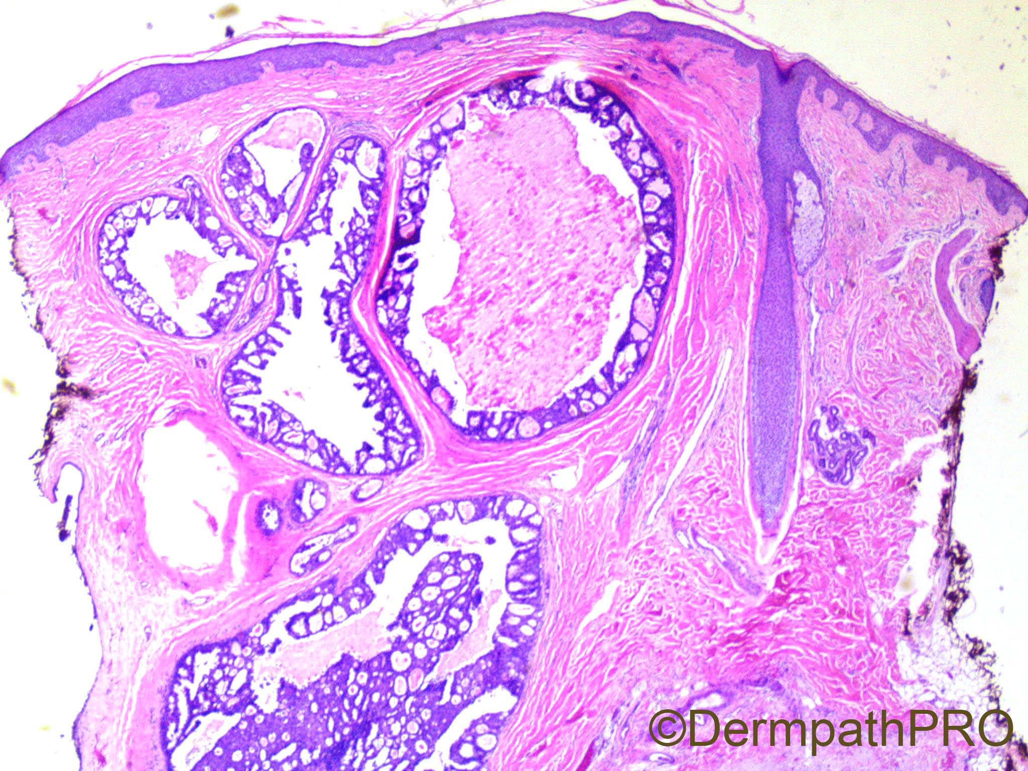
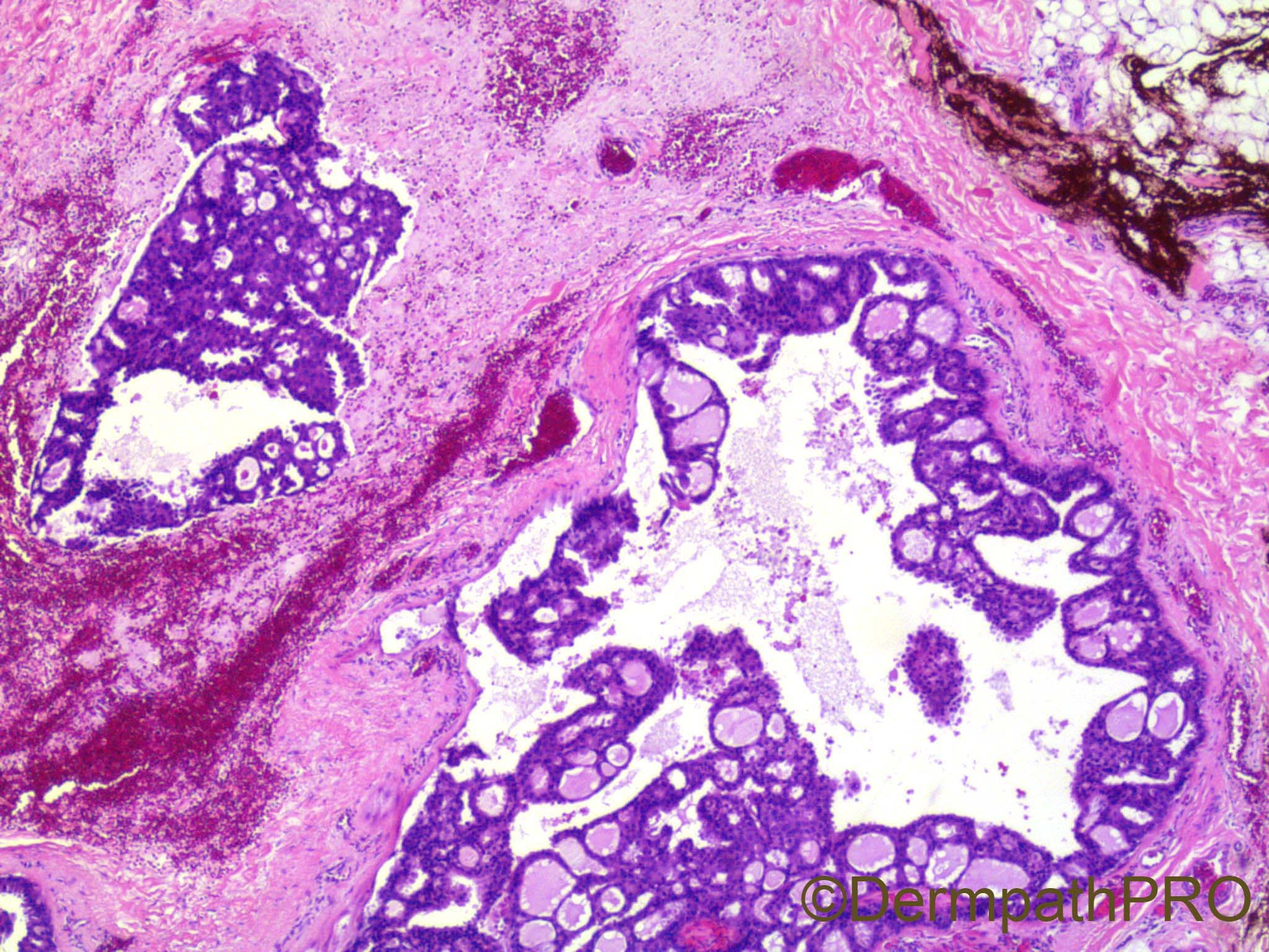
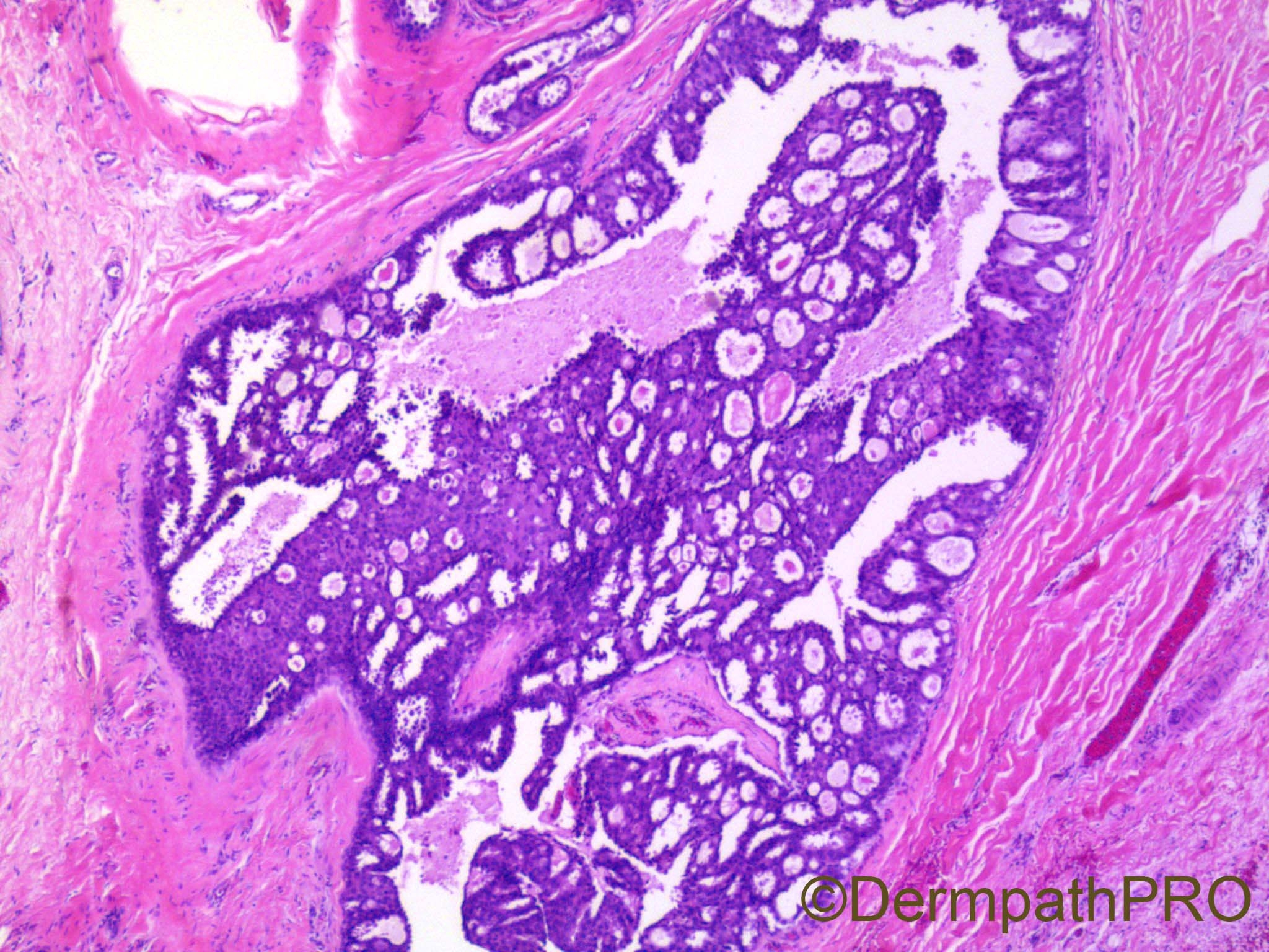
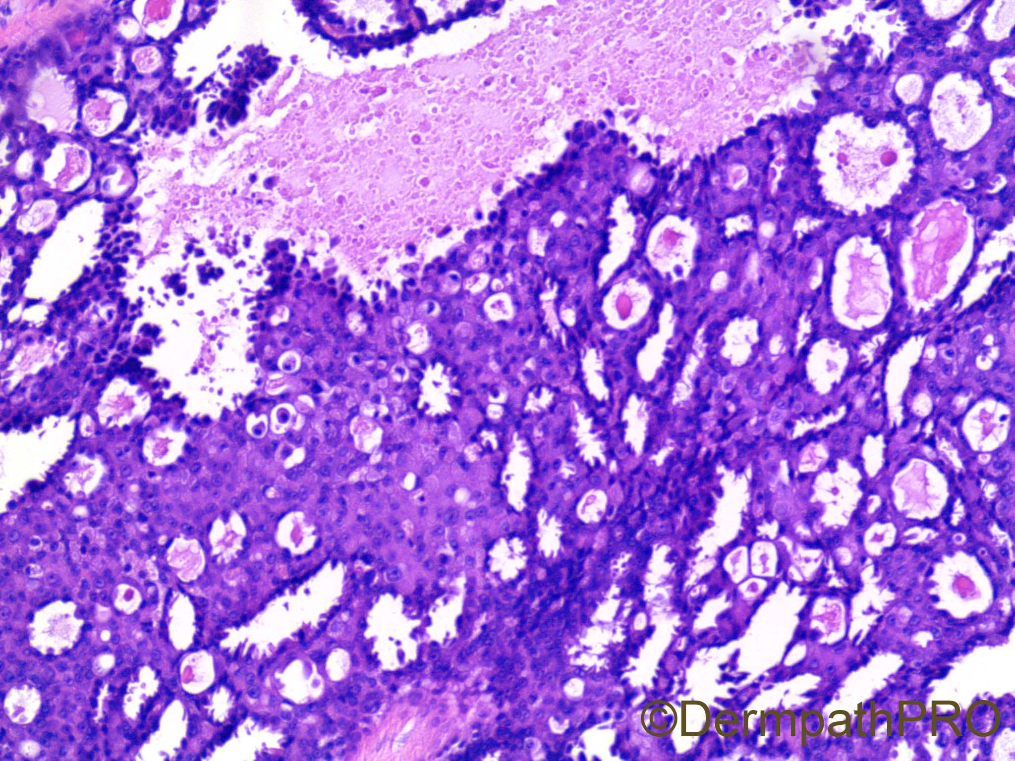
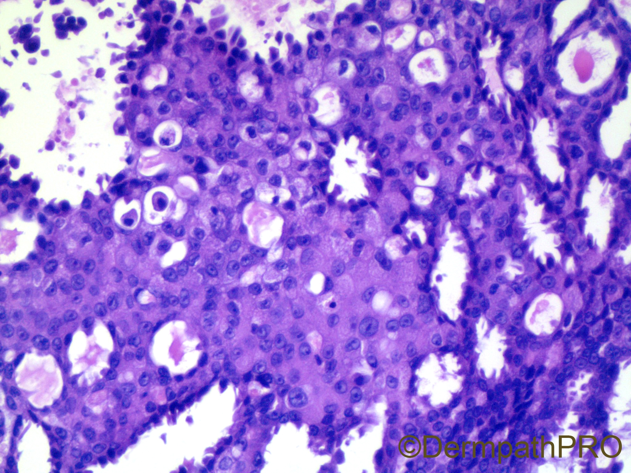
65 year old man with lesion on scalp. Rule out pilar cyst.


  Report Record

User Feedback


Mets vs Eccrine Ductal Ca vs Primary Cutaneous Mammary Analog Secretory Carcinoma With ETV6-NTRK3 Translocation ( rare )

Share this comment


Link to comment
share_externally

Krishnakumar subramanian

Posted

apocrine adenocarcinoma primary versus mets

Share this comment


Link to comment
share_externally

This is an in situ lesion, as marked by myoepithelial peripheral layer. Odd site for an EMPSGC but this is my first spot for now. 

Share this comment


Link to comment
share_externally

vincenzo

Posted (edited)

...unless this is a focal dermal component of a syringocystoadenocarcinoma...But I don't see other diagnostic findings of SCAC in this pictures. Apocrine changes are strict, but they don't definitely oppose to a diagnosis of EMPSGC, considering an apocrine origine is admitted...waiting for other comments|

Edited by vincenzo

Share this comment


Link to comment
share_externally

Alex-Ventura-Leon

Posted

Difficult. Apocrine Carcinoma is my first thought. Second, EMPSGC, and third Metastatic Carcinoma.

Share this comment


Link to comment
share_externally

Looks like some kind of in situ carcinoma first. But could it be that it's just a tubulopapillary hidradenoma?

Share this comment


Link to comment
share_externally

Uma Sundram

Posted

Great comments, everyone! This was a difficult case. We first did our best to rule out a metastasis. The patient's clinical work up was negative and markers like CDX2 and TTF1 were also negative. p63 is positive for a myoepithelial layer. With apocrine differentiation, it's very difficult to use cytology to make a diagnosis of malignancy. We finally settled on 'apocrine tumor with atypical features' and recommended a re excision. Neuroendocrine markers were negative. The patient went to another institution where a diagnosis of in situ carcinoma was made, which points to how difficult the case is, and how variable expert opinions can be.

Share this comment


Link to comment
share_externally

Dr. Richard Carr

Posted

Nice idea about mammary analogue secretary carcinoma.

Share this comment


Link to comment
share_externally


Join the conversation

You can post now and register later. If you have an account, sign in now to post with your account.

Guest
Add a comment...

×   Pasted as rich text.   Paste as plain text instead

  Only 75 emoji are allowed.

×   Your link has been automatically embedded.   Display as a link instead

×   Your previous content has been restored.   Clear editor

×   You cannot paste images directly. Upload or insert images from URL.

Loading...

×
×
  • Create New...