Jump to content
In this section we have spot diagnoses posted on a daily basis since June 2010, now over 4000! You can review the archived cases and read the suggested diagnoses by users and the final comment by the contributors.
Case are uploaded each week day by 10 am UK time with the correct diagnosis will generally be posted at 8 pm UK time. Why not view the most recent spot diagnosis and proffer a diagnosis?

Case Number : Case 2212 - 30 November 2018 Posted By: Dr. Richard Carr

Please read the clinical history and view the images by clicking on them before you proffer your diagnosis.
Submitted Date :
   (0 reviews)

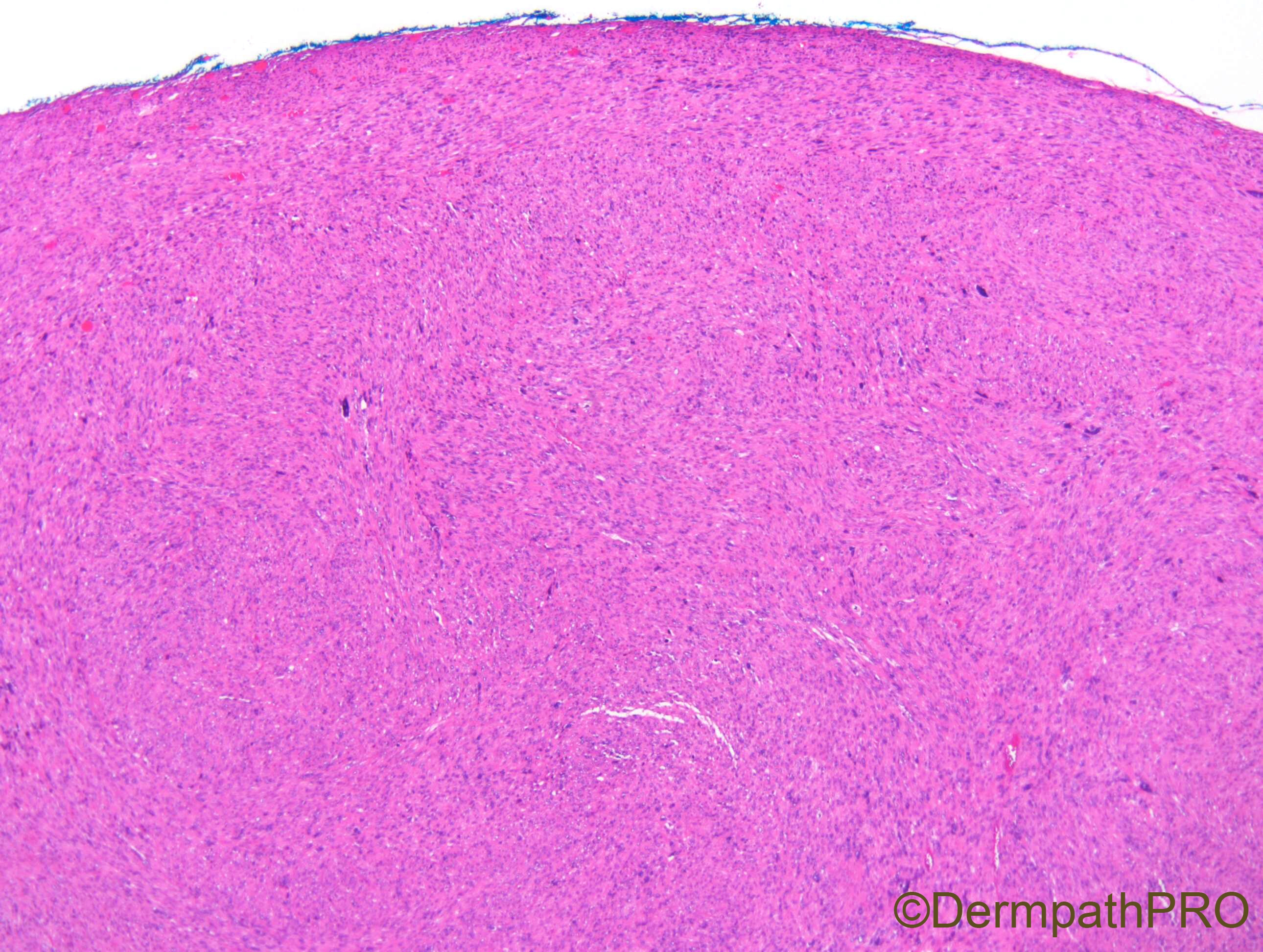
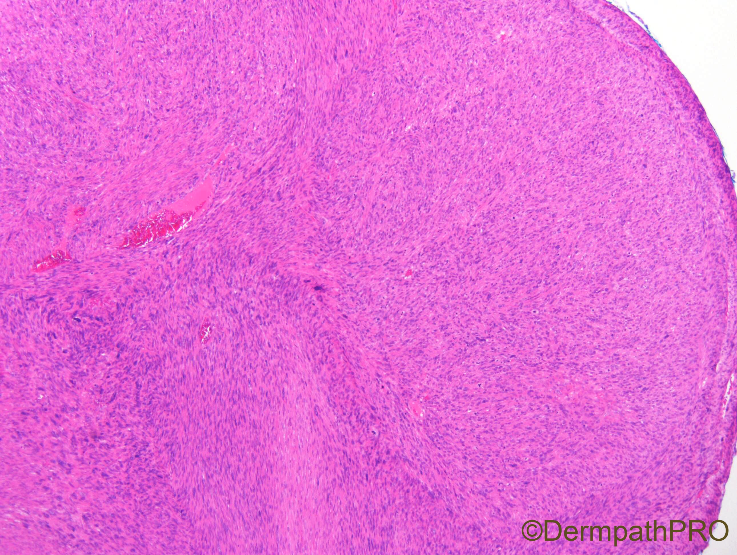
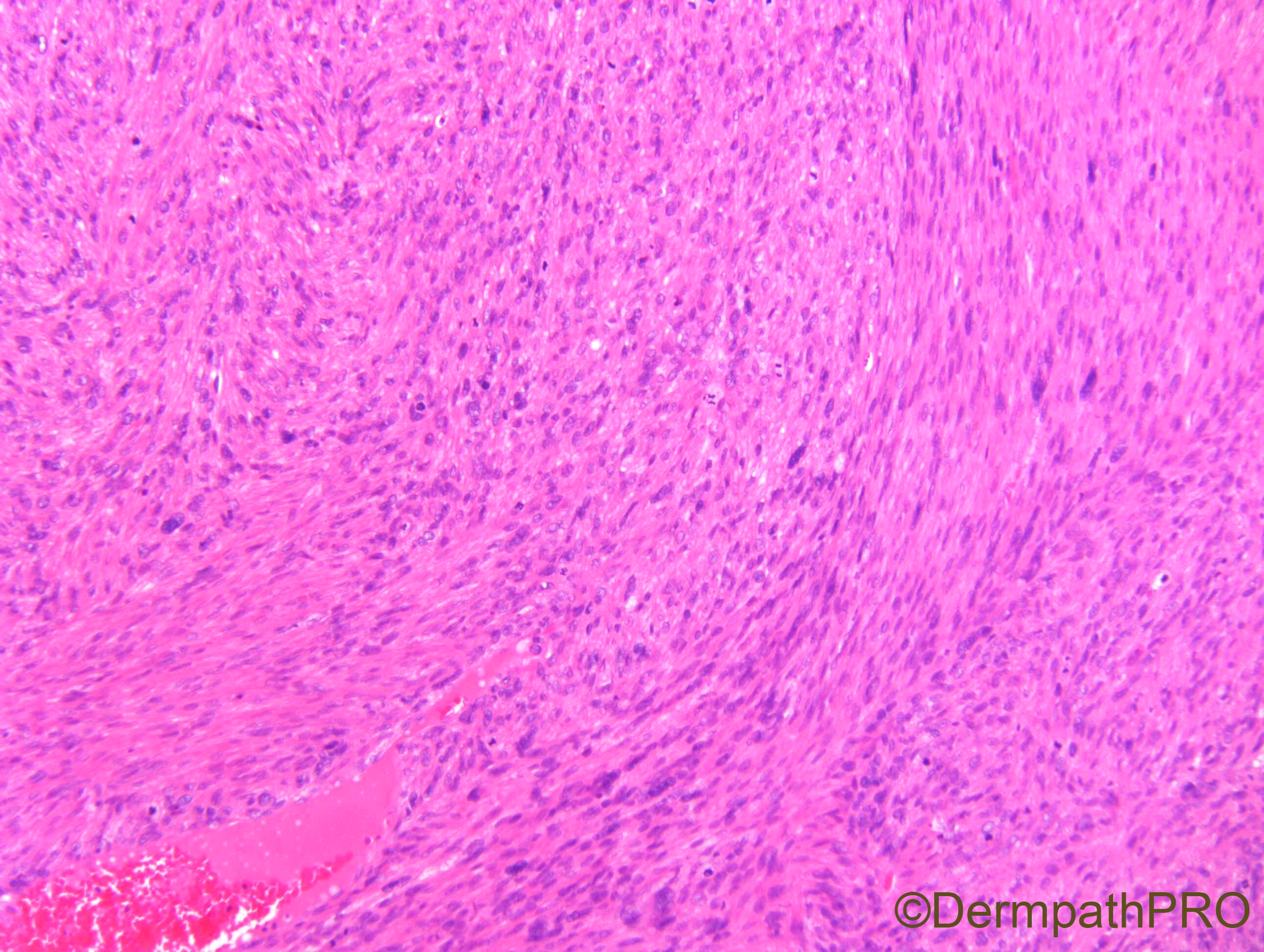
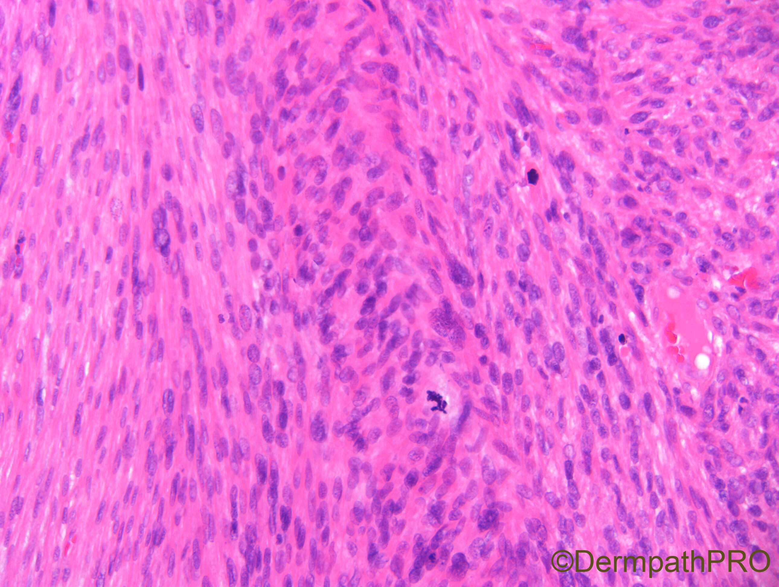
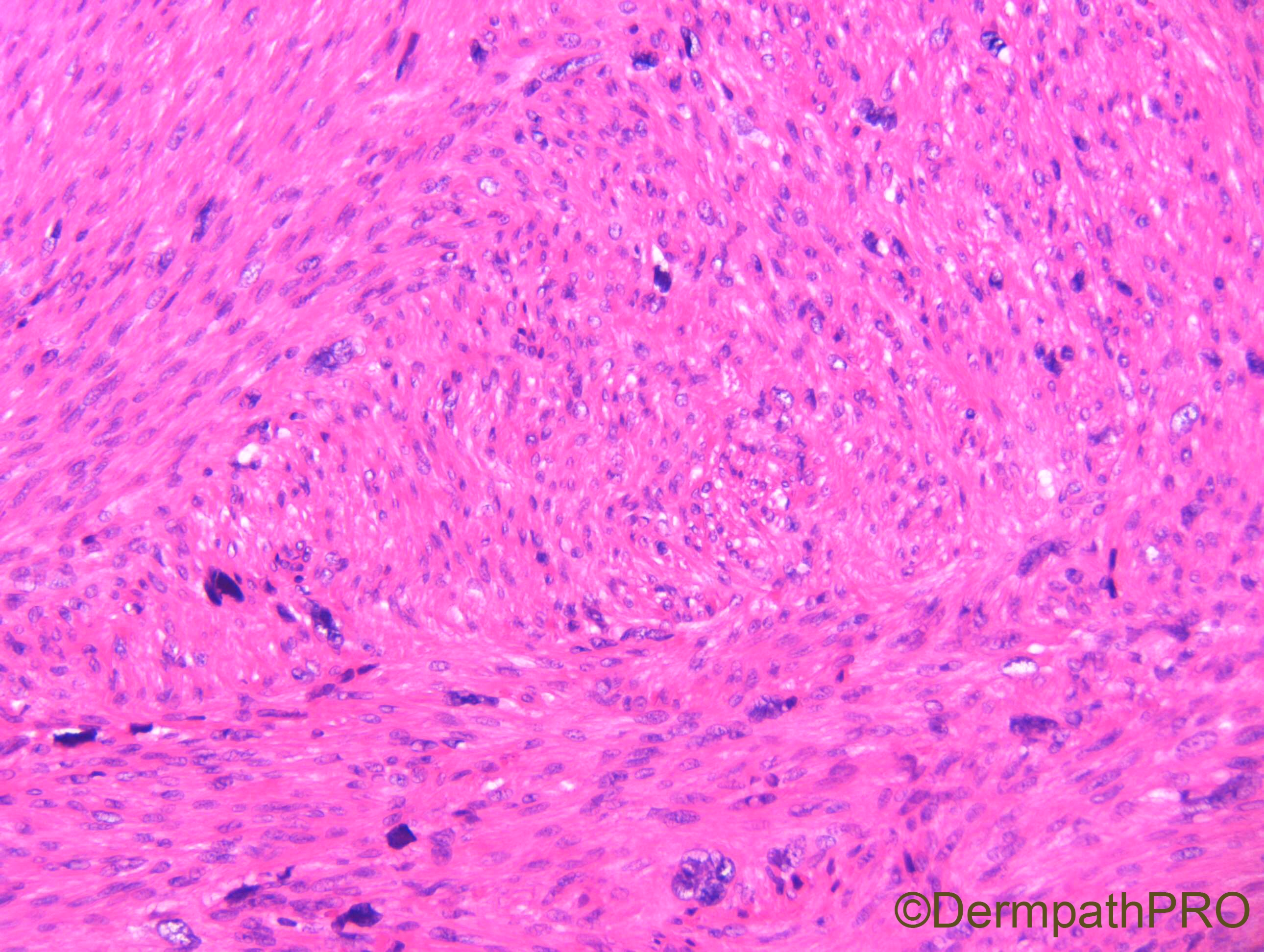
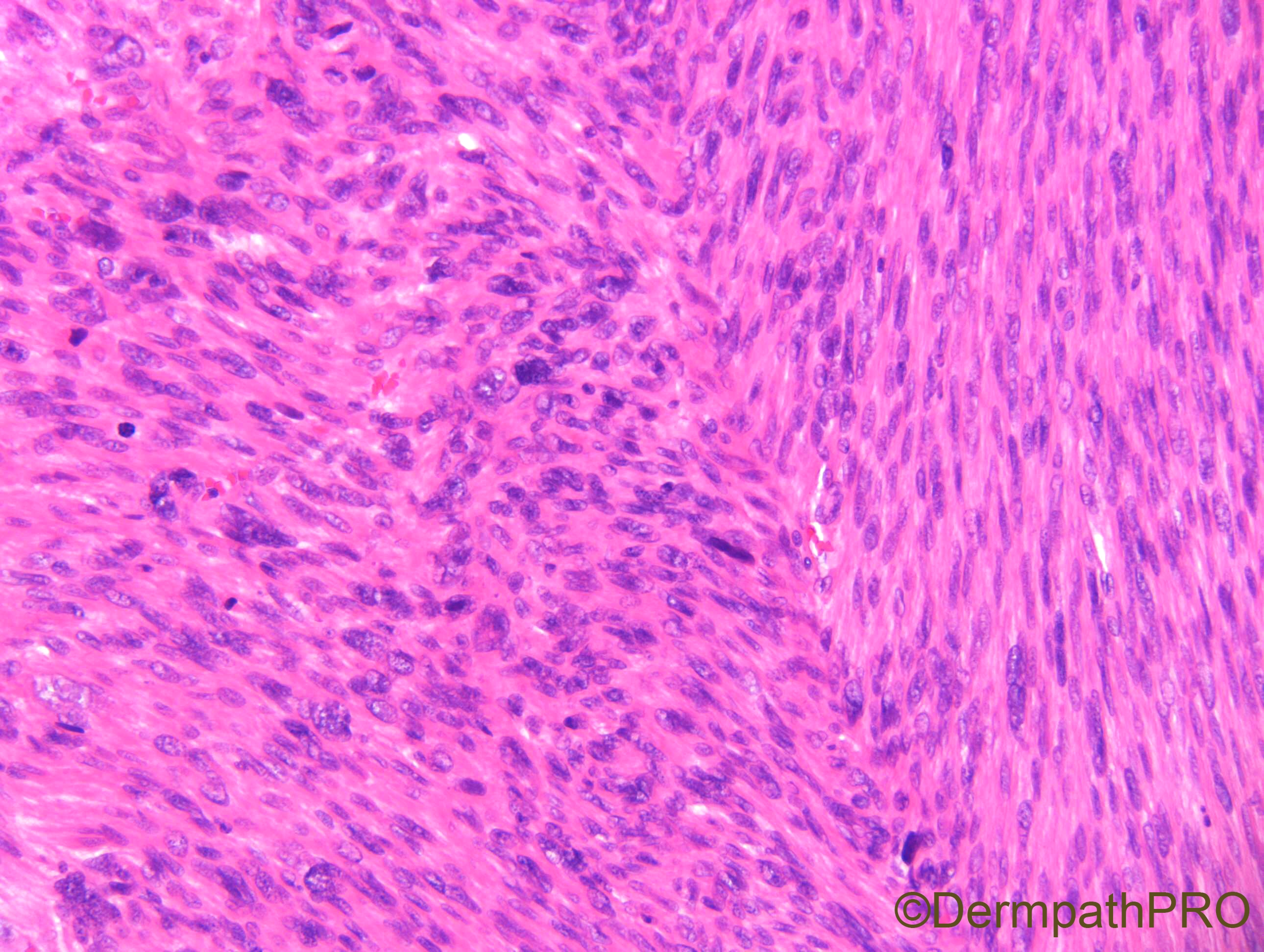
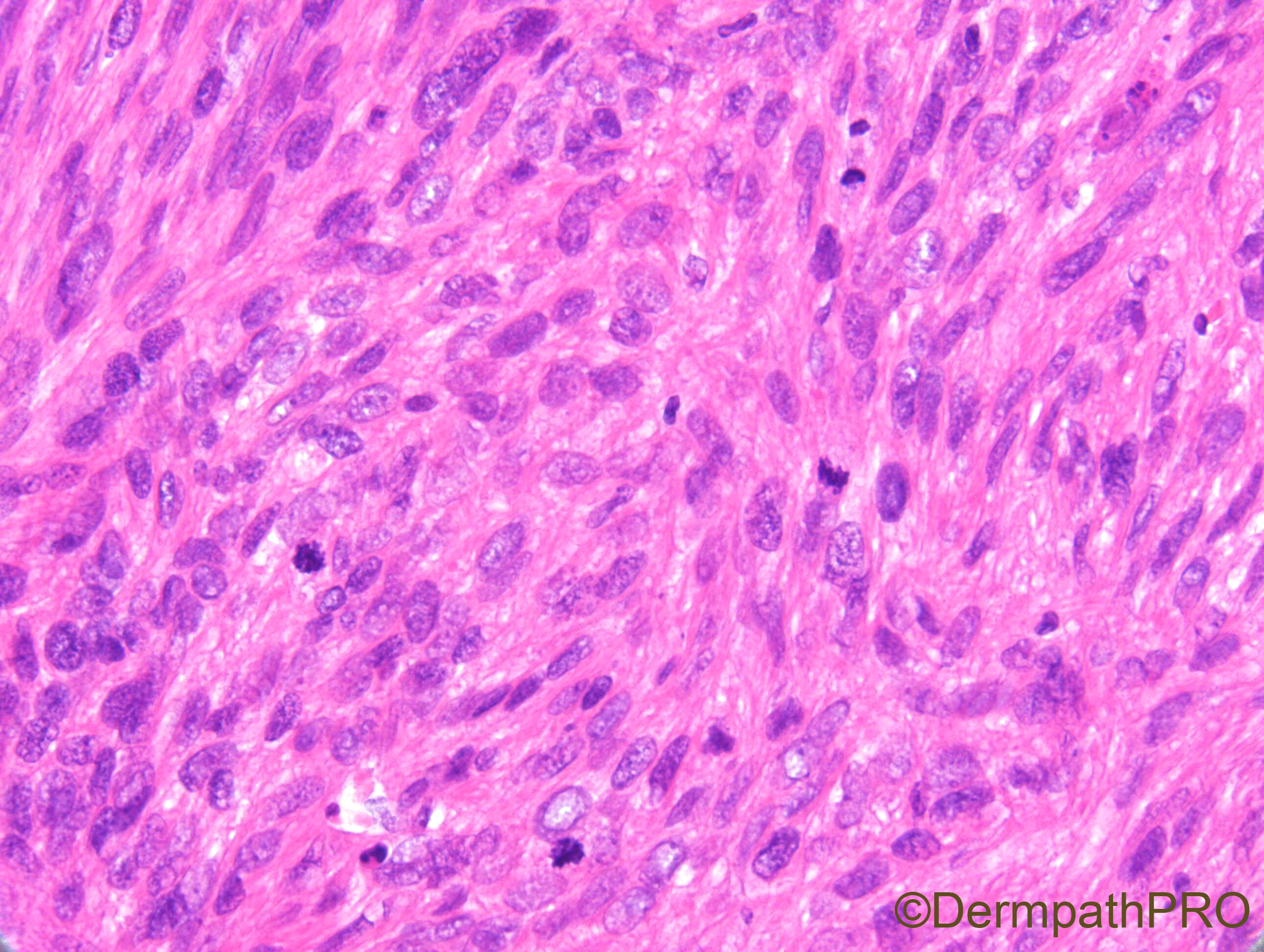
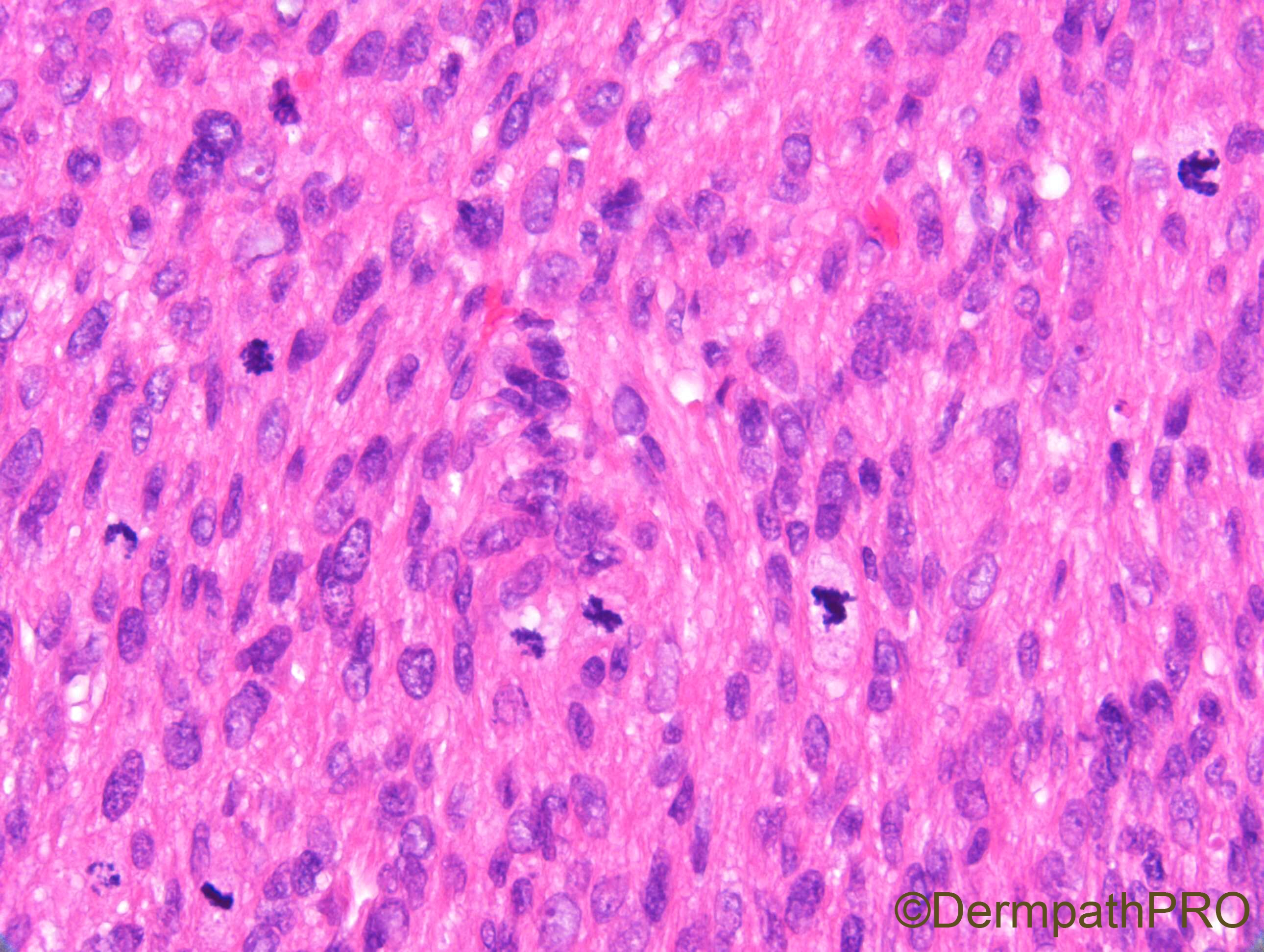
F70. Vulval cystic nodule.


  Report Record

User Feedback


Krishnakumar subramanian

Posted

malignant spindle cell tumor-Leiomyosarcoma

Share this comment


Link to comment
share_externally

Alex-Ventura-Leon

Posted

Atypical smooth muscle neoplasm. It seem very well circunscribed and i would like to know the total size of the lesión.

But Agree. My first possibility is Leiomyosarcoma.

Share this comment


Link to comment
share_externally

Only two criteria for leiomyosarcoma: atypia and mitosis. Unknown size and margins. But there are atypical mitosis also, so agree with Collegues. 

Share this comment


Link to comment
share_externally

Dr. Richard Carr

Posted

It was about 2.5cm if that changes things?

Share this comment


Link to comment
share_externally

Krishnakumar subramanian

Posted

atypical intradermal smooth muscle neoplasm can it be called like this instead of a sarcoma

Share this comment


Link to comment
share_externally

To answer Dr. Carr's question, size does change things dependent on what system you use to classify vulvar smooth muscle neoplasms. Remember smooth muscle neoplasms of the OB/GYN tract are classified based on a different set of histologic criteria (uterus or vulva) compared to non OB/GYN smooth muscle neoplasms. Some smooth muscle neoplasms of the uterus that are called "leiomyoma" might be classified as leiomyosarcoma in soft tissue or other extrauterine site / organs. So you need to know exactly what criteria you are using for these tumors.

I read this article not too long ago and you might find it useful when dealing with smooth muscle neoplasm of the vulva. Dermatopathologists usually don't do non-skin anatomic pathology so sometimes it's useful to show the cases to your colleagues of other subspecialties.

Criteria for Risk Stratification of Vulvar and Vaginal Smooth Muscle Tumors: An Evaluation of 71 Cases Comparing Proposed Classification Systems

Smooth muscle tumors (SMTs) are the most common mesenchymal neoplasms of the vulva and vagina,1 yet their infrequency is demonstrated by the prevalence of published reports as individual cases or small series. The rarity of these tumors makes development of a prognostic classification system challenging. However, accurate risk stratification of tumors according to pathologic findings is critical for appropriate management of patients.

To date, 3 site-specific classification methods have been proposed: 2 for vulvar SMTs and 1 for vaginal SMTs (Table 1). The first sets of criteria were published in 1979 by Tavassoli and Norris2,3, 1 for vulvar SMTs and 1 for vaginal SMTs. Their review of 32 vulvar SMTs led to identification of 3 main risk features based on tumor recurrence: gross size ≥5 cm, mitotic index≥5 figures/10 high power fields (HPFs) and infiltrative tumor interface. Finding 2 of 3 features qualifies a tumor as low-grade leiomyosarcoma and the presence of all 3 features warrant a diagnosis of leiomyosarcoma. A similar review of 60 vaginal SMTs established varying combinations of risk features that included degree of cytologic atypia, mitotic index and infiltrative tumor interface. Tumors with moderate or severe cytologic atypia and mitotic index ≥5 figures/10 HPFs are designated leiomyosarcoma. Further, of the 5 recurrent tumors in their series, the 1 tumor that metastasized had infiltrative margins in contrast to the 4 other locally recurrent tumors that were circumscribed. It was suggested by the authors that the presence of infiltration alone warranted classification as leiomyosarcoma in vaginal SMTs until proven otherwise due to infiltration as a general indicator of more aggressive behavior in mesenchymal tumors.


  Capture.PNG.5f4d3eac8954979fe0df4cccb86bc47b.PNG 

Prognostic criteria were expanded in 1996 by Nielsen et al4 following review of 25 SMTs of the vulva. A set of 4 criteria incorporating gross and microscopic findings were proposed: gross size (≥5 cm), presence of infiltrative margin, mitotic index (≥5 mitoses/10 HPFs) and degree of cytologic atypia (moderate to severe). If 0 or 1 criterion was met, the tumor should be interpreted as leiomyoma; if 2 criteria were satisfied, the tumor should be interpreted as atypical leiomyoma; and if 3 or 4 criteria were fulfilled, the tumor should be interpreted as leiomyosarcoma.

Despite these proposed site-specific classification systems, it is our experience that many pathologists tend to apply criteria for SMTs of the uterus5 when evaluating vulvovaginal SMTs. The 2014 World Health Organization (WHO) Classification of Tumors of Female Reproductive Organs1 outlines 3 morphologic features to assess in uterine SMTs: degree of cytologic atypia, mitotic index and the presence of tumor cell necrosis. Most practitioners diagnose a tumor as leiomyosarcoma when at least 2 of these 3 features are identified, requiring that the degree of cytologic atypia be at least moderate and the mitotic index be a minimum of 10 figures/10 HPFs. Other combinations of these 3 components, whether due to a lesser degree of cytologic atypia, lower mitotic index or presence/absence of tumor cell necrosis, fall within a spectrum of SMTs such as SMT of uncertain malignant potential (STUMP), leiomyoma with bizarre nuclei and mitotically active leiomyoma, which have been correlated with potential for aggressive behavior from outcome-based studies of uterine SMTs.6–9

In addition, before 2004, morphologic criteria for risk stratification of ovarian SMTs were nonexistent. A review of 54 SMTs of the ovary by Lerwill et al10 led to validation of diagnostic categories and prognostic criteria of uterine SMTs. The authors concluded that, as in the uterus, ovarian leiomyosarcoma may be diagnosed when at least 2 of 3 criteria are satisfied: moderate to severe cytologic atypia, mitotic index in excess of 10 figures/10 HPFs and the presence of tumor cell necrosis.


Share this comment


Link to comment
share_externally

Dr. Richard Carr

Posted

The mitotic rate was >14/mm2 (~28/10 hpf) and there was moderate to severe cellular pleomorphism. Although this lesion is currently small I don't expect it would have stayed that way.

I called it leiomyosarcoma (it was pretty circumscript).

Great discussion - thanks.

Also I realised I had not posted response for Case 2202 - it was not BFH as most of you thought.

Share this comment


Link to comment
share_externally


Join the conversation

You can post now and register later. If you have an account, sign in now to post with your account.

Guest
Add a comment...

×   Pasted as rich text.   Paste as plain text instead

  Only 75 emoji are allowed.

×   Your link has been automatically embedded.   Display as a link instead

×   Your previous content has been restored.   Clear editor

×   You cannot paste images directly. Upload or insert images from URL.

Loading...

×
×
  • Create New...