Jump to content
In this section we have spot diagnoses posted on a daily basis since June 2010, now over 4000! You can review the archived cases and read the suggested diagnoses by users and the final comment by the contributors.
Case are uploaded each week day by 10 am UK time with the correct diagnosis will generally be posted at 8 pm UK time. Why not view the most recent spot diagnosis and proffer a diagnosis?

Case Number : Case 2172 - 5 October 2018 Posted By: Dr. Richard Carr

Please read the clinical history and view the images by clicking on them before you proffer your diagnosis.
Submitted Date :
   (0 reviews)

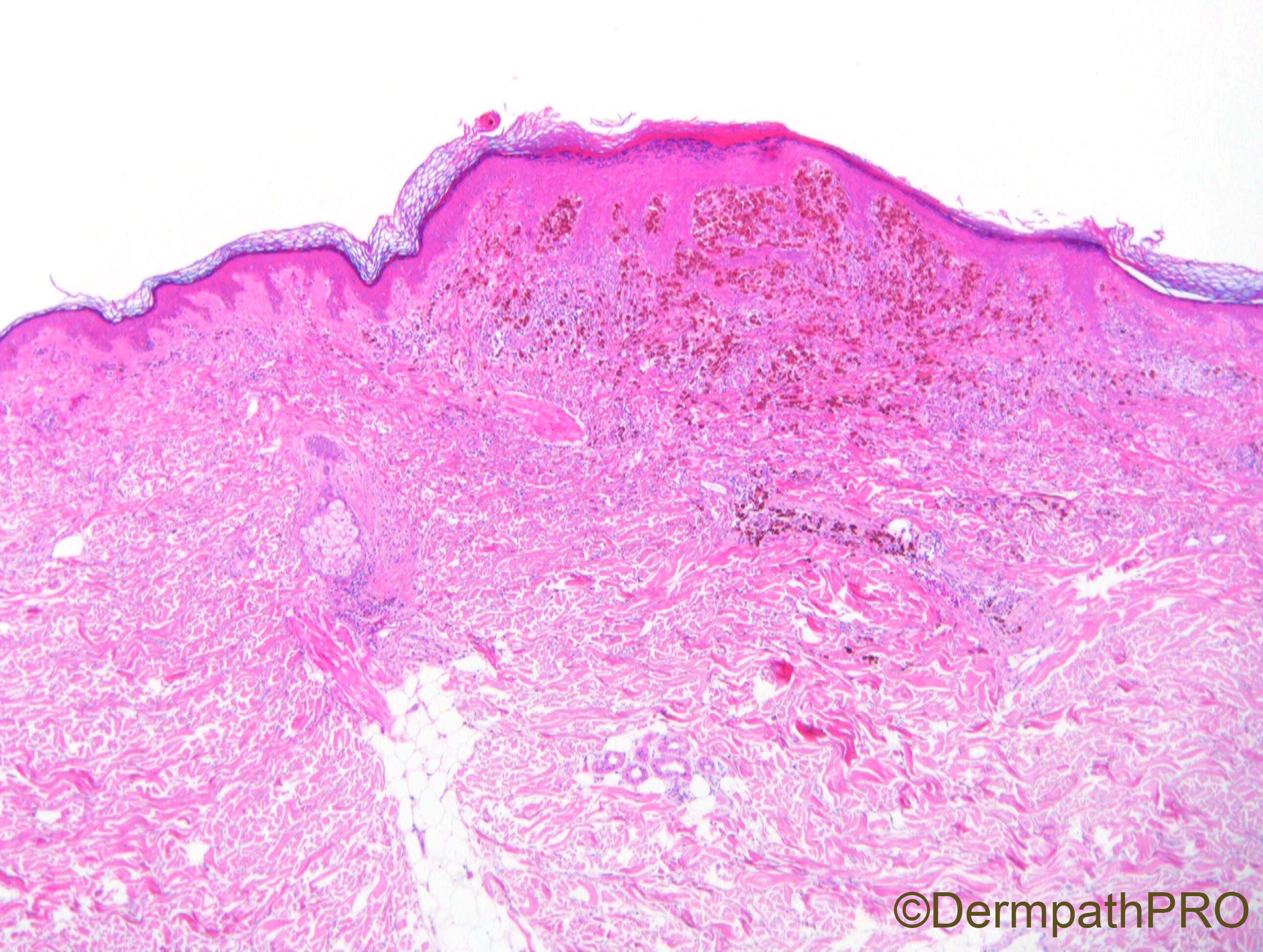
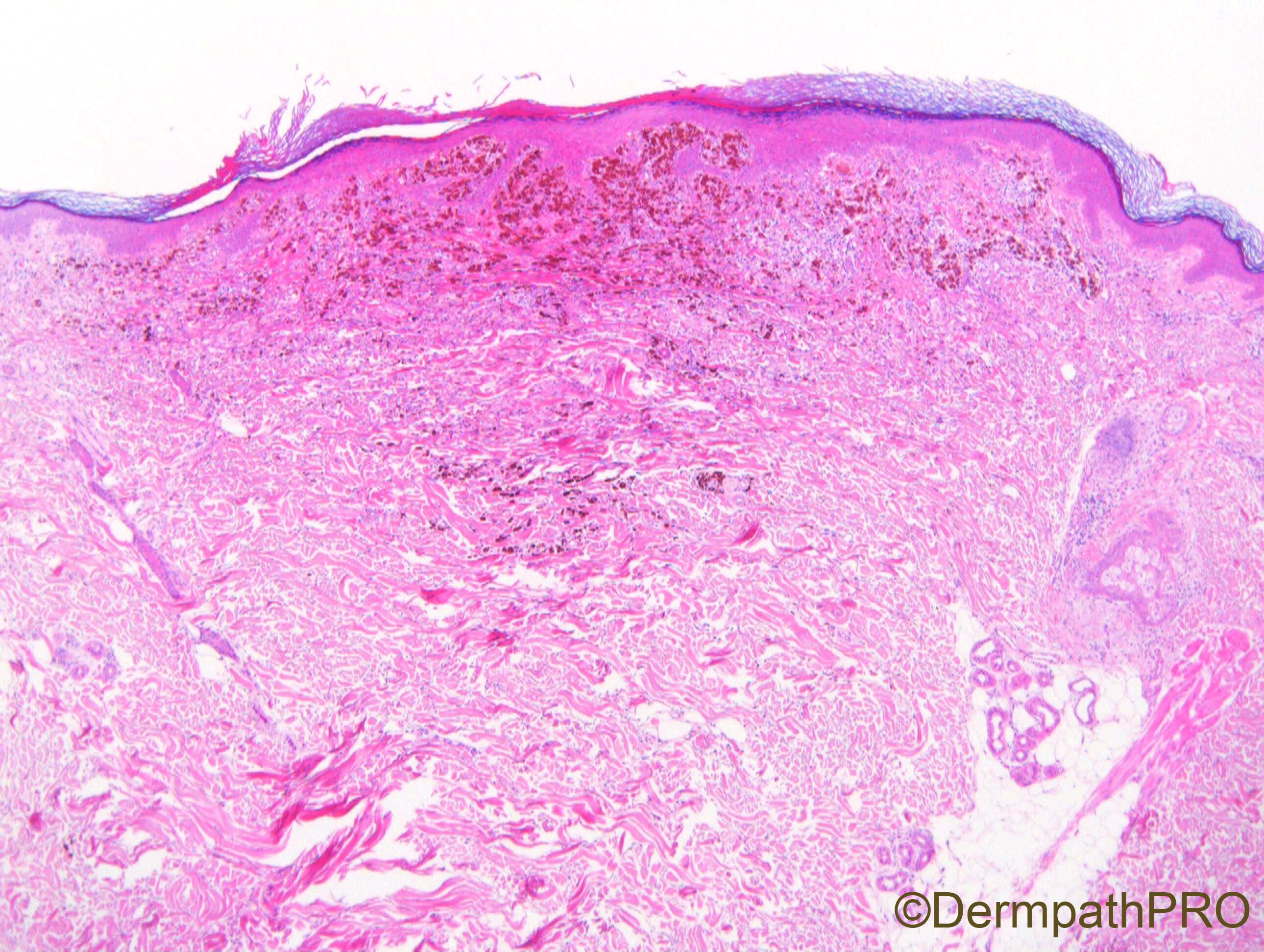
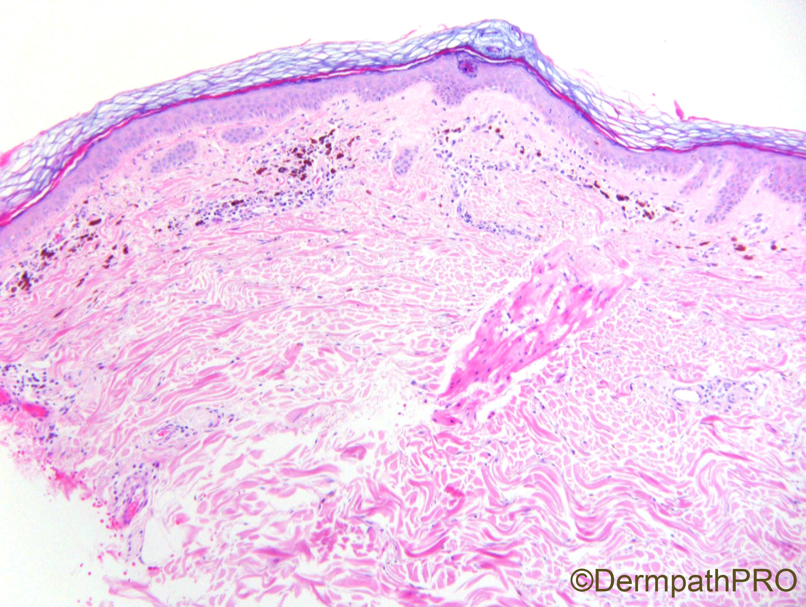
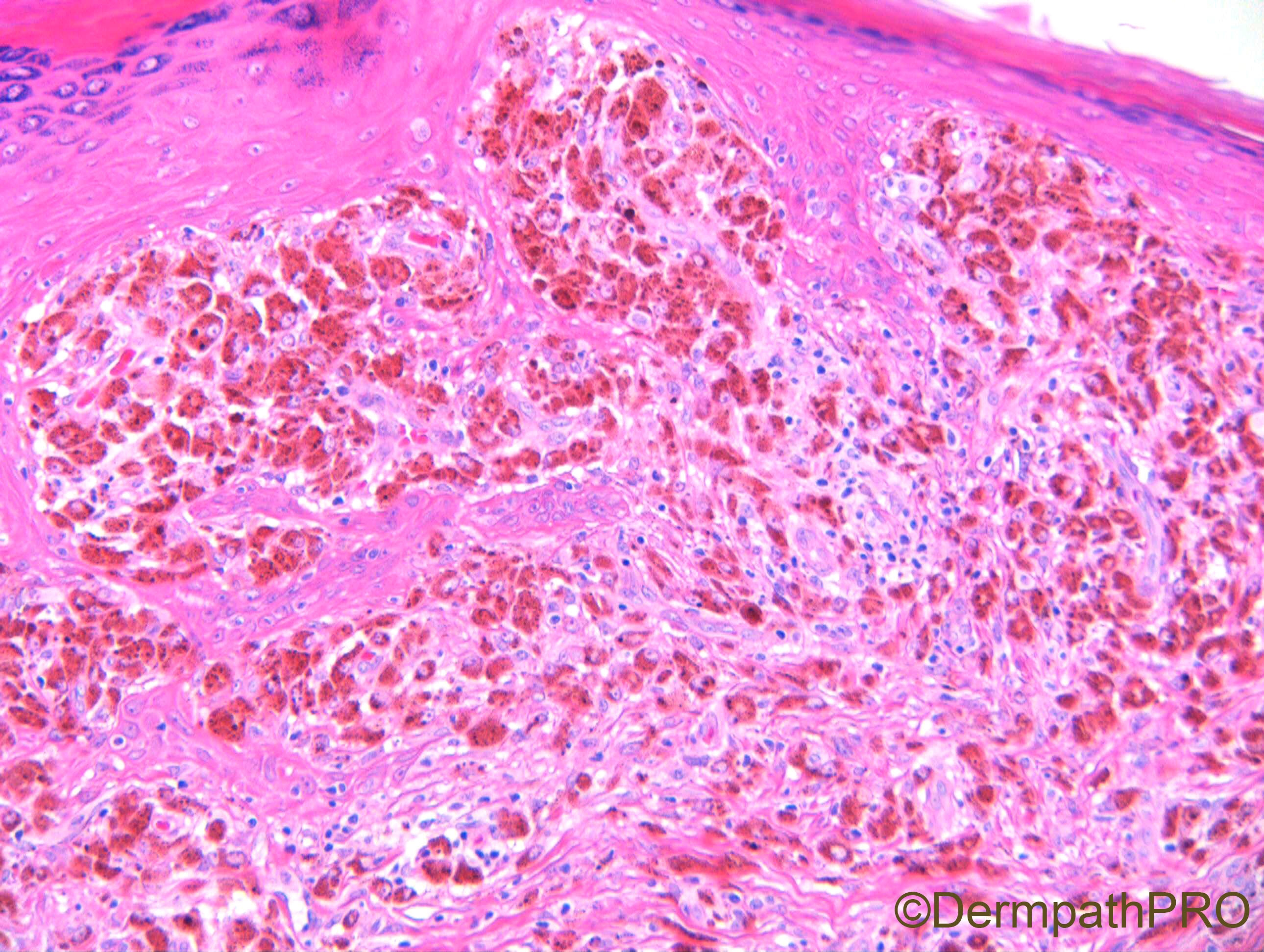
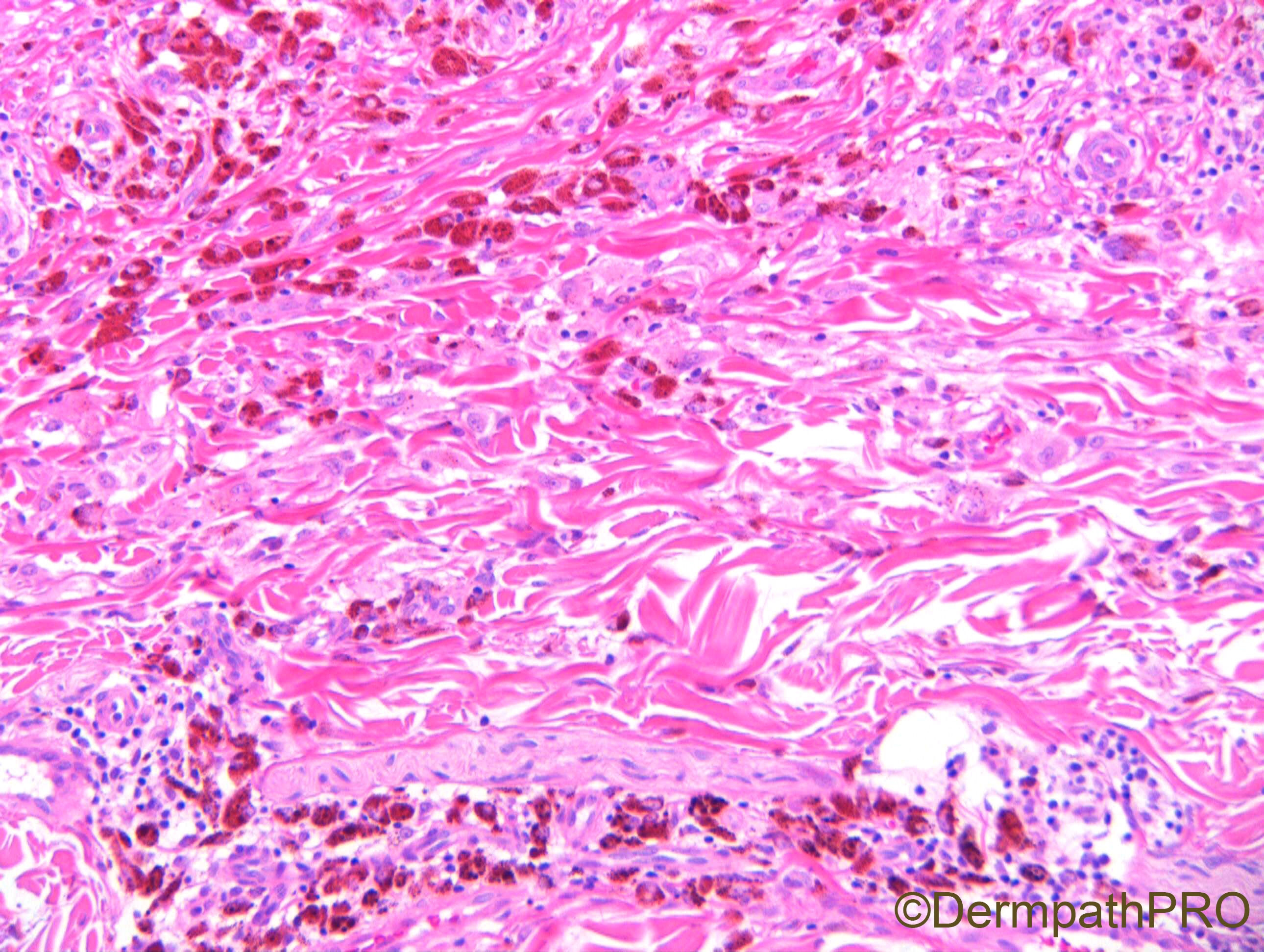
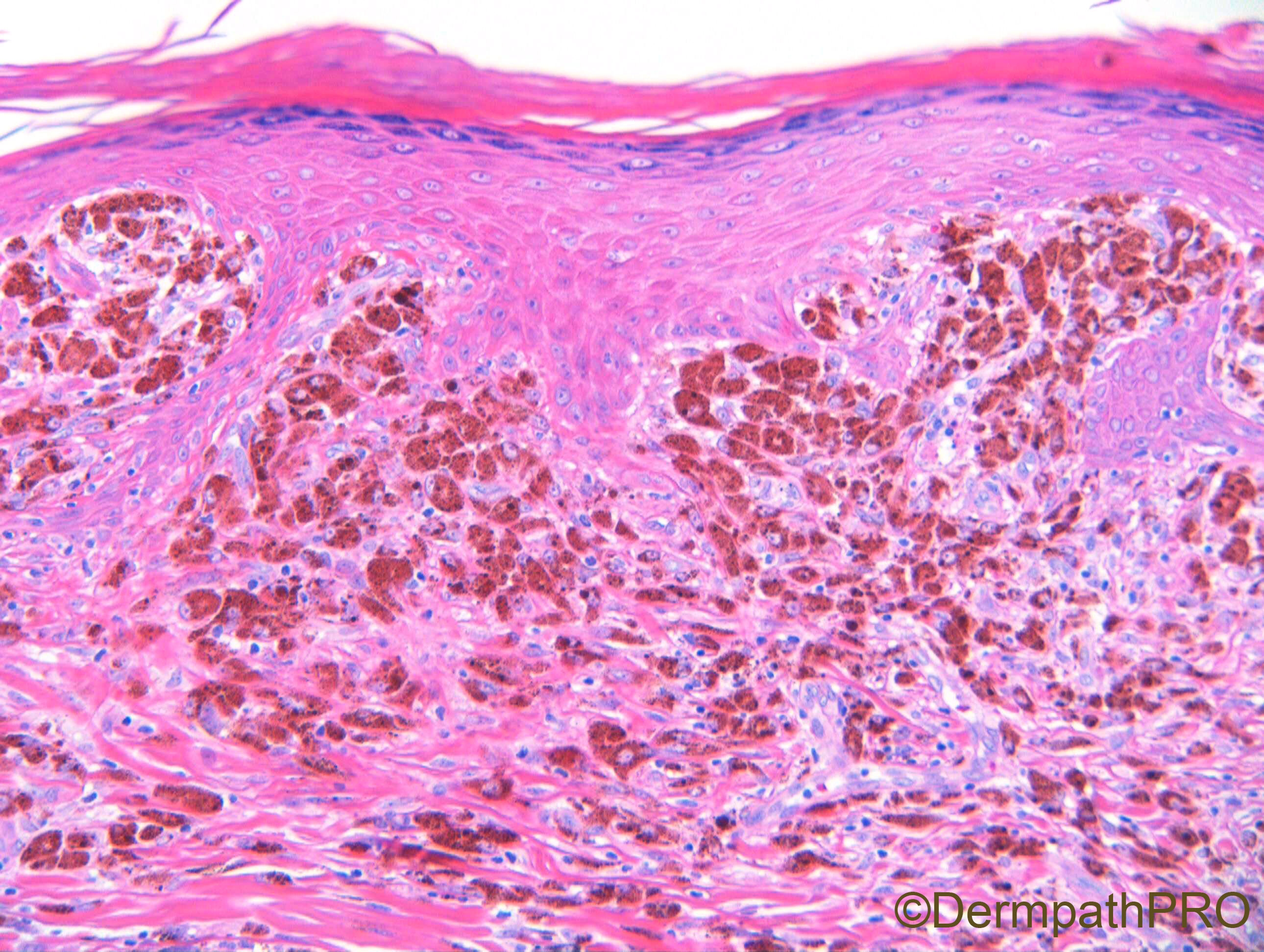
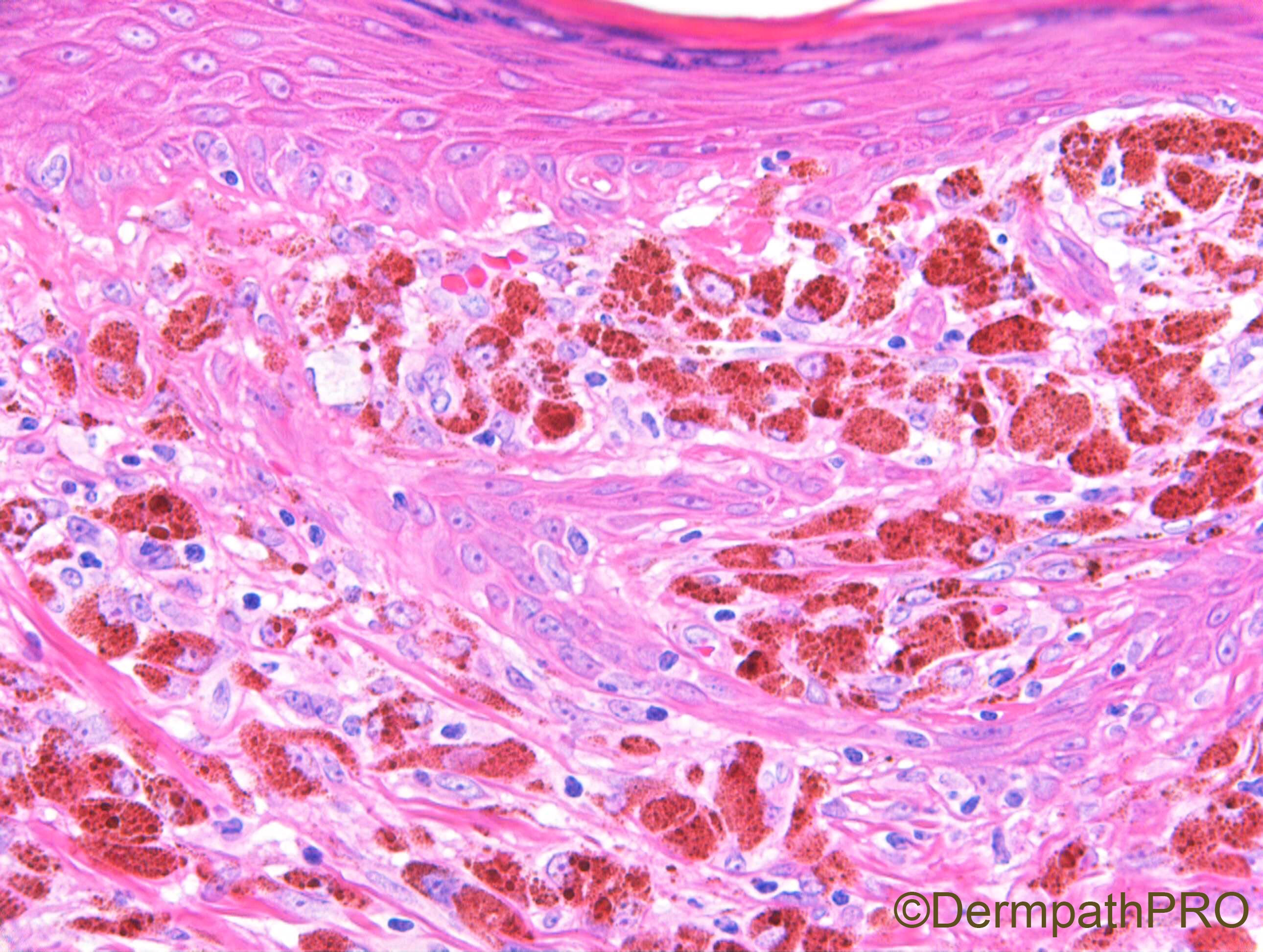
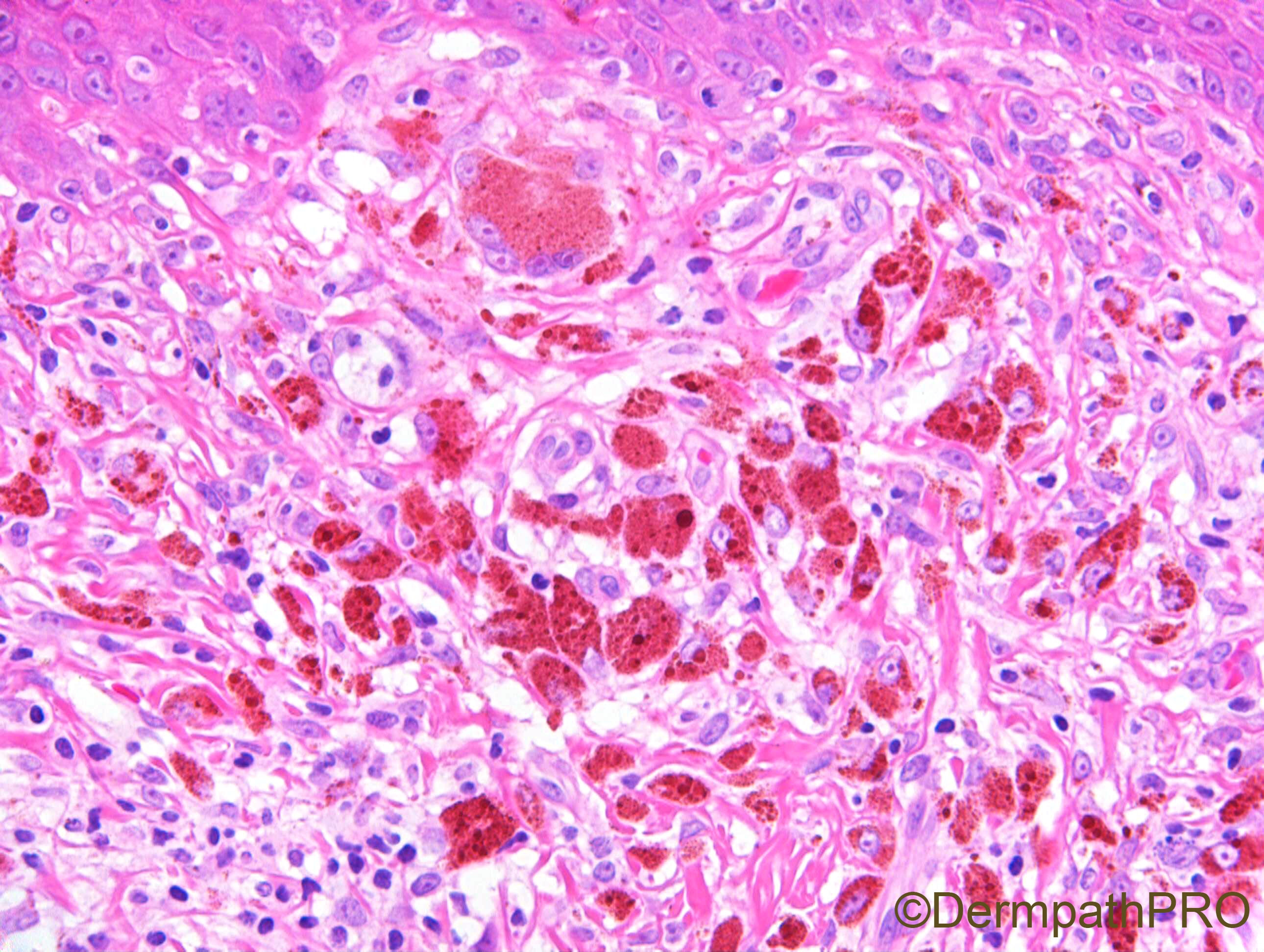
F65. 7 x 4mm lesion on back. ?naevus ?melanoma


  • Like 2
  Report Record

User Feedback


Dr. Richard Carr

Posted

19 views already but only 1 comment - not counting this one!

Share this comment


Link to comment
share_externally

Anil Patki

Posted

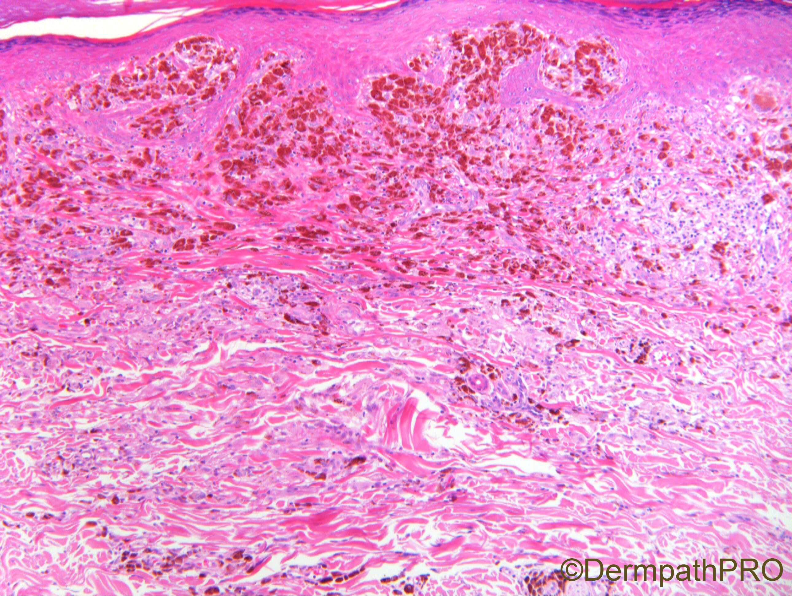
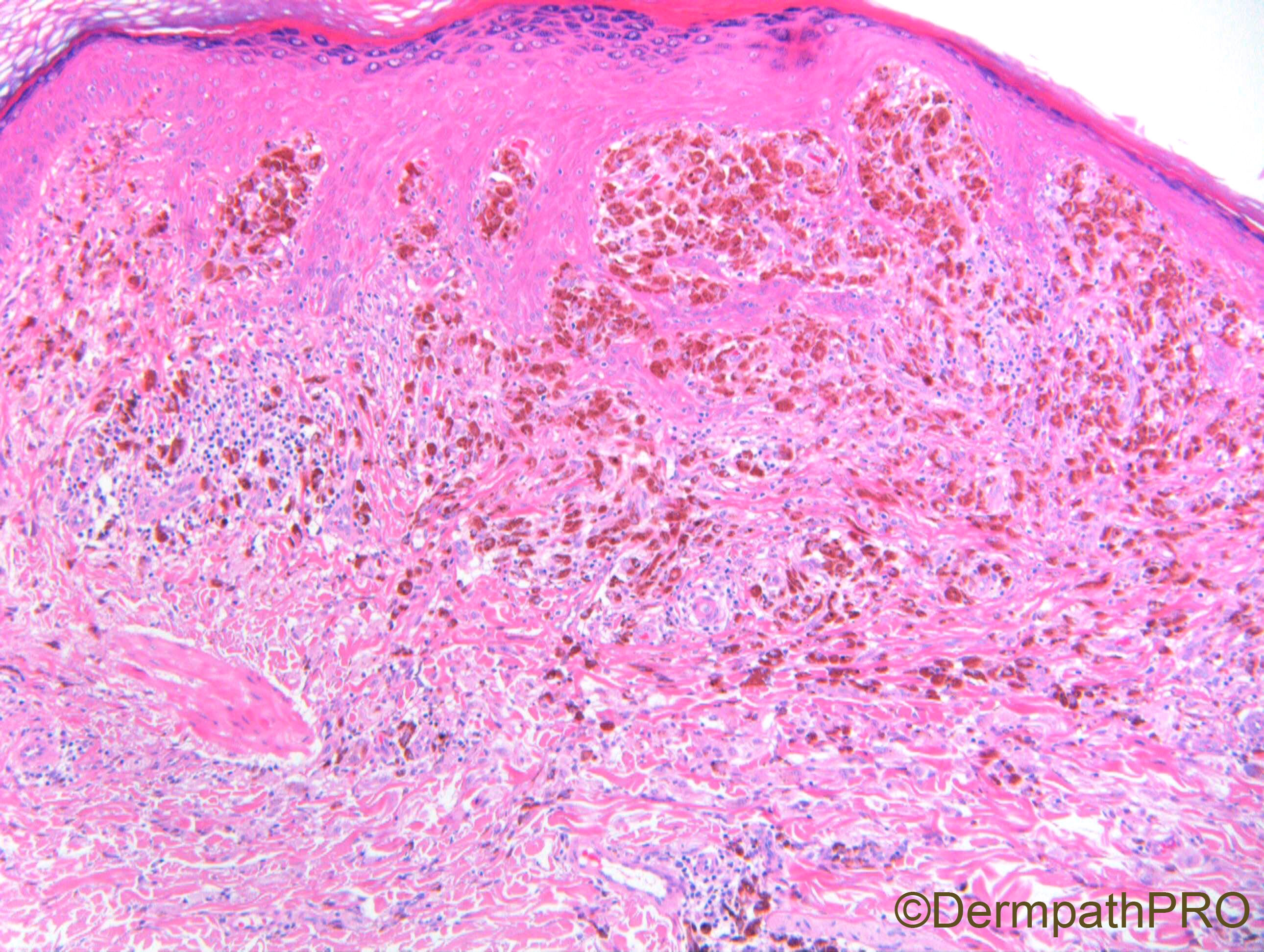
Large intensely pigmented and smaller polygonal melanocytes in the papillary dermis and those interspersed in collagen in the lower dermis suggest epithelioid blue nevus. 

Share this comment


Link to comment
share_externally

At first, I thougt all those cells were melanophages, so my diagnosis was Tumoral melanosis, but then I realize that some of them are isolates atypical melanocytes in the bases of the epidermis, some of them pagetoid. Thus, my diagnosis is an almost complete regressed melanoma. But first I would perform immunostains to be sure all those cells are melanophages (with red chromogen).

Share this comment


Link to comment
share_externally

Krishnakumar subramanian

Posted

tumoral melanosis

does the patient have any lymph nodes 


Share this comment


Link to comment
share_externally

Regressed Melanoma with Tumoral Melanosis, or PEM? This is the question!

I favor tumoral melanosis because of 1) a lateral extension of regression ( unexpected in a PEM ) and 2) not found epithelioid spindle cell component.

Share this comment


Link to comment
share_externally

Dr. Richard Carr

Posted

Yes well done those who suggested tumoural melanosis. I think this is an important case. Of course you would do immunostaining to determine if these are all macrophages/melanophages or whether they are melanocytic.

I normally find I can moderately confidently tell melanophages from melanocytic cells but a nice thin H&E is most helpful. If you can't use a red chromagen you can use a brown chromagen with a Giemsa counterstain (melanin goes green). I personally don't prefer bleaching slides prior to the immunostaining. Given the apparent lichenoid reaction here I'd expect a few reactive melanocytes in the epidermis to look plump but I don't think we saw any nests or confluence of cells.

We are not seeing the biphasic pattern of pigmented epithelioid melanocytoma (PEM) - those tumours have roughly speaking 50% melanophages and 50% melanocytic cells with more dusty melanin. PEM are also very rare (but of course not a good criteria on dermpathpro. The lateral pigment incontinence is against PEM and PEM are often combined lesions with an identifiable (BRAF, NRAS) precursor naevocellular naevus.

The lateral superficial pigment incontinence suggests regression of a pigmented thin / in situ lesion and may be what we see as an end-stage lichen planus-like reaction / keratosis. Theoretically the thin part is compatible with a host inflammatory reaction (post lichen planus-like reaction) to any thin pigmented lesion including solar lentigo, seborrhoeic keratosis, pigmented actinic keratosis or bowenoid actinic keratosis, superficial BCC, junctional naevus & dyplastic naevus and of course melanoma in situ. We suspect most cases of solitary lichen planus-like reaction / keratosis are responses to a solar lentigo / thin seborrhoeic keratosis but I'm always a little guarded in partial biopsies as the more sinister possibilities exist and may not be sample. When the reaction is lichenoid colloid bodies will ususally be readily identifiable in the papillary dermis and can be highlighted by pan-keratin stains but they would still not distinguish the potential diagnoses above as by-stander keratinocytes will drop of in reactions to melanocytic lesions too (quite common in atypical Spitz tumours and can be mis-construed as Kamino bodies that are type IV collagen and limited to the dermo-epidermal junction only).

However in this case we also have the highly dense ("tumour" forming) nodular melanophages with melanophages that extend  in depth >1mm (Breslow). Given the density and location of the deeper of melanophages we have to conclude that on the balance of probability (given the rarity of benign heavily pigmented lesions in Caucasiians aged over 60 years) this patient had at least a thin melanoma with adnexal extension or possibly a locally invasive melanoma over 1mm in thickness that has fully regressed. I advised follow-up as for an invasive melanoma up to 1.5mm thickness that would include close watchful waiting of the "primary" site and examination for lymph nodes. A sentinel lymph node biopsy could be considered if available. Given the primary excited sufficient immune response to undergo complete regression I'd expect the tumour to have a low risk for later clinically apparent metastatic disease but obviously not zero.

I have seen deeper pigmented melanophages in a completely regressed (previously biopsied pigmented BCC). In that case a good clue was dense clustering of colloid bodies (can be keratin derived amyloid) below the papillary dermis and intermixed with the melanophages.

Share this comment


Link to comment
share_externally

Very interesting case, Dr Richard! Thank you!

Share this comment


Link to comment
share_externally


Join the conversation

You can post now and register later. If you have an account, sign in now to post with your account.

Guest
Add a comment...

×   Pasted as rich text.   Paste as plain text instead

  Only 75 emoji are allowed.

×   Your link has been automatically embedded.   Display as a link instead

×   Your previous content has been restored.   Clear editor

×   You cannot paste images directly. Upload or insert images from URL.

Loading...

×
×
  • Create New...