Jump to content
In this section we have spot diagnoses posted on a daily basis since June 2010, now over 4000! You can review the archived cases and read the suggested diagnoses by users and the final comment by the contributors.
Case are uploaded each week day by 10 am UK time with the correct diagnosis will generally be posted at 8 pm UK time. Why not view the most recent spot diagnosis and proffer a diagnosis?

Case Number : Case 2176 - 11 October 2018 Posted By: Raul Perret

Please read the clinical history and view the images by clicking on them before you proffer your diagnosis.
Submitted Date :
   (0 reviews)

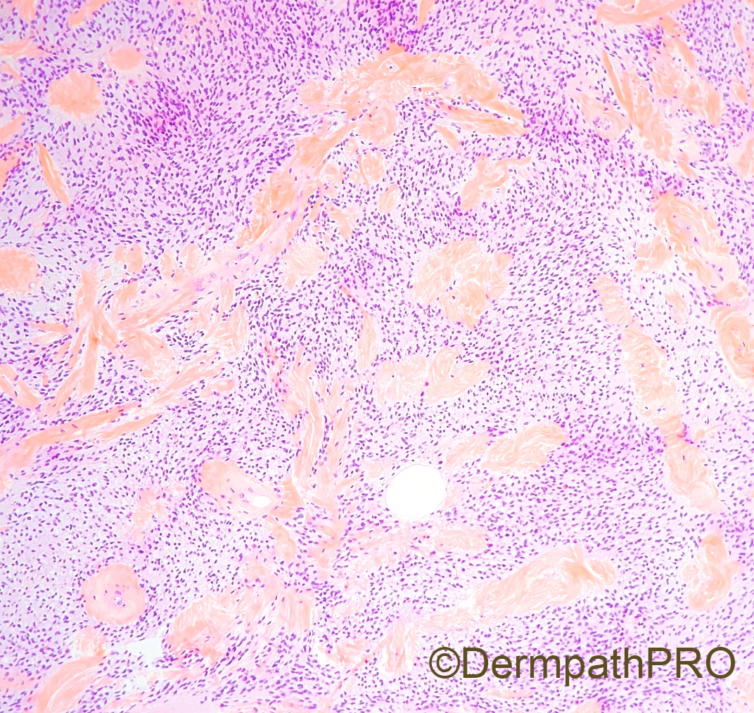
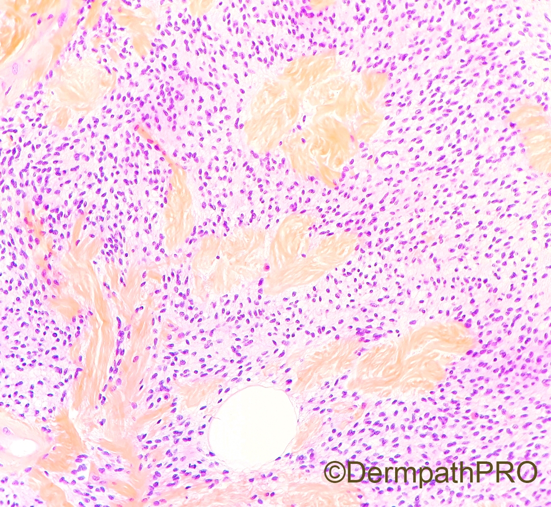
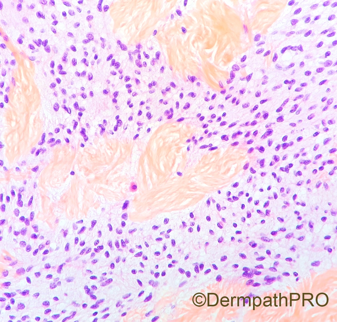
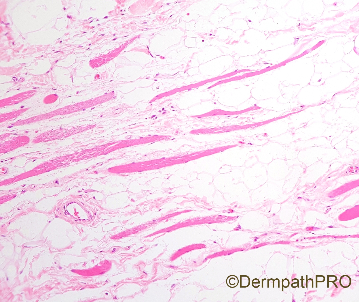
10 cm forearm tumor (superficial and deep) of a 60 year old male.


  • Like 1
  Report Record

User Feedback


What is the relationship between the last picture and the other figures. The last figure appears to show an intramuscular lipomatous tumor. The other figures appear to show a spindle cell lesion with a lot a hyalinization. Are they 2 components of the same tumor like a dedifferentiated liposarcoma (although the lipomatous component does not really look atypical). Maybe do mdm2 or Ck4 to see whether the lipomatous and spindle components are the same tumor. Of course a lot more IHC on the spindle cell component but at least MUC4.

Share this comment


Link to comment
share_externally

Thanks Paul. The lipomatous component does not look like a myxoid liposarcoma so the other component is most likely not a round cell liposarcoma. Probably will need to do MDM2 or CK4. Maybe RB1.

Share this comment


Link to comment
share_externally

After ruling out myxoid liposarcoma

I am thinking of dendritic fibromyxoid lipoma

Share this comment


Link to comment
share_externally

vincenzo

Posted (edited)

Unusual size and unusual site, but favor Spindle Cell Lipoma. Maybe a plexiform variant.

Edited by vincenzo

Share this comment


Link to comment
share_externally

Dr. Richard Carr

Posted

I'm starting to think Thursdays are the new Friday! Can't wait to find out what this is. For those who don't know the french like to use Saffron in their H&E hence the orange collagen! That is about as useful as I can be but I'm going for the atypical lipoma-dedifferentiated hypophesis although in my training days there were no (dediff) cases reported in the superficial / subcutaenous soft tissue (although this one is into skeletal muscle).

Share this comment


Link to comment
share_externally

Thanks for the information. The collagen indeed looks very orange and I had a hard time to figure out what it is.

Share this comment


Link to comment
share_externally

vincenzo

Posted (edited)

Let me add another spot: hyalinizing spindle cell tumor with giant rosettes (aka LGFMS). But my  first is SCL. 

Edited by vincenzo

Share this comment


Link to comment
share_externally

Dr. Richard Carr

Posted

Is Raul playing the waiting game?  I'll nudge him. I did consider intra-muscular, partly spindle cell lipoma today.


Share this comment


Link to comment
share_externally

Raul Perret

Posted

Dear all, thank you for the comments. As many of you suggested, this is a spindle cell lipoma. The morphology is suggestive of this diagnosis as we see spindle cells without atypia on a rather myxoid backgroud with numerous mastocytes. Please note that the broad collagen bundles are not the ones classically seen in SCL (ropey, similar to grated carrots), instead they look like the starburst collagen pattern seen in low grade endometrial stromal sarcomas (I also see it more frequently in mammary type myofibroblastoma a lesion closely related to SCL) . An important point is the one remarked by Vincenzo, the clinical an morphological context of this particular lesion is wrong because SCL never depelops from deep soft tissues and is typically seen in the back of adult males. For this reason, we performed a CGH analysis to rule out other neoplasms with different biologic potential like an atypical lipomatous tumor, atypical spindle cell lipomatous tumor or a dedifferentiated liposarcoma. CGH showed a simple genomic profile with a classic 13q deletion (including rb1 locus) and a deletion of 16q which is also frequently seen in SCL. In conclusion, this is a spindle cell lipoma which originated on the subcutaneous tissue and secondarily infiltrated the deep compartment (probably due to its size and topography). Please do remember that rb1 loss by IHC can be seen in all the differentials mentioned above so if you have something that looks like spindle cell lipoma but is located deeply you should be cautious before rendering such a diagnosis. Hope you liked the case, sorry for the late response but i have been quite busy lately. Cheers

Share this comment


Link to comment
share_externally

Paul,

Great case. I suggested RB1 but i was thinking more of atypical spindle cell lipomatous tumors because of the unusual location and the high cellularity. Fletcher's study on atypical spindle cell lipomatous tumor did not have molecular backup so just because the tumor has deletion of chromosome 13, it is definitely a spindle cell lipoma? In addition, most pathologists don't have molecular testing so I would say the this lesion may be classified by most pathologists as atypical spindle cell lipomatous tumor based on RB1 loss , high cellulaity, lack of ropey collagen, and unusual location. What is your thought about that?

Share this comment


Link to comment
share_externally

Raul Perret

Posted

Dear Anh, thank your for the interest on the case. Atypical spindle cell lipomatous tumor is a controversial entity with a high interobserver variability. In my department, for performing such a diagnosis we search for a component of atypical cells (at least focally) and univacuolated lipoblasts (ice cream cone like, PS100 +) and CGH would show a more complex profile than spindle cell lipoma or in some cases a flat profile. Although the latter is based solely in our experience.

As you remark, the absence of CGH analysis in the paper of Marino Enriquez et al. raises some issues as we cannot exclude that many cases of the series could actually correspond to spindle cell lipomas or rare atypical lipomatous tumors/dedifferentiated liposarcomas with amplification of alternative genes (MDM4, etc. seen in around 1% of cases).

On the issue of spindle cell lipoma/atypical lipomatous tumor i recommend you the following papers: DOI 10.1007/s00428-014-1568-8  and  10.1007/s00428-014-1568-8. As you will see, the team of Thomas Mentzel reports deletions of RCBTB2 and ITM2B (flanking genes of RB1) in atypical spindle cell lipomatous tumors compared to spindle cell lipoma. I must confess that I do not know if those genes are deleted in this case (will try to see if the CGH that we perform includes them in the analysis). Once again, in our department, the CGH profiles that we see in cases fitting the morphological criteria of ASLT are rather different.

To sum up, in rare cases showing the morphological and immunohistochemical features depicted in this case I think we should perform more complex studies (particularly CGH) for a better understanding of these rare neoplasm. In particular it helps to support that the lesion is benign with a small chance of local recurrence and that the surgical excision is sufficient.

By the way, my name is Raul not Paul ;)

Share this comment


Link to comment
share_externally


Join the conversation

You can post now and register later. If you have an account, sign in now to post with your account.

Guest
Add a comment...

×   Pasted as rich text.   Paste as plain text instead

  Only 75 emoji are allowed.

×   Your link has been automatically embedded.   Display as a link instead

×   Your previous content has been restored.   Clear editor

×   You cannot paste images directly. Upload or insert images from URL.

Loading...

×
×
  • Create New...