Jump to content
In this section we have spot diagnoses posted on a daily basis since June 2010, now over 4000! You can review the archived cases and read the suggested diagnoses by users and the final comment by the contributors.
Case are uploaded each week day by 10 am UK time with the correct diagnosis will generally be posted at 8 pm UK time. Why not view the most recent spot diagnosis and proffer a diagnosis?

Case Number : Case 2179 - 16 October 2018 Posted By: Uma Sundram

Please read the clinical history and view the images by clicking on them before you proffer your diagnosis.
Submitted Date :
   (0 reviews)

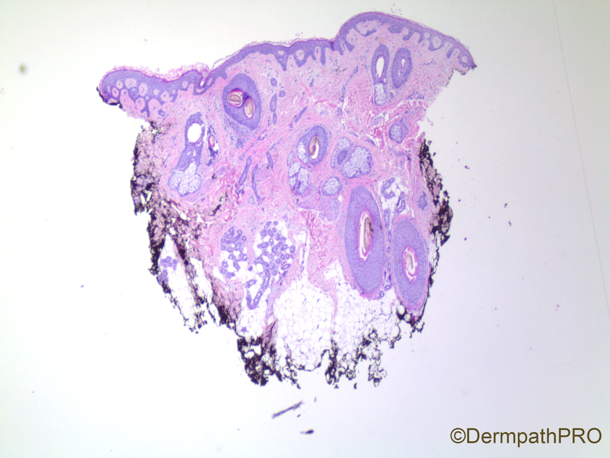
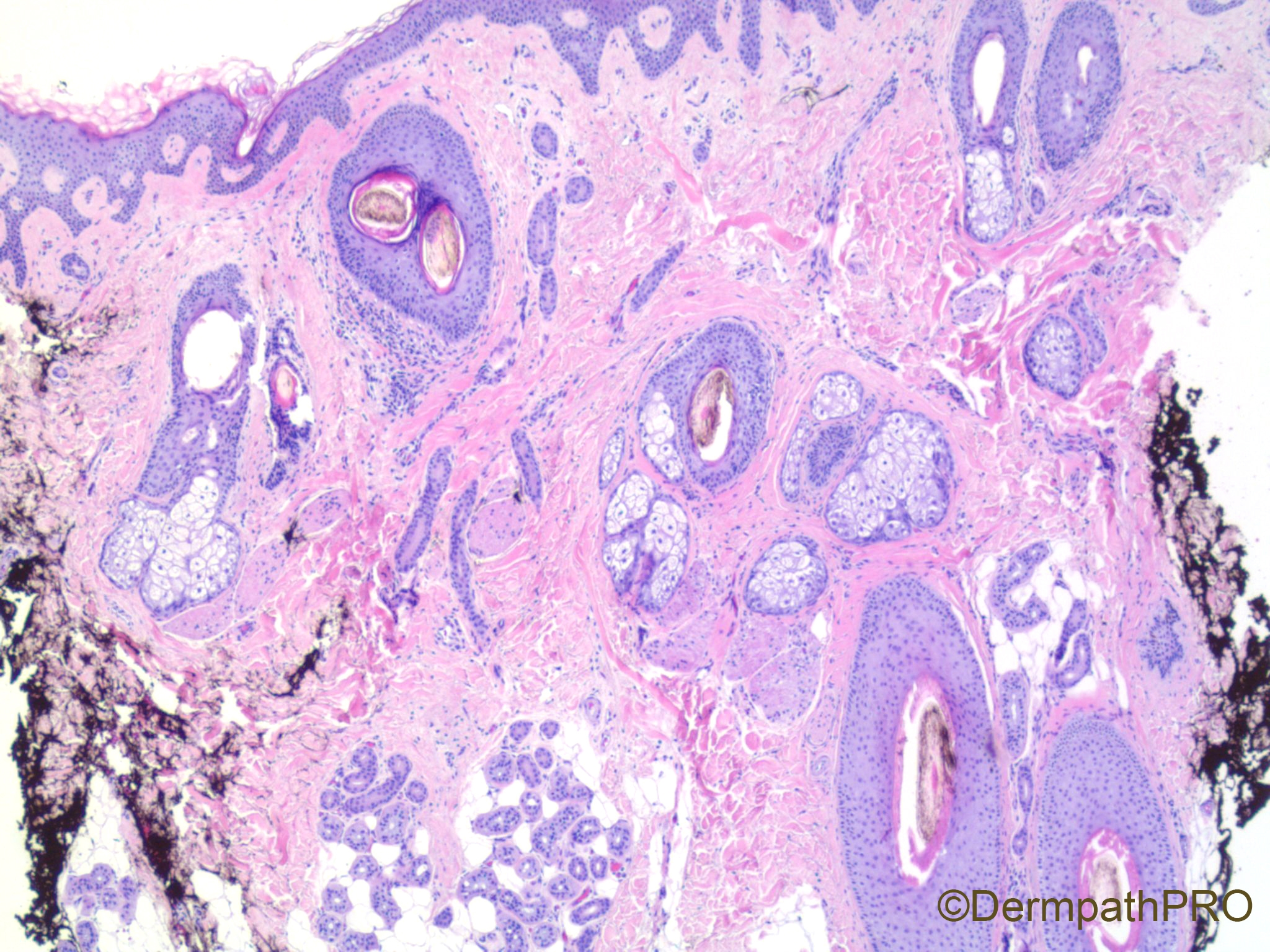
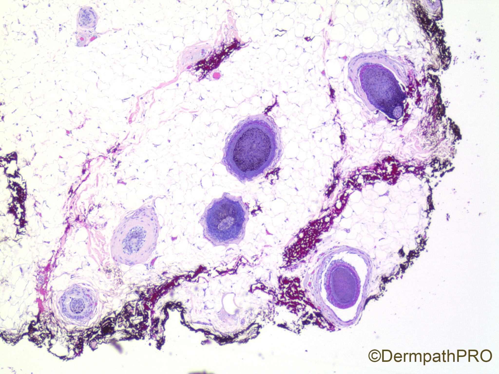
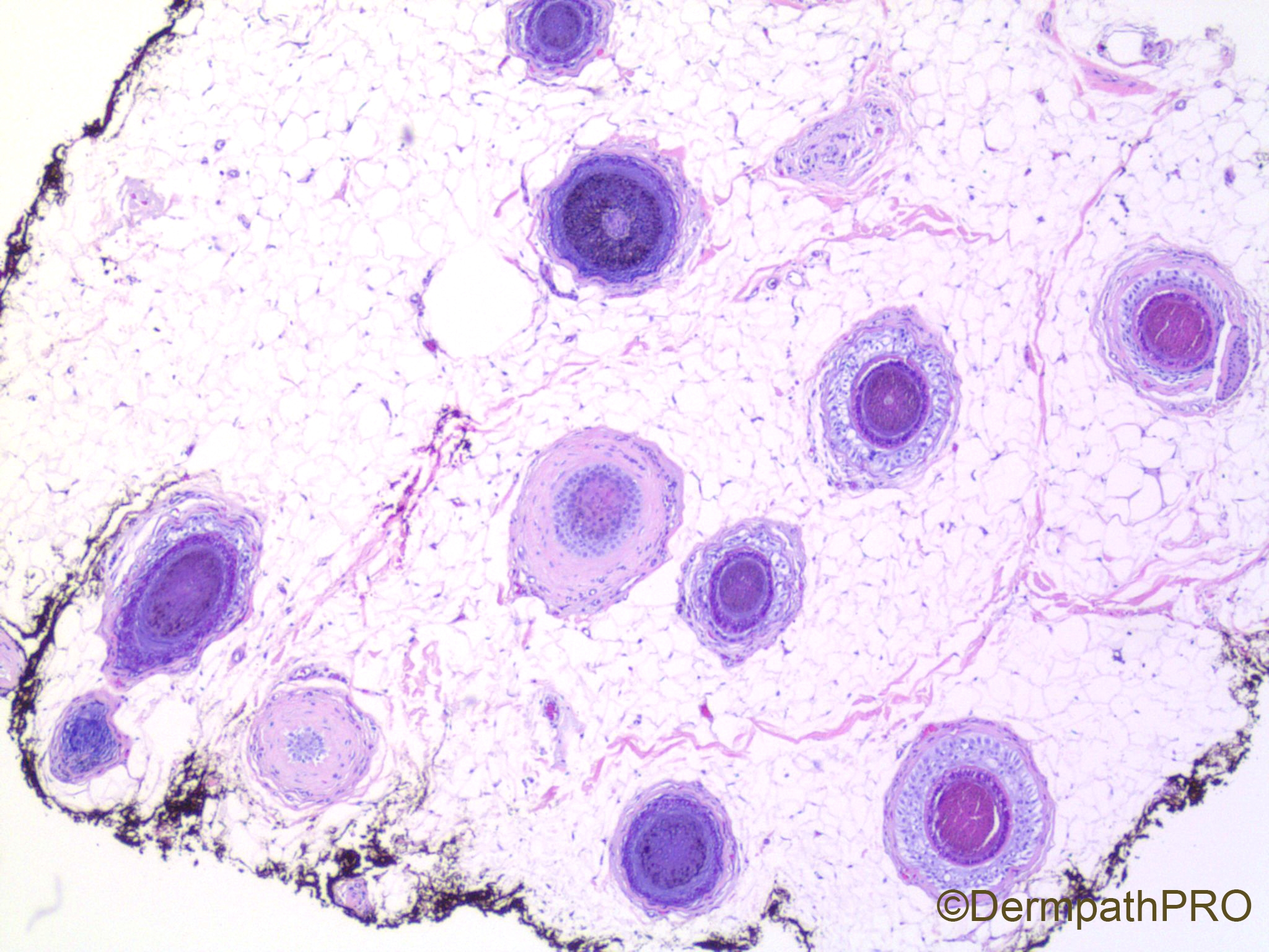
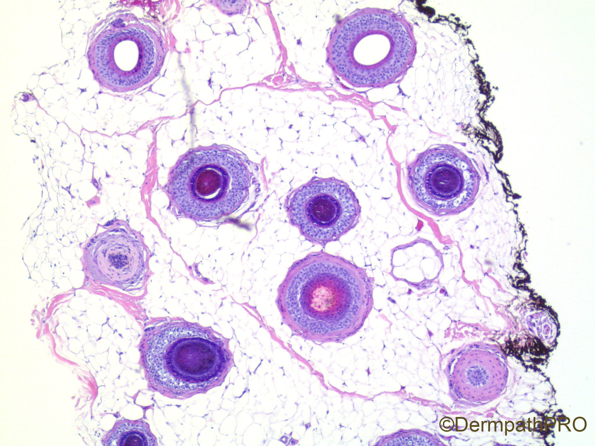
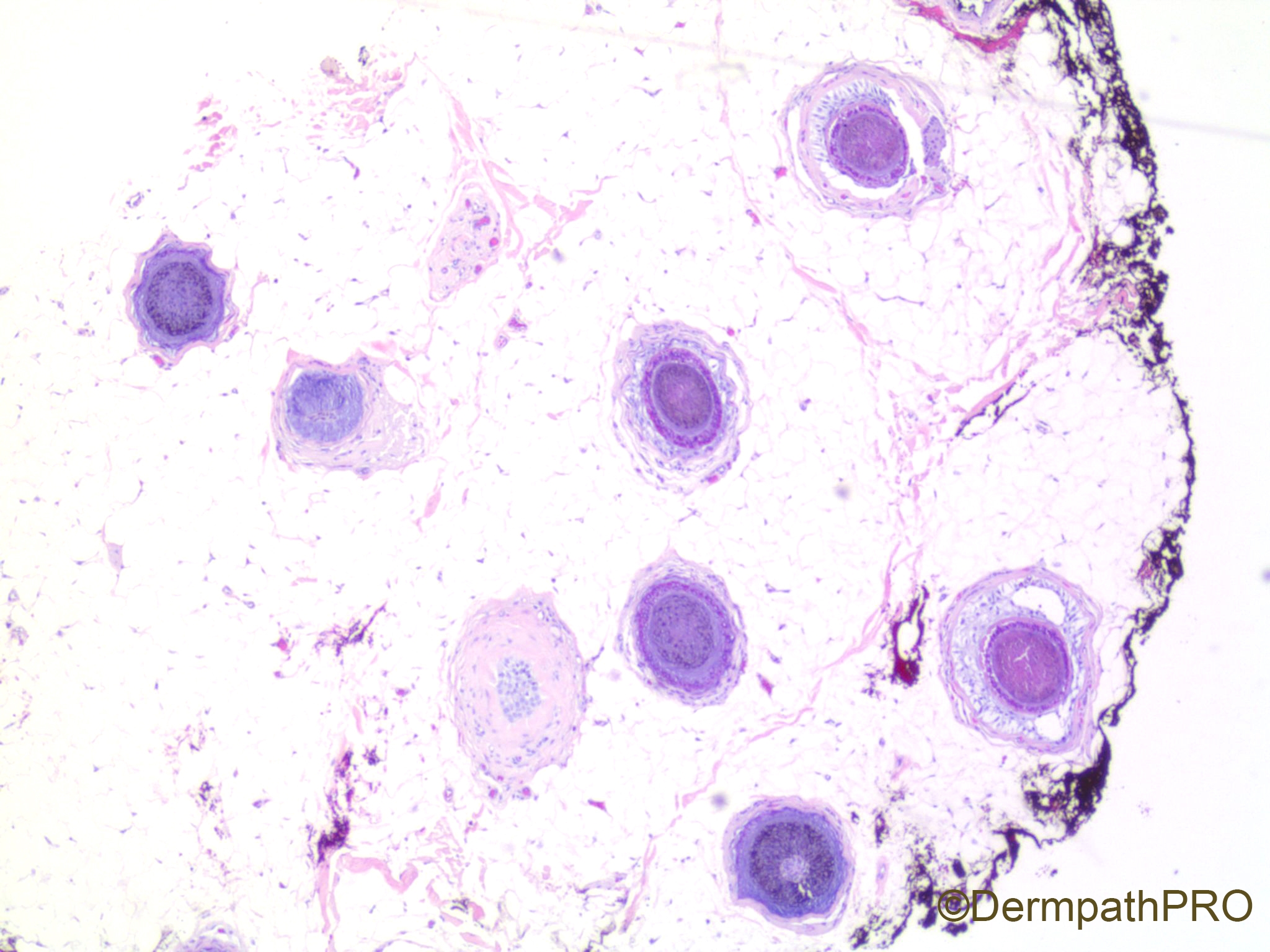
40 year old woman with diffuse hair loss.


  Report Record

User Feedback


Variation in follicle size with miniaturization,  preserved adnexae and absence of inflammation and fibroplasia suggest female pattern baldness. 


Share this comment


Link to comment
share_externally

Alex-Ventura-Leon

Posted

Im not an expert in Alopecia but mi first impression is Female Androgenetic Alopecia (FAGA)

Share this comment


Link to comment
share_externally

Krishnakumar subramanian

Posted

yes Female Androgenetic alopecia

But I am surprised why biopsy was done

am I missing something

Share this comment


Link to comment
share_externally

Uma Sundram

Posted

Yes, androgenetic alopecia. It was such a classic that I wanted to post it.  SInce we don't get many alopecia cases, I thought this would be a good teaching case! The biopsy was performed to exclude other causes of alopecia.

Share this comment


Link to comment
share_externally

Dr. Richard Carr

Posted

Thanks Uma. Yes agree we should be posting alopecia cases a bit more as our clinicians here only biopsy rarely so we all learn from the relatively few and the somewhat challenging cases we see. I prefer "androgenic" personally! "androgenetic" sounds like a genetic disorder but I guess you could argue it is of course.

Good talk from Tim McCalmont on the alopecias in Sau Paulo. I asked if we could basically drop the vertical sections and he pretty much agreed once you''ve done your learning curve with the horizontals there is very little need for the vertical - bearing in mind you'd occasionally have to level up and down as appropriate. We tend to look at the first 3 levels and can often give a diagnosis in 50% and only do the more extensive step levels (I do L4-12 at 150 micron steps) as required.

I've heard "experts" in public talking about serial sections and looking at 100's of levels which I've never been inclined to do as the horizontals give you every follicle in the biopsy different levels. You either know what it is or you don't. Obsessively close levels/serial sections rarely help in any situation in dermatopathology (sprotrichosis perhaps could be an exception to find the microorganism in the centre of the granuloma) but even then culture is the gold standard and the diagnosis should be suspected with the appropriate pathologoy and a negative fungal stain.

Share this comment


Link to comment
share_externally


Join the conversation

You can post now and register later. If you have an account, sign in now to post with your account.

Guest
Add a comment...

×   Pasted as rich text.   Paste as plain text instead

  Only 75 emoji are allowed.

×   Your link has been automatically embedded.   Display as a link instead

×   Your previous content has been restored.   Clear editor

×   You cannot paste images directly. Upload or insert images from URL.

Loading...

×
×
  • Create New...