Jump to content
In this section we have spot diagnoses posted on a daily basis since June 2010, now over 4000! You can review the archived cases and read the suggested diagnoses by users and the final comment by the contributors.
Case are uploaded each week day by 10 am UK time with the correct diagnosis will generally be posted at 8 pm UK time. Why not view the most recent spot diagnosis and proffer a diagnosis?

Case Number : Case 2182 - 19 October 2018 Posted By: Dr. Richard Carr

Please read the clinical history and view the images by clicking on them before you proffer your diagnosis.
Submitted Date :
   (0 reviews)

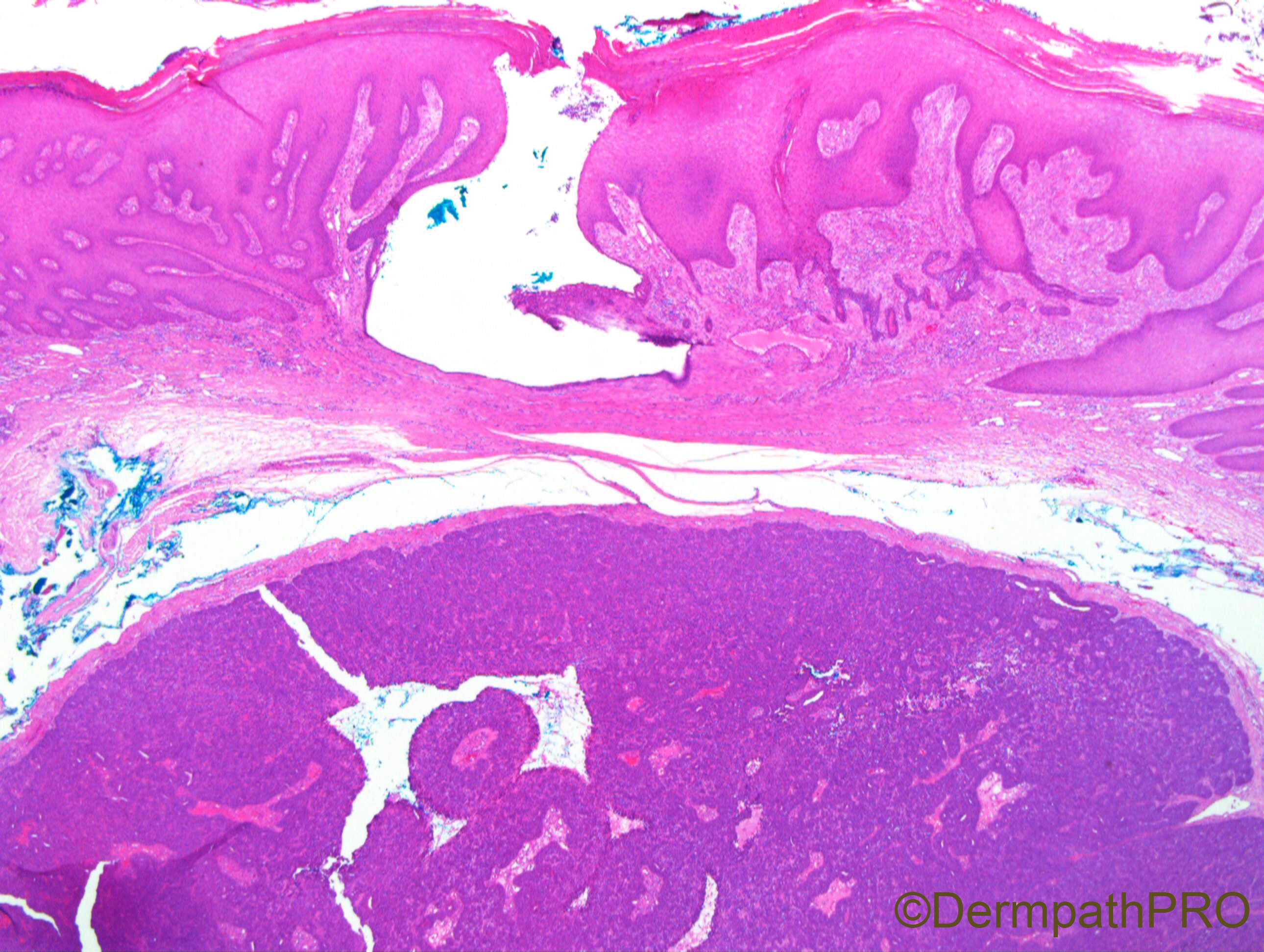
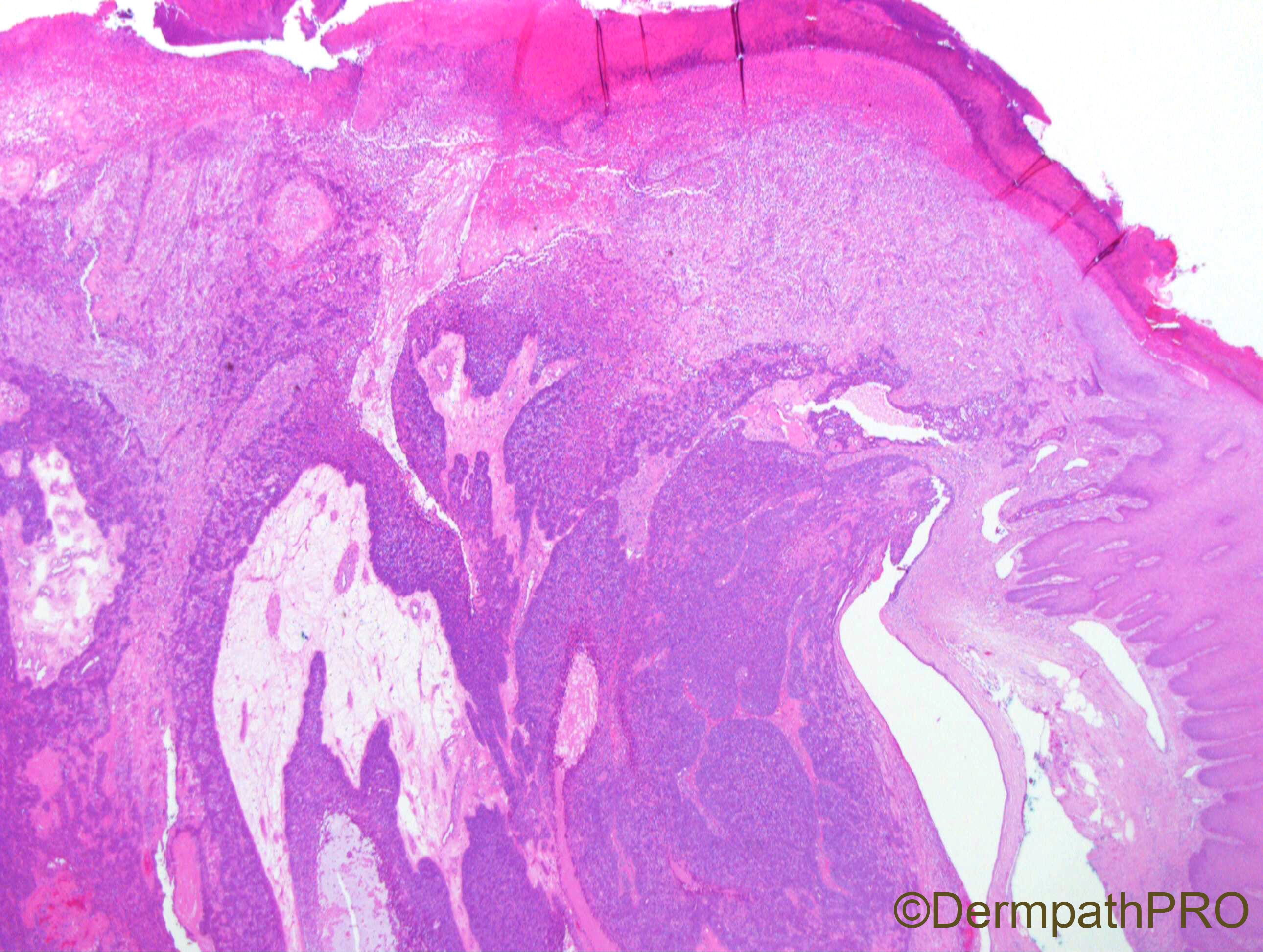
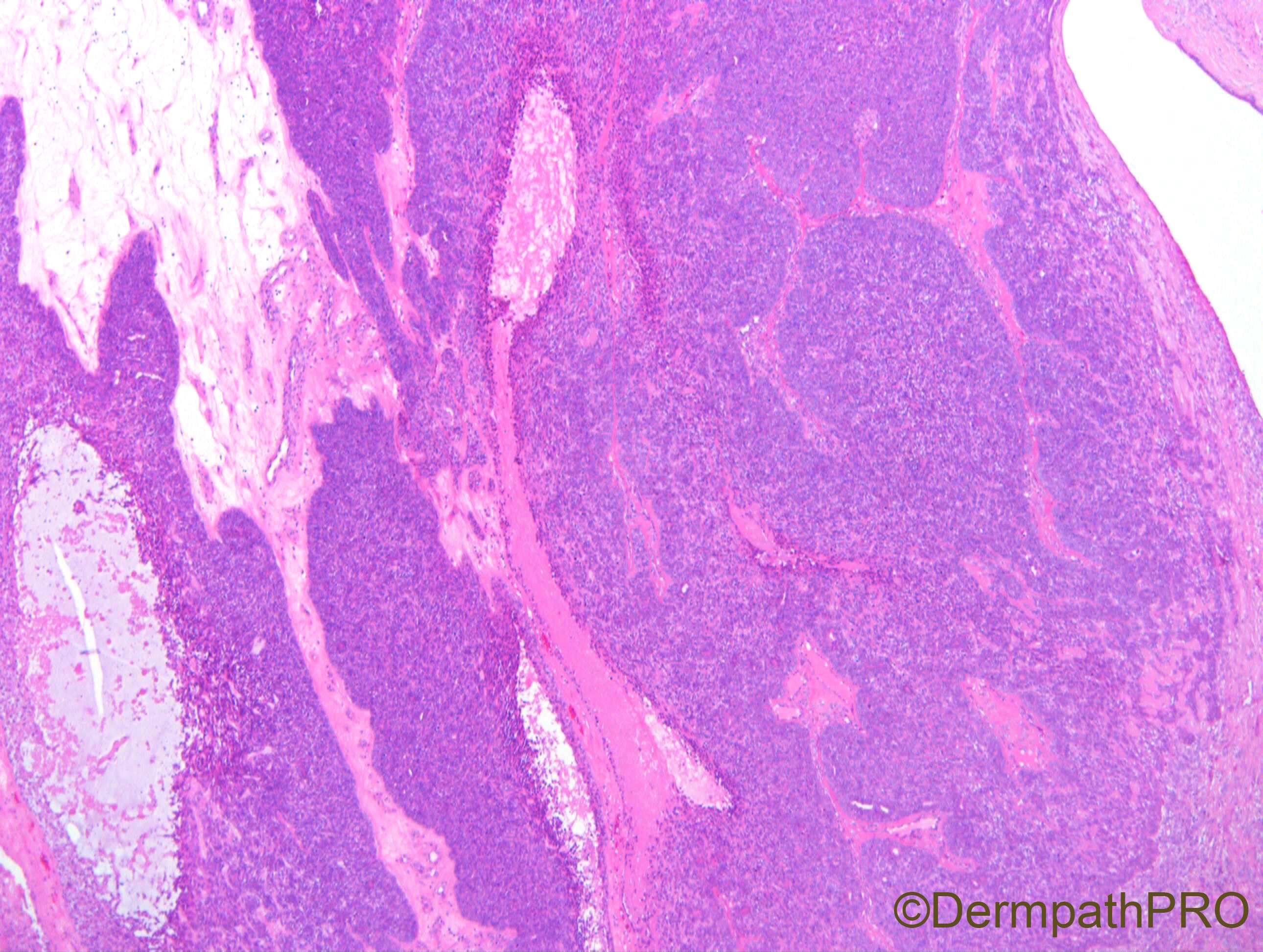
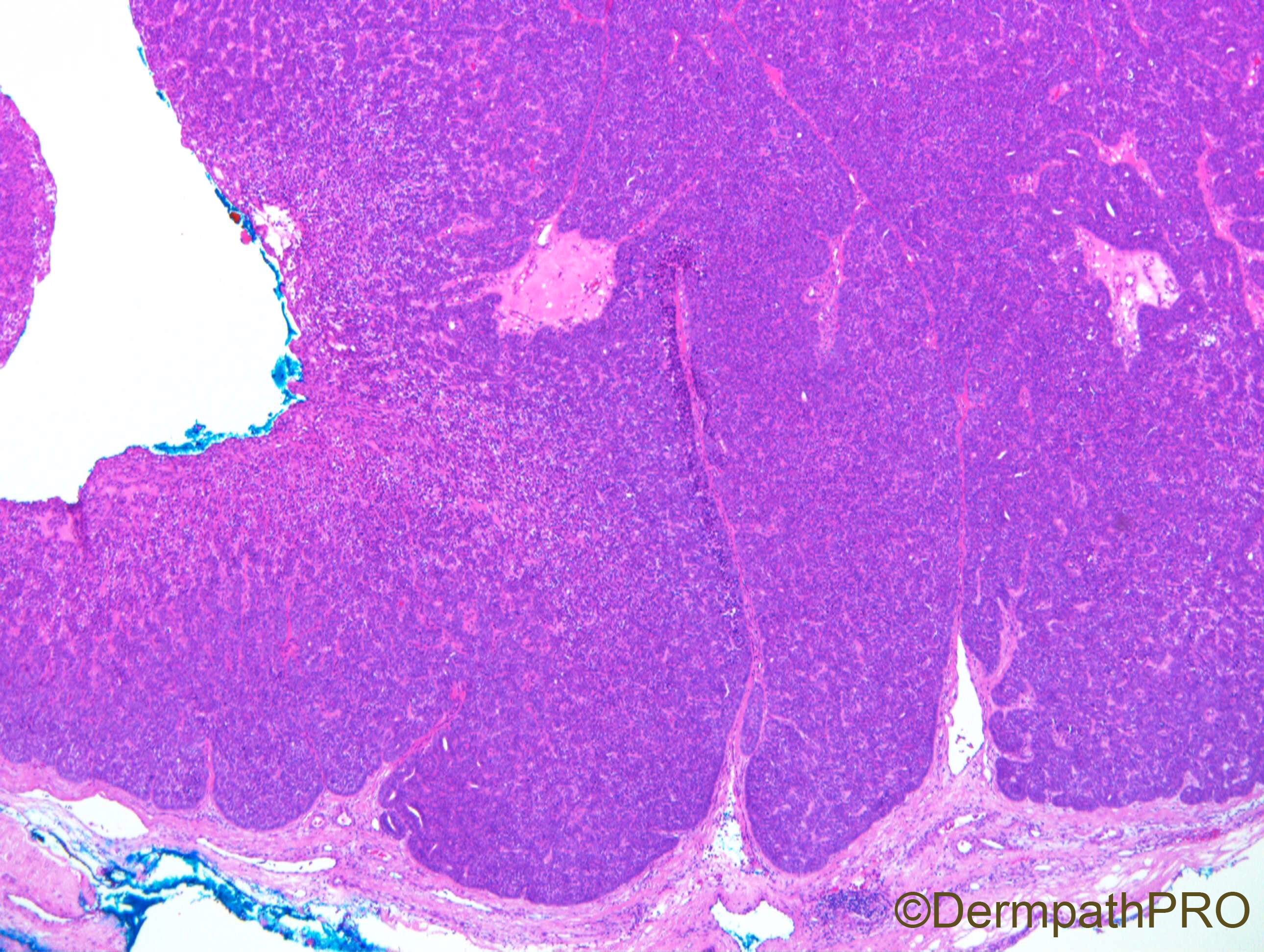
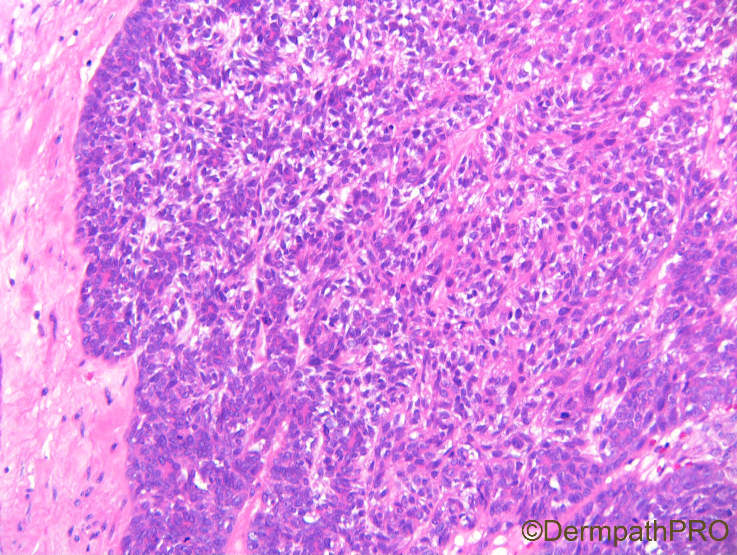
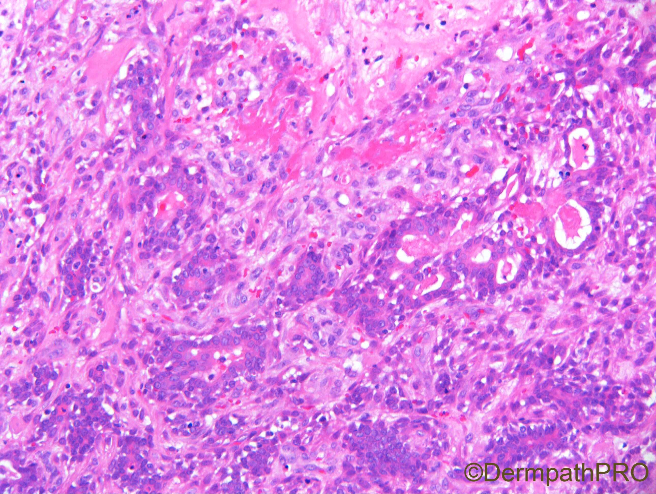
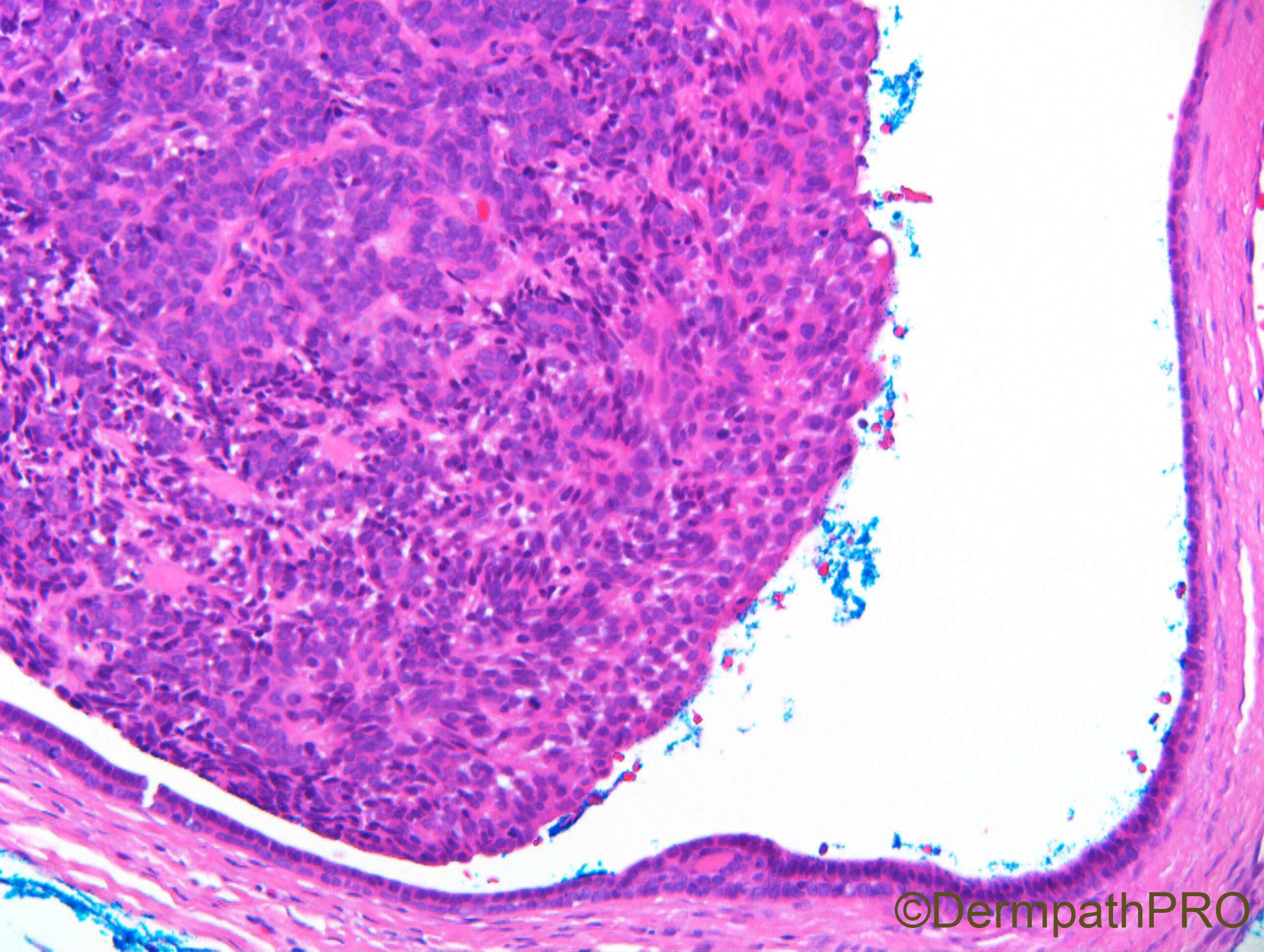
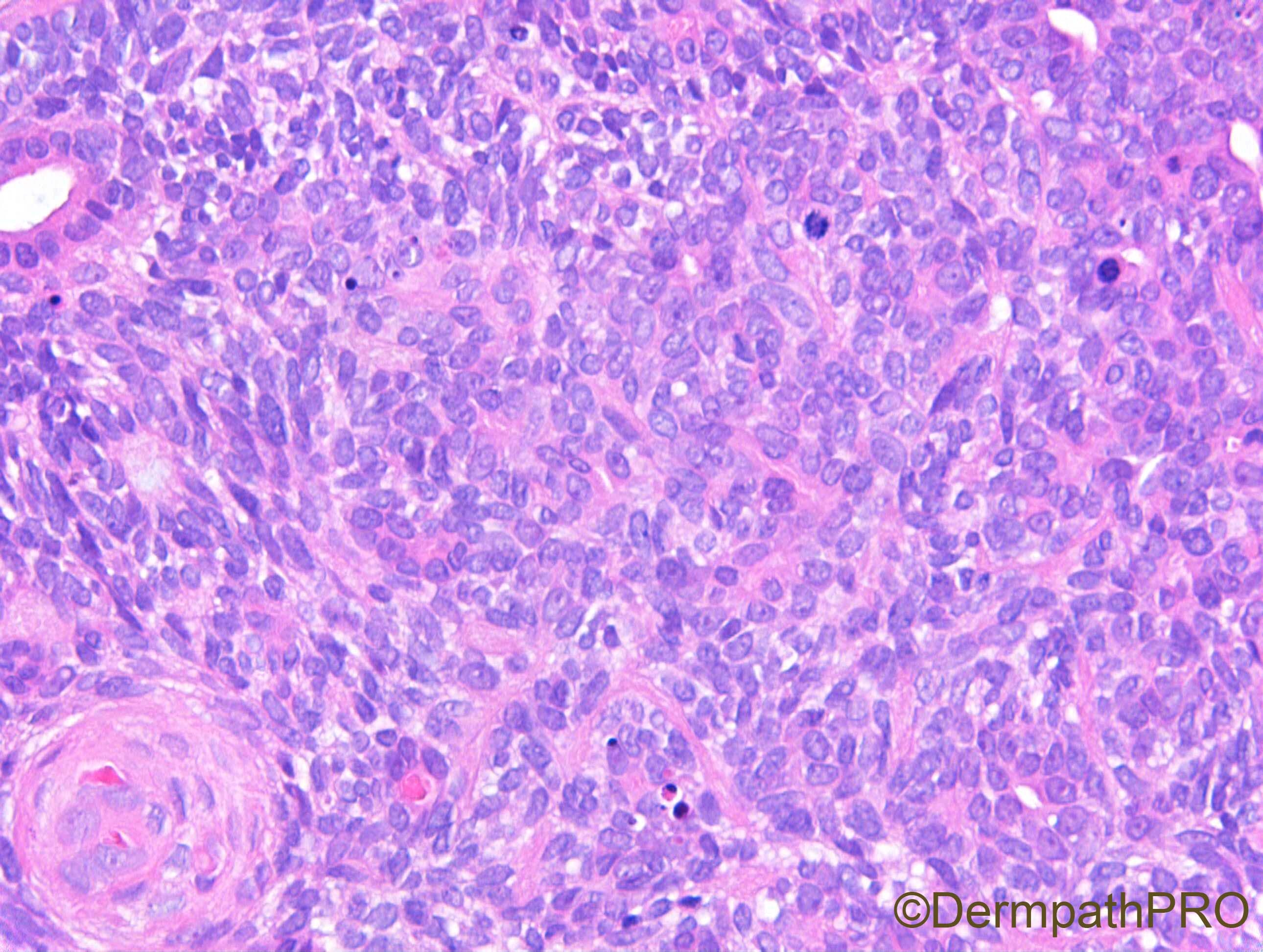
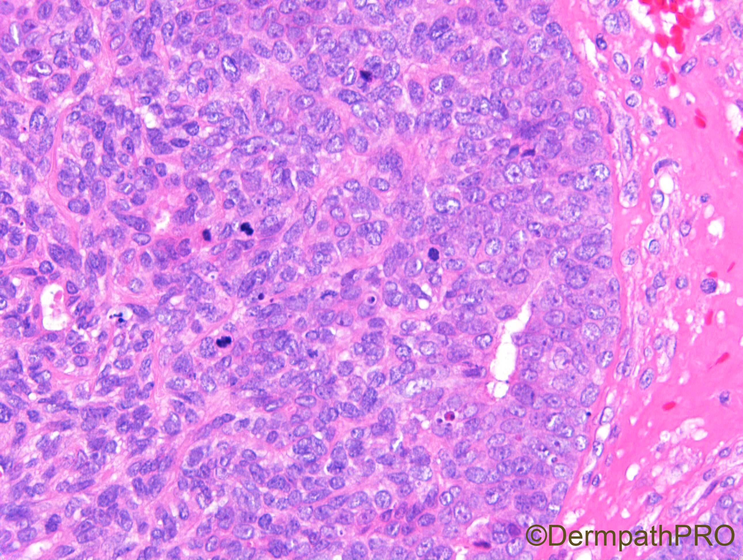
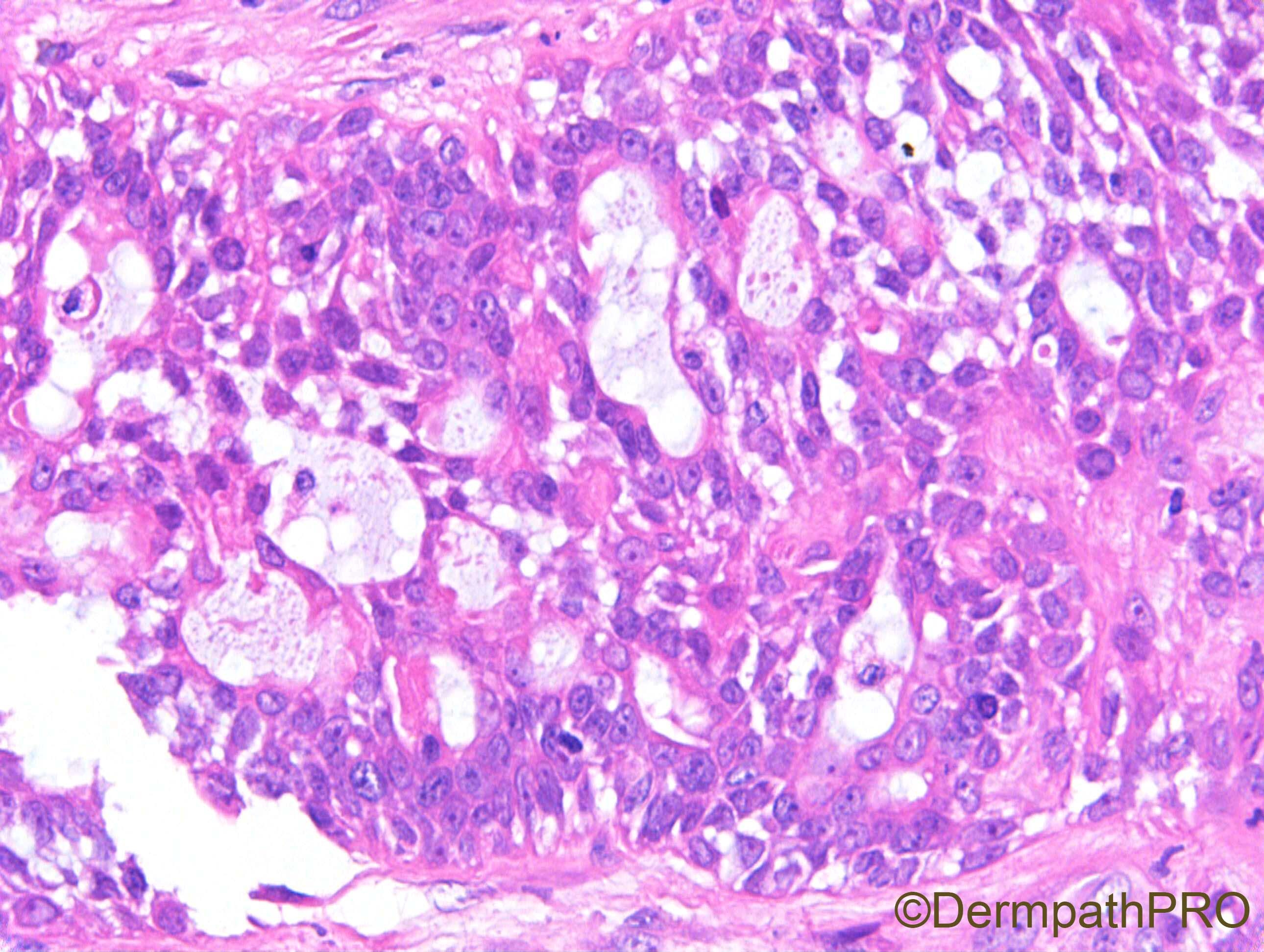
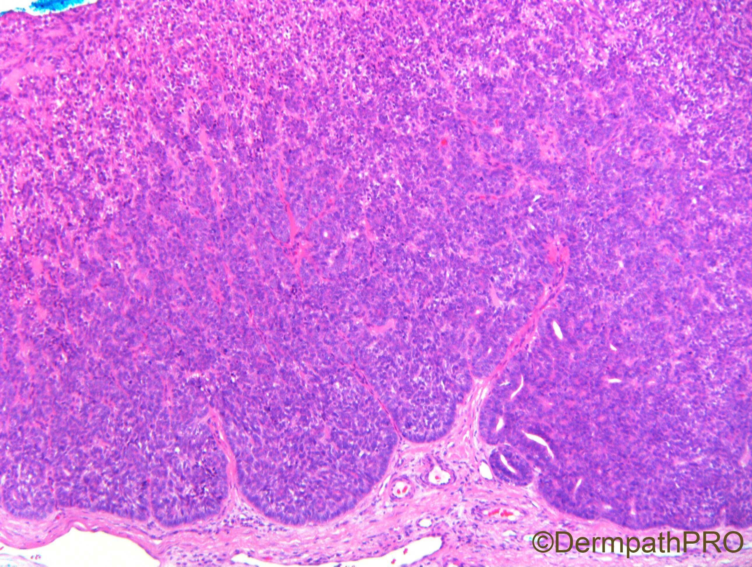
M55. Middle finger. ?neurofibroma

  • Like 1
  Report Record

User Feedback


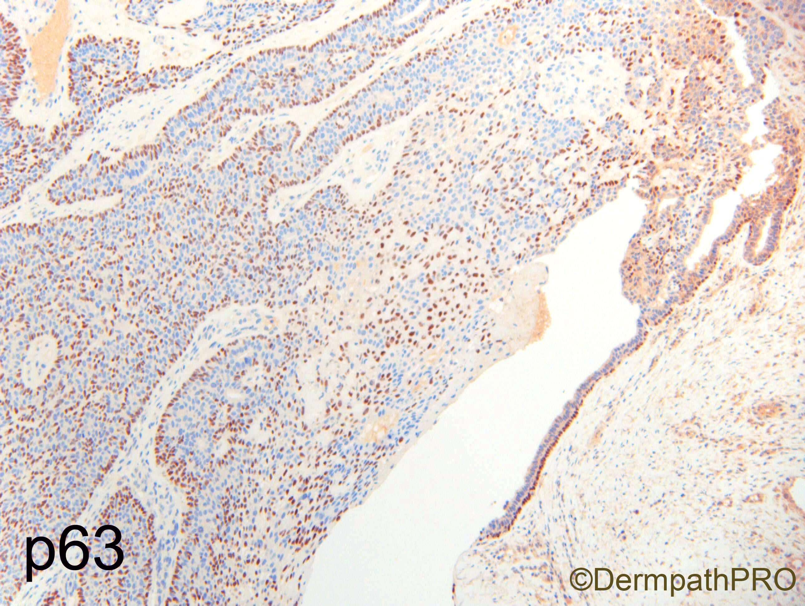
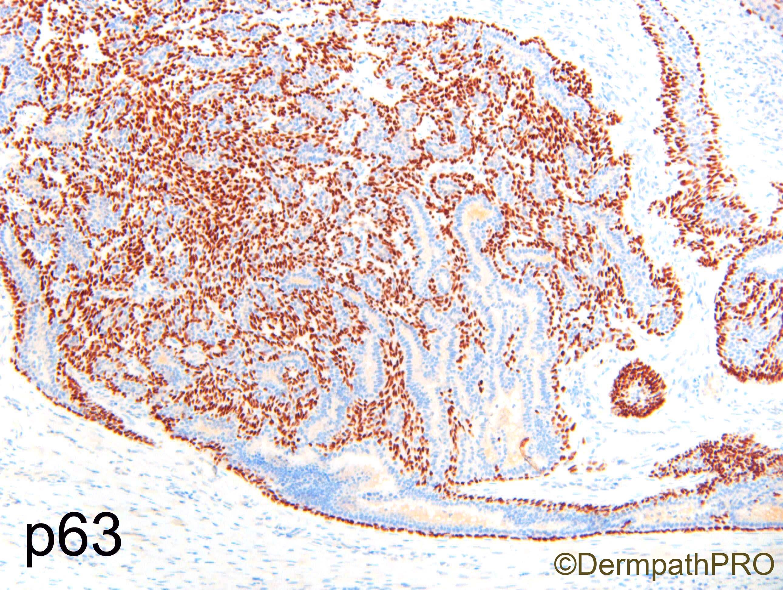
Even with this predominantly solid growth admixed with cystic spaces the diagnosis is Digital papillary carcinoma. The basal tumor cells are typically p63-positive wich can be used to differentiate from metastatic carcinomas as well. We should always think about this cancer when we see adnexial carcinomas in the fingers.

Share this comment


Link to comment
share_externally

I would like to make an instinctively diagnosis of low grade malignant spiradenoma, but can only agree with Igor: yes, this is an aggressive digital papillary adenocarcinoma.

Share this comment


Link to comment
share_externally

I guess most dermatopathologists would agree that this is an aggressive digital papillary adenocarcinoma, mainly because of the tumor location. I will try to describe this lesion just from a morphologic stand point. I think the superficial part of the tumor is attached to the epidermis and for the most part, the tumor looks like a poroma (and probably would be classify as a poroma in other parts of the body, at least at the superficial aspect of the tumor). Honestly I do not see papillary structures in this lesion. Instead I see in the atypical or malignant part of the tumor (the last few H&E pictures) a neoplasm with dual myoepithelial and epithelial differentiation, evidenced by both morphologic and immunohistochemical findings. Such a biphasic tumor is called epithelial-myoepithelial carcinoma in the salivary glands and lung and adenomyoepithelioma in the breast.  Primary cutaneous adenomyoepithelioma does exist although very rare (only 6 cases reported). There has been no case of malignant adenomyoepithelioma reported in the skin. Since adenomyoepitheliomatous hyperplasia is described in adnexal tumor / spiradenoma ( although I found only in the textbook Cutaneous Adnexal Tumors from Dmitry V. Kazakov), I would argue that this is a case of a malignant adenomyoepithelioma arising in a poroma (Dr. Carr might be able to see on the real slides a transition from poroma to malignant adenomyoepithelioma via adenomyoepitheliomatous hyperplasia because it is not easy to see on the figures although Figure 6 appears to show an area of dual differentiation with not a lot of atypia and mitotic activity, to wit, adenomyoepitheliomatous hyperplasia).

Share this comment


Link to comment
share_externally

Dr. Richard Carr

Posted

Some really great responses. Anh I really liked your discussion and suggestion (yes this lesion is an "adeno-myoepithelial" lesion)

I called it a digital papillary adenocarcinoma (we can drop the "aggressive" as that was the designation attached to adenoma in the past). I agree morphologically lesions appear deceptively low-grade. The anatomic location is of course the main clue. The lesion has a predominantly spiradenoma-like pattern without the "papillary" component. The p63 helps to distinguish the lesion from poromas and hidradenoma which lack the myoepithelial cells and as has been mentioned metastatic adenocarcinoma. In my experience DPA show this spiradenoma-like pattern not infrequently. The presence of myoepithelial component does not indicate a lack of metastatic potential (Suchak et al is the best reference).

In this series of 31 cases, 6 lacked papillary formations, "grade" did not predict behaviour, 4 patients had agressive clinical course. Histopathologic features were not found to be predictive of outcome. The presence of tumor-associated myoepithelial cells histologically and immunohistochemically was not synonymous with benignity. 

https://www.ncbi.nlm.nih.gov/pubmed/23026931

The main learning point is to excise completely with clear margin (usually requires some form of amputation) and this appears to reduce the risk of local recurrence and metastasis. The course may be indolent & protracted and ultimately end in fatal outcome.

Share this comment


Link to comment
share_externally

Alex-Ventura-Leon

Posted

I'm late, but my impression was malignant Poroma (or Acrospiroma) or something arising in a Poroma.

Great description by Anh, and excellent explanation and final remarks by Dr Carr.

I learn something new today.

Share this comment


Link to comment
share_externally


Join the conversation

You can post now and register later. If you have an account, sign in now to post with your account.

Guest
Add a comment...

×   Pasted as rich text.   Paste as plain text instead

  Only 75 emoji are allowed.

×   Your link has been automatically embedded.   Display as a link instead

×   Your previous content has been restored.   Clear editor

×   You cannot paste images directly. Upload or insert images from URL.

Loading...

×
×
  • Create New...