Jump to content
In this section we have spot diagnoses posted on a daily basis since June 2010, now over 4000! You can review the archived cases and read the suggested diagnoses by users and the final comment by the contributors.
Case are uploaded each week day by 10 am UK time with the correct diagnosis will generally be posted at 8 pm UK time. Why not view the most recent spot diagnosis and proffer a diagnosis?

Case Number : Case 2379 - 8 August 2019 Posted By: Arti Bakshi

Please read the clinical history and view the images by clicking on them before you proffer your diagnosis.
Submitted Date :
   (0 reviews)

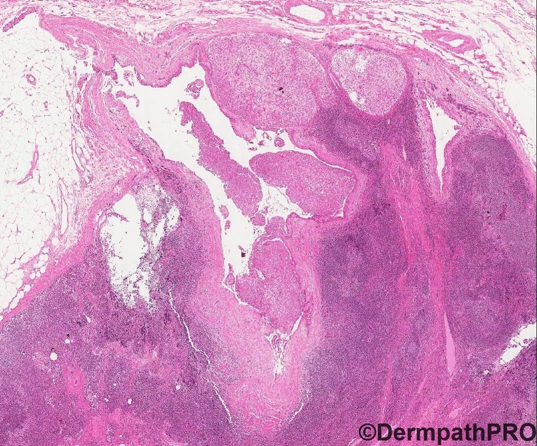
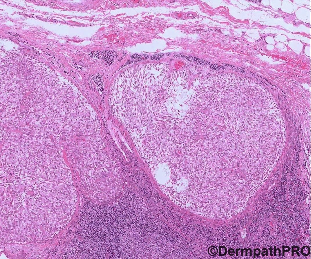
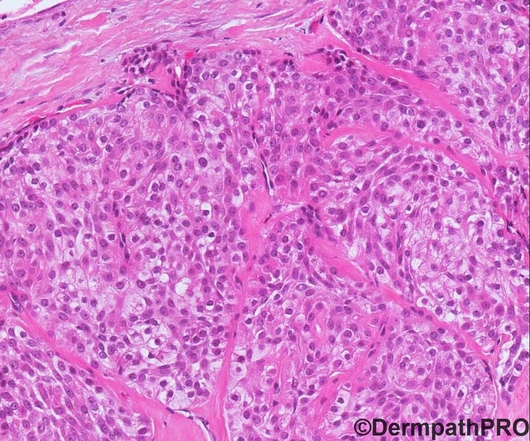
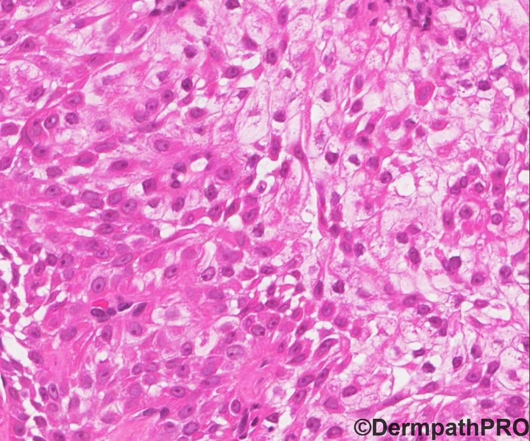
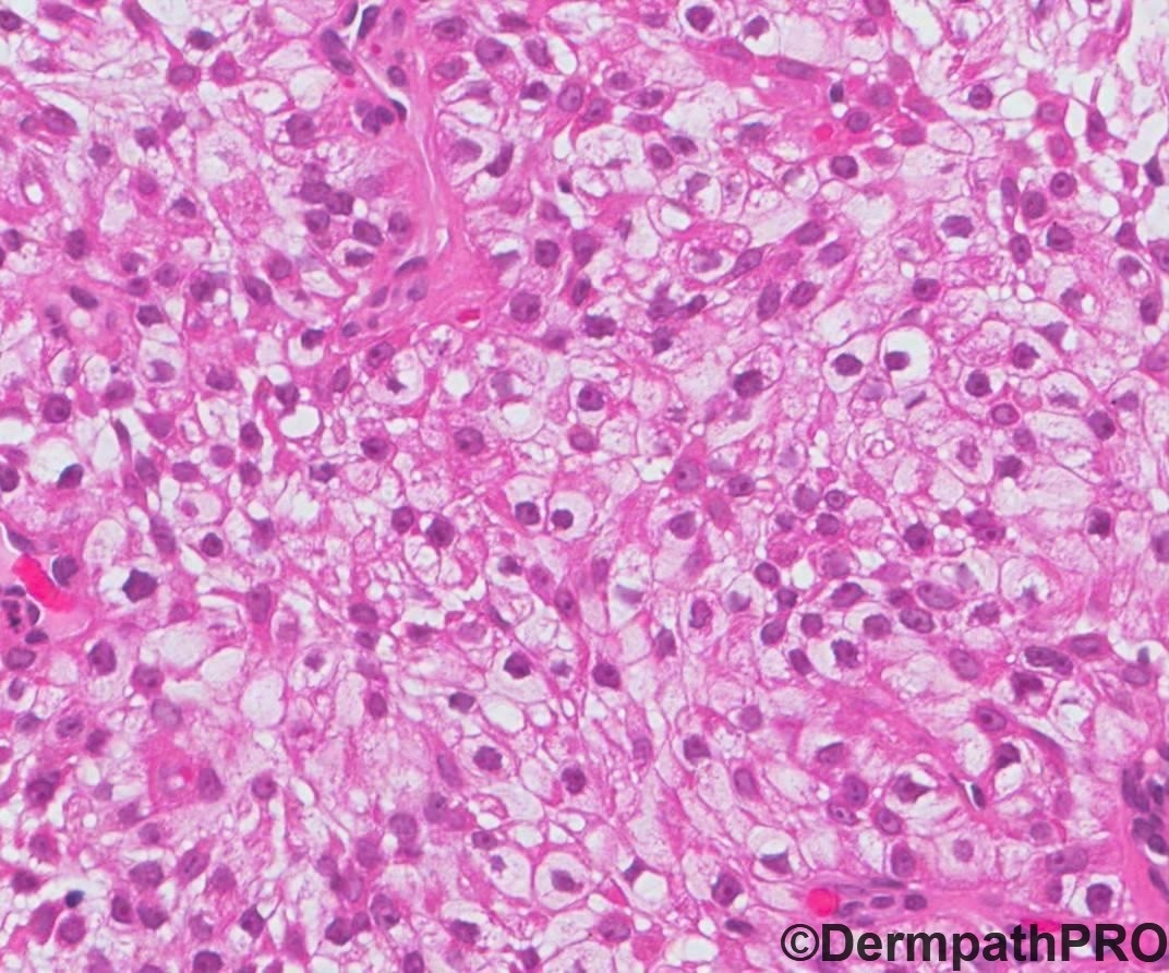
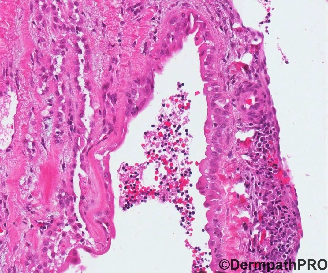
37 year old male – right axillary lymph node biopsy.Previous history of partial nephrectomy (left kidney) for clear cell renal cell carcinoma. Also history of benign adnexal tumour excised from axilla, 3 years back.


  • Like 1
  Report Record

User Feedback


Krishnakumar subramanian

Posted

IHC data on CAIX and CK 7 needed with a history of Clear cell RCC

Share this comment


Link to comment
share_externally

At first glance, it seems obviously a metastatic clear cell renal cell carcinoma. The clinical history and the histological pictures are pretty suggestive of RCC metastasis. But there is a history of a benign adnexial tumor in the axilla 3 years ago. Looking closer, we do not see the classical vasculature of RCC. There are reports of a rare nodal clear cell hidradenoma wich could be primary nodal or secondary to a "benign" metastasis. IHQ with PAX-8, RCC, p63 and keratin 5/6 could help in this differentiation.

Share this comment


Link to comment
share_externally

Meenakshi Batrani

Posted

4 hours ago, IgorSC said:

At first glance, it seems obviously a metastatic clear cell renal cell carcinoma. The clinical history and the histological pictures are pretty suggestive of RCC metastasis. But there is a history of a benign adnexial tumor in the axilla 3 years ago. Looking closer, we do not see the classical vasculature of RCC. There are reports of a rare nodal clear cell hidradenoma wich could be primary nodal or secondary to a "benign" metastasis. IHQ with PAX-8, RCC, p63 and keratin 5/6 could help in this differentiation.

Agree. Lymph node involvement by clear cell nodular hidradenoma is known. In last image, there appears to be a cystic/duct like structure with apocrine type lining. IHC needed to differentiate from RCC. 

Share this comment


Link to comment
share_externally

Agree with above.  There aren’t erythrocytes between clear cell nests and there is an obvious apocrine cystic change in last fig.  Thinking of a Low grade hydradenocarcinoma.  

Share this comment


Link to comment
share_externally

Dr. Richard Carr

Posted

Agree looks like hidradenoma in a LN.

Share this comment


Link to comment
share_externally


Join the conversation

You can post now and register later. If you have an account, sign in now to post with your account.

Guest
Add a comment...

×   Pasted as rich text.   Paste as plain text instead

  Only 75 emoji are allowed.

×   Your link has been automatically embedded.   Display as a link instead

×   Your previous content has been restored.   Clear editor

×   You cannot paste images directly. Upload or insert images from URL.

Loading...

×
×
  • Create New...