Jump to content
In this section we have spot diagnoses posted on a daily basis since June 2010, now over 4000! You can review the archived cases and read the suggested diagnoses by users and the final comment by the contributors.
Case are uploaded each week day by 10 am UK time with the correct diagnosis will generally be posted at 8 pm UK time. Why not view the most recent spot diagnosis and proffer a diagnosis?

Case Number : Case 2462 - 10 December 2019 Posted By: Uma Sundram

Please read the clinical history and view the images by clicking on them before you proffer your diagnosis.
Submitted Date :
   (0 reviews)

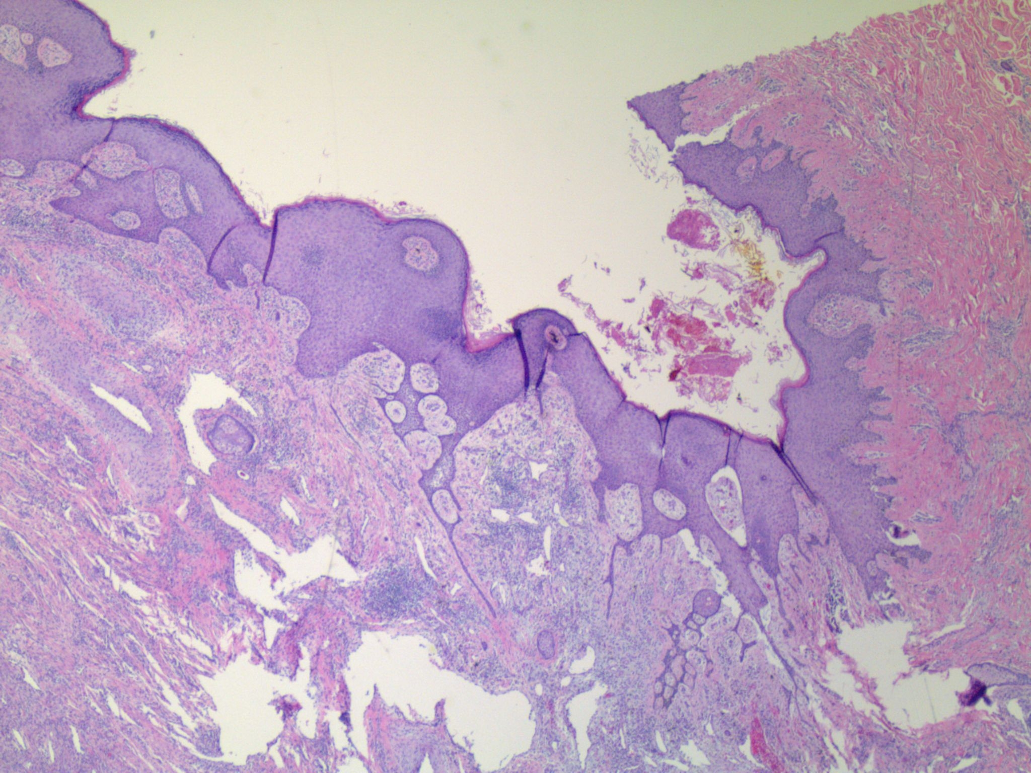
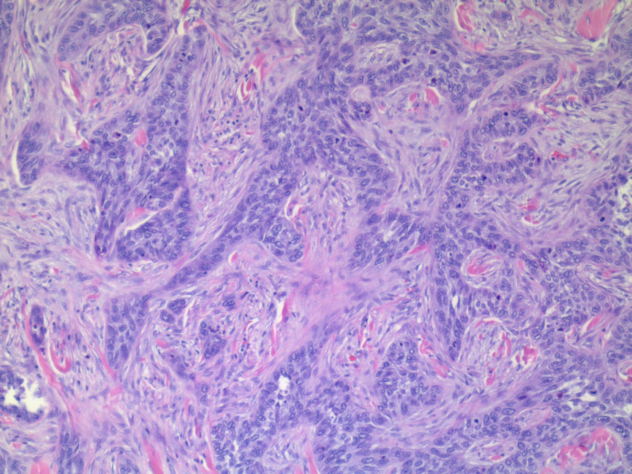
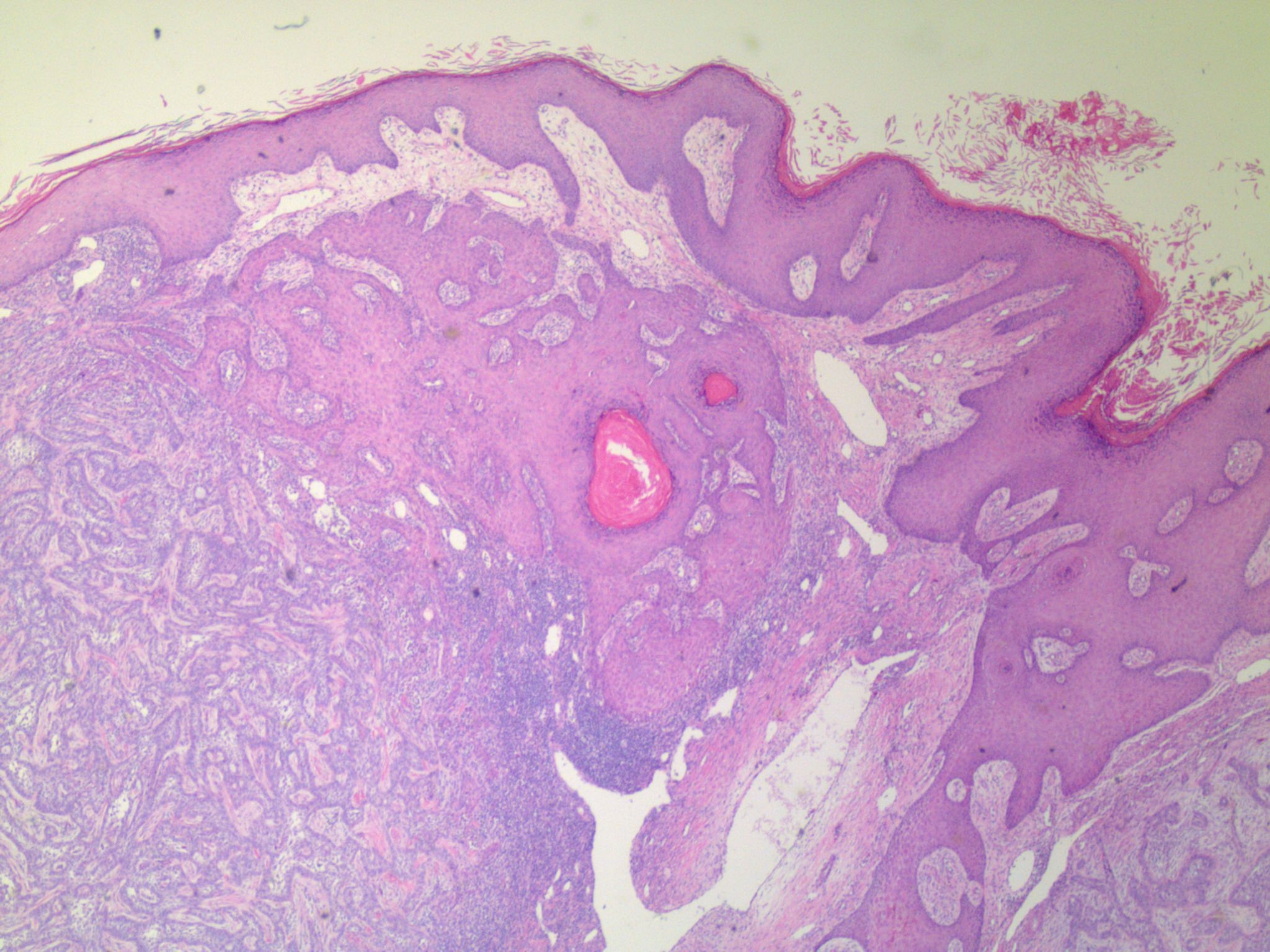
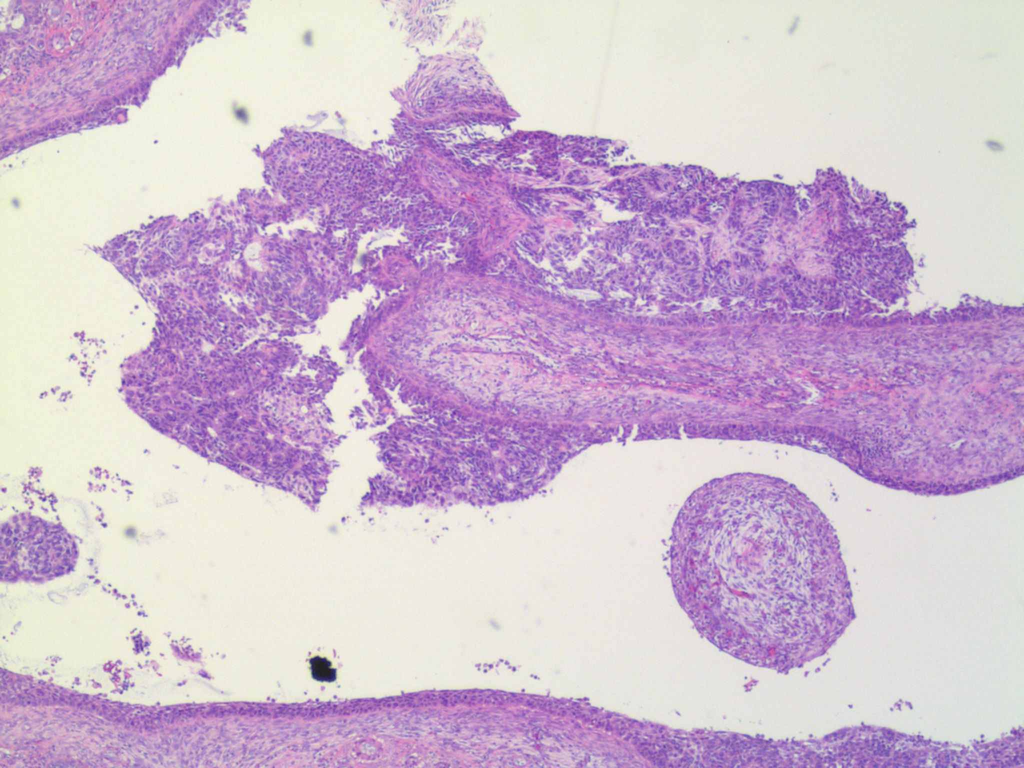
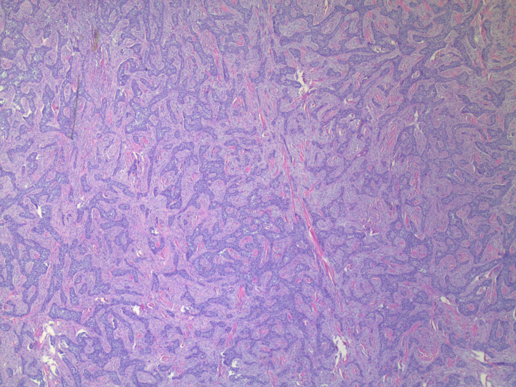
55 year old male with growing lesion on shoulder.


  Report Record

User Feedback


Krishnakumar subramanian

Posted

infiltrative BCC

can  we have BerEP4. IHC

Share this comment


Link to comment
share_externally

I think that this is a basal cell carcinoma which shows features of infiltrative pattern, fibroepithelioma of Pinkus and in one field syringocystadenoma papilliferum like differentiation. 

Share this comment


Link to comment
share_externally

Uma Sundram

Posted

BEREP4 is negative. Any thoughts other than BCC?

Share this comment


Link to comment
share_externally

Uma Sundram

Posted

Sorry, I note the diagnosis of syringocystadenocarcinoma as well.

Share this comment


Link to comment
share_externally

Eman El-Nabarawy

Posted

? SCC arising from syringocystadenoma papilliferum.

Share this comment


Link to comment
share_externally

Dr. Mona Abdel-Halim

Posted

Looking at images 2,3,5, and with the information of negative BEREP-4, I am thinking in basaloid SCC, but honestly I can not understand the orientation of image 4! Putting it with image 1, may be the tumor is arising in a cyst?! 

Share this comment


Link to comment
share_externally

?Hidroadenocarcinoma ( malignant hidroadenoma papilliferum )...

Share this comment


Link to comment
share_externally

The problem is: this lesion is malignant, so I can't think of a desmoplastic trichilemmoma...and trichilemmal carcinoma is a dark entity for me...but I'd like this diagnosis!!!

Share this comment


Link to comment
share_externally

Dr. Mona Abdel-Halim

Posted

12 minutes ago, vincenzo said:

The problem is: this lesion is malignant, so I can't think of a desmoplastic trichilemmoma...and trichilemmal carcinoma is a dark entity for me...but I'd like this diagnosis!!!

Had the same thoughts too!

Share this comment


Link to comment
share_externally

Dr. Richard Carr

Posted

Very difficult. The basaloid tumours seems to have a specialised stroma so I suppose we could consider a true trichoblastic carcinoma / trichoblastoma (mitotic++ but odd infiltrative-like growth) I'd also expect at least some BerEP4 staining. I think EMA is important here. If it's completely negative BCC with negative BerEP4 is back on the agenda. Could add CD10 for stromal condensations and CK20 for reactive merkel cells (in trichoblastoma). 

p53 and p16 might be useful if they show a null or a diffuse positive (supports malignant diagnosis). Agree the other cystic part does look like syringocystadenocarcinoma again the p53 and p16 & Ki67 might help with that and a p63 (for the myoepithelial layer. Nice challenge would like to see the glass! There is follicular pseudoepitheliomatous hyperplasia here. 

Share this comment


Link to comment
share_externally

Dr. Richard Carr

Posted

BTW please check out my case from last friday: Clinical & dermoscopic images.

Share this comment


Link to comment
share_externally

Dr. Richard Carr

Posted

Case 2455 is also open / undiagnosed from 29/11/19.

Share this comment


Link to comment
share_externally

Uma Sundram

Posted

Well, I'm glad I wasn't the only one challenged here! I have been having some trouble posting on my account, so apologize in advance for the tardiness in posting. EMA is negative so I did include BerEP4 negative BCC in my differential (common things being common). p53 was positive and Ki67 was relatively high, so I did not think of something benign. I was also intrigued by the cystic component so I did think of both syringocystadenocarcinoma as well as hidradenocarcinoma. Importantly, I alerted the clinician about the possibility of hidradenocarcinoma (I think the most likely fit, with pseudoepitheliomatous hyperplasia), since they can be more clinically aggressive than a typical (albeit unusual) BCC. We did go around and around about lineage though!

Share this comment


Link to comment
share_externally


Join the conversation

You can post now and register later. If you have an account, sign in now to post with your account.

Guest
Add a comment...

×   Pasted as rich text.   Paste as plain text instead

  Only 75 emoji are allowed.

×   Your link has been automatically embedded.   Display as a link instead

×   Your previous content has been restored.   Clear editor

×   You cannot paste images directly. Upload or insert images from URL.

Loading...

×
×
  • Create New...