Jump to content
In this section we have spot diagnoses posted on a daily basis since June 2010, now over 4000! You can review the archived cases and read the suggested diagnoses by users and the final comment by the contributors.
Case are uploaded each week day by 10 am UK time with the correct diagnosis will generally be posted at 8 pm UK time. Why not view the most recent spot diagnosis and proffer a diagnosis?

Case Number : Case 2464 - 12 December 2019 Posted By: Saleem Taibjee

Please read the clinical history and view the images by clicking on them before you proffer your diagnosis.
Submitted Date :
   (0 reviews)

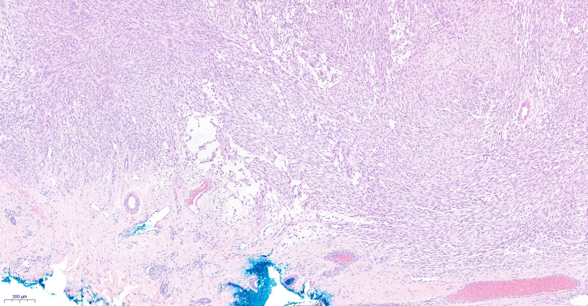
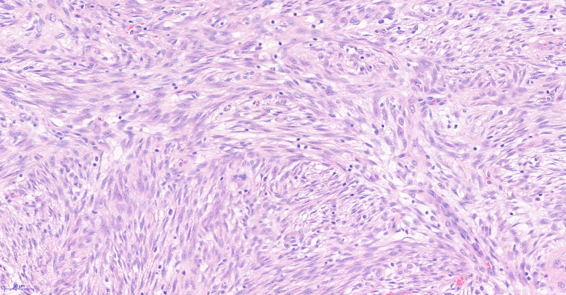
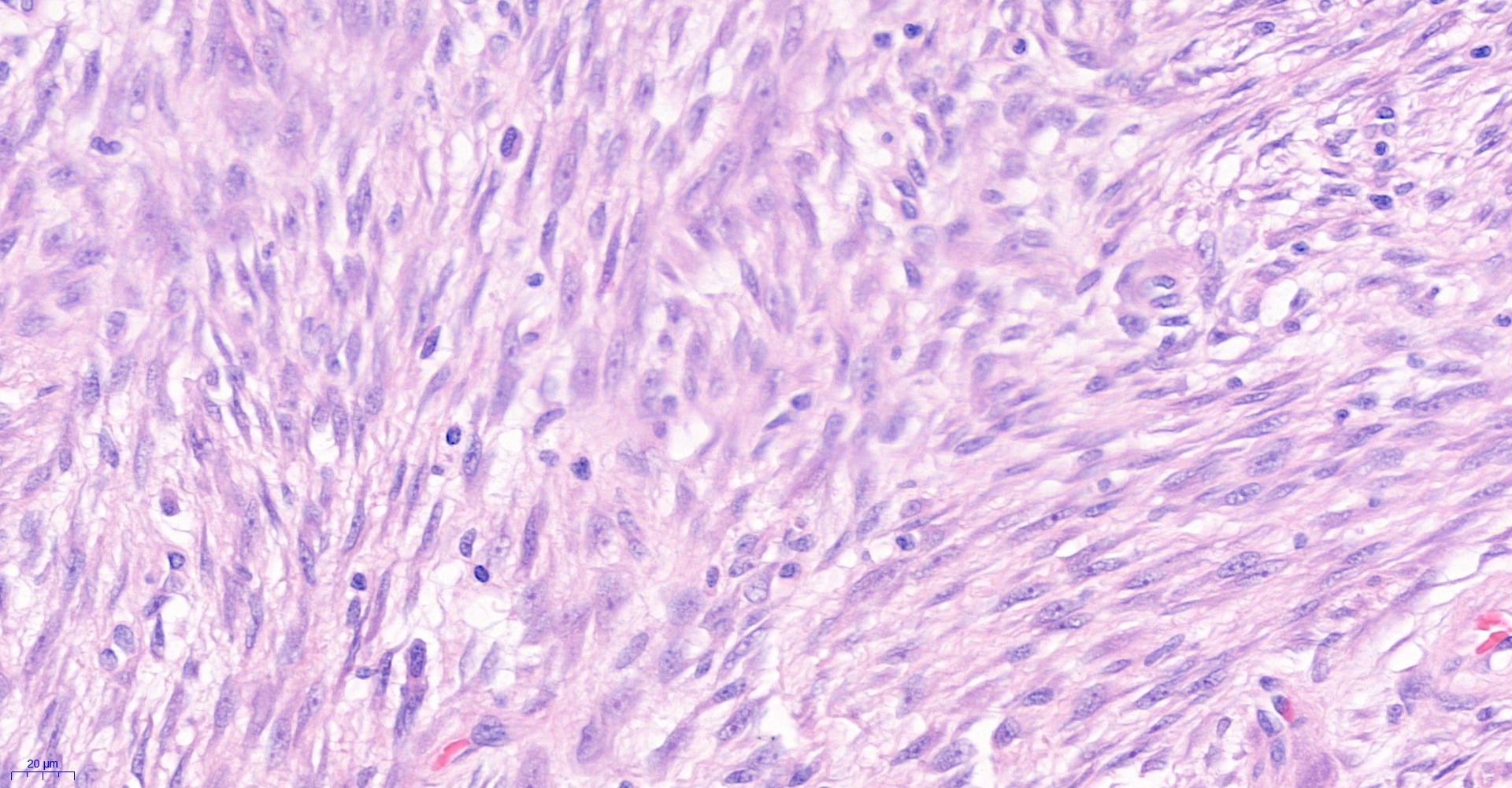
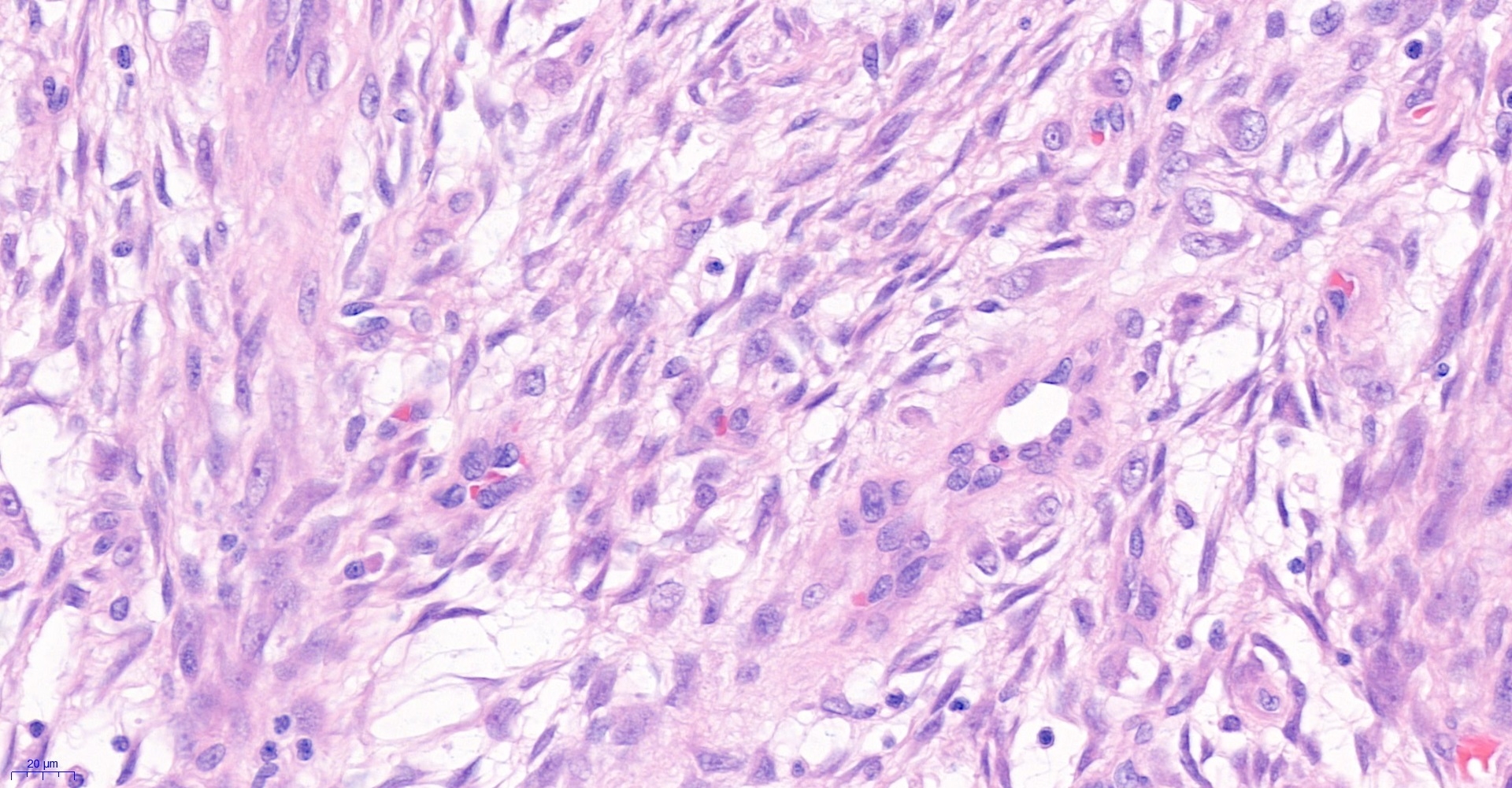
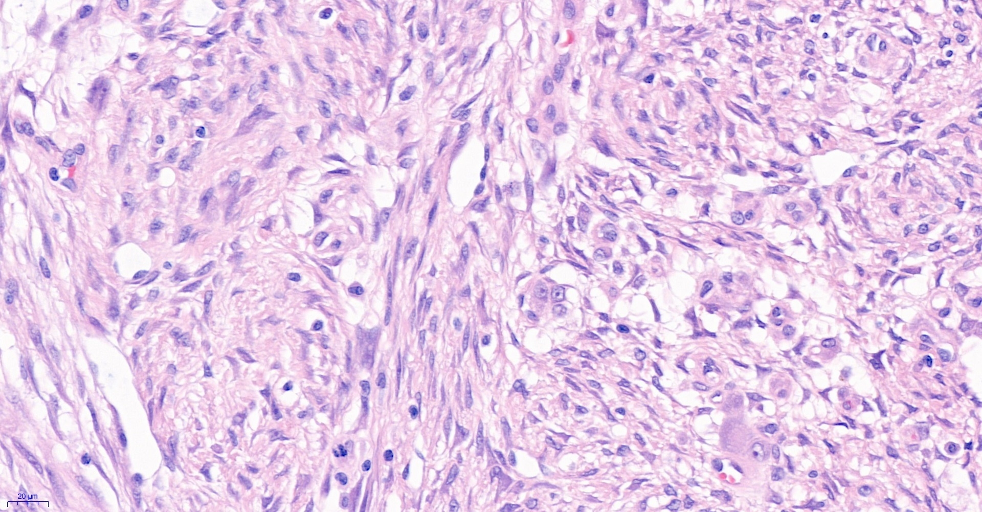
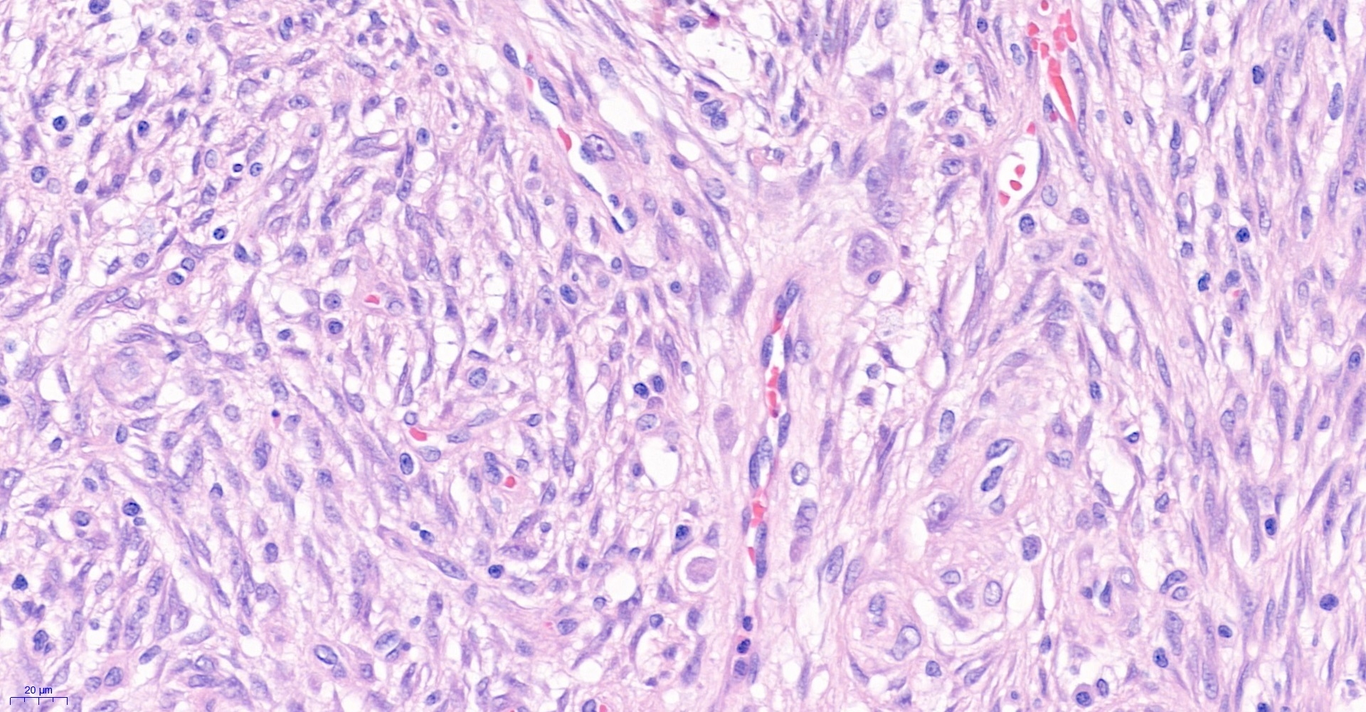
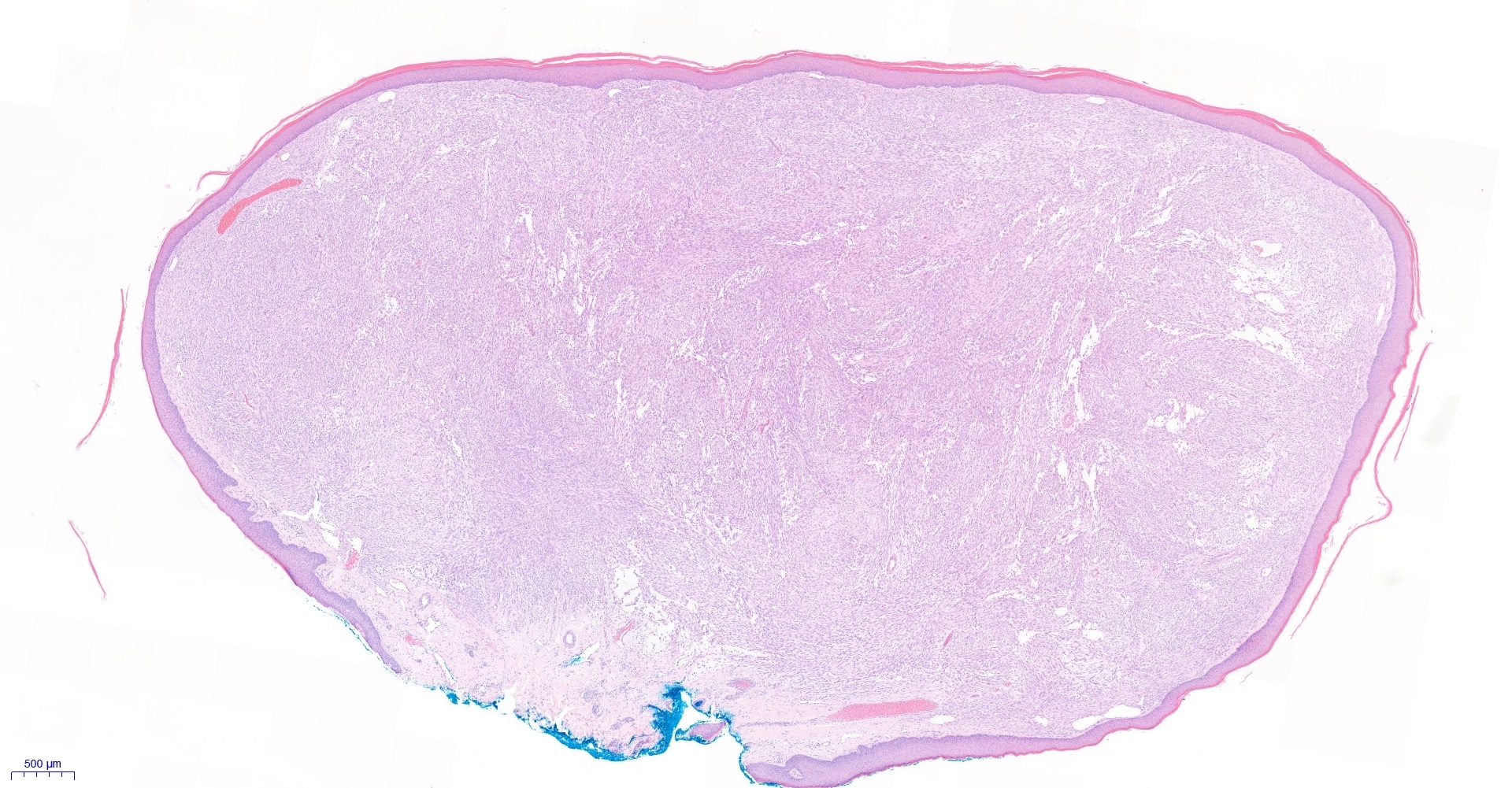
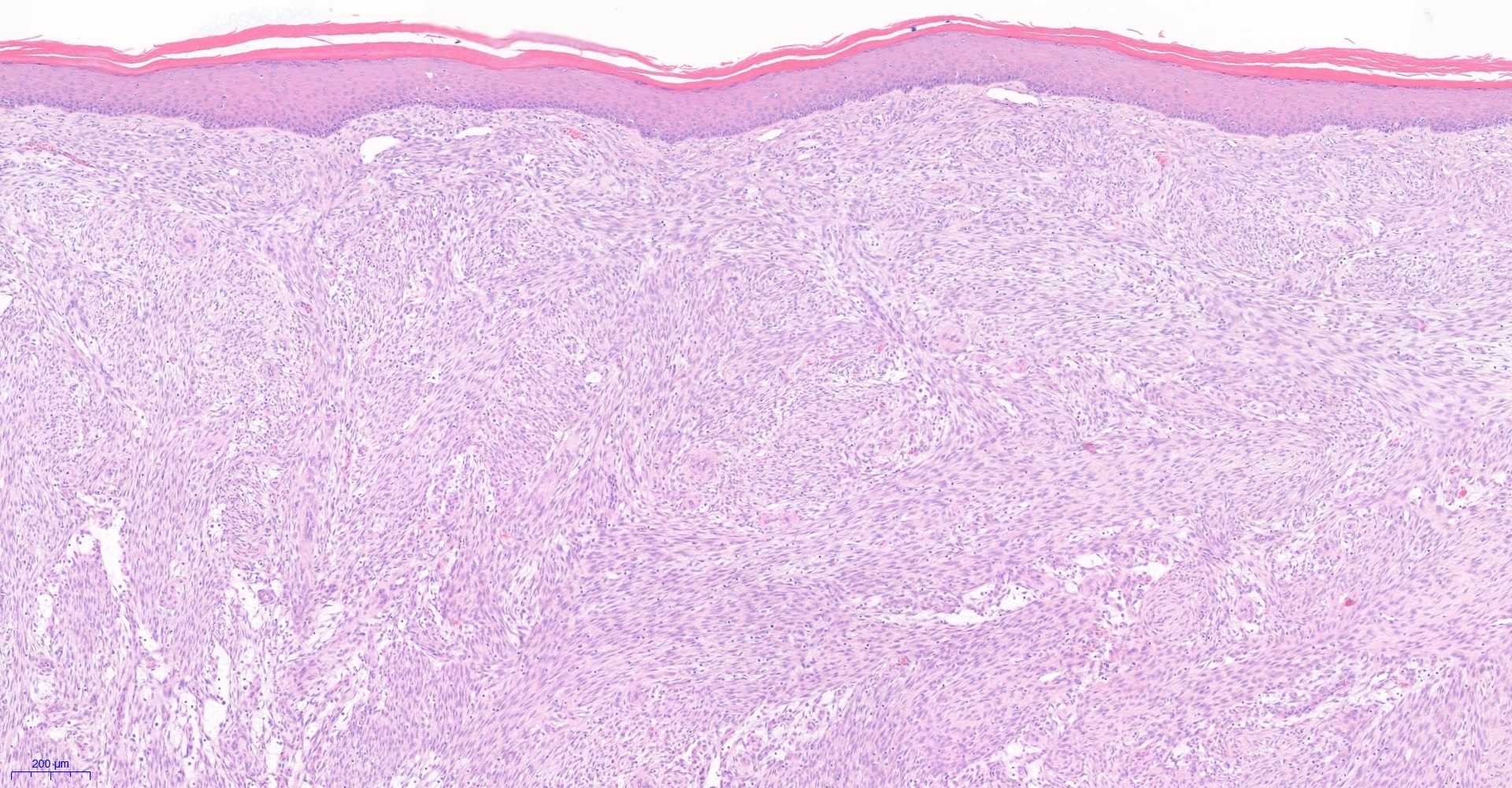
7 year old boy. Shave of lesion on mid back ?pyogenic granuloma


  Report Record

User Feedback


Victor Delgado

Posted

Agree with Mona, I would take into account BFH vs. Leiomyoma, as well.

Share this comment


Link to comment
share_externally

17 hours ago, Dr. Mona Abdel-Halim said:

Dermal nodular fasciitis?


Yes! Dermal NF, polypoid variant

Share this comment


Link to comment
share_externally

Krishnakumar subramanian

Posted

spindle cell  juvenile xanthogranuloma to be also considered. I am not able to see giant cells

Share this comment


Link to comment
share_externally

Saleem Taibjee

Posted

Nodular fasciitis was considered in the differential diagnosis, but in the end was not favoured. The silhouette is the clue.

Share this comment


Link to comment
share_externally

OK! The silhouette would be typical of epithelioid histiocytoma... but, sailing like a salmon, against the current, I favor a polypoid nodular fasciitis ( Bracey TS, Wharton S, Smith ME.J Cutan Pathol. 2009 Sep;36(9):980-2. doi: 10.1111/j.1600-0560.2008.01159.x.).


Share this comment


Link to comment
share_externally

There are some large cells with vesicular nucleus, prominent nucleolus, and abundant basophilic cytoplasm, reminiscent of ganglion cells. I agree with everybody that the lesion looks like nodular fasciitis, but since we see ganglion-like cells, the correct term is probably something starting with "proliferative" such as proliferative fasciitis or myositis, But those lesions are usually deep located. I checked Pubmed and there are rare cases of intradermal proliferative fasciitis which I think this case might represent an example of that entity. Don't know whether the lesion would have a fusion gene involving USP6 or not?

Share this comment


Link to comment
share_externally

Saleem Taibjee

Posted

Thank you for the interesting discussion on this case, which is probably not clear-cut. I agree that there are nodular fasciitis like features, and am interesting to learn about the polypoid variant described. However, the ALK was convincingly positive in this case (see below), and many of you will be aware of the relatively recent literature regarding ALK fusions (with concomitant ALK expression on immuno) now identified in epithelioid fibrous histiocytoma e.g. https://www.ncbi.nlm.nih.gov/pubmed/25413595. Admittedly this is not an entirely specific finding, but as far as I am aware, has not been shown in nodular fasciitis (please correct me if I am wrong). I did share the case with Eduardo Calonje, and his opinion was also of EFH. He tells me he has seen other instances of this rather 'spindle cell variant' of EFH which often causes confusion. I also shared the case with Raul Perrett. His comments as follows: "Yes Saleem it did cross my mind and after reviewing the case I think you are right. I have never seen an EFH with a predominant spindle cell component very closely resembling myofibroblasts + myxoid areas. However, this has been already published! (https://www-ncbi-nlm-nih-gov.gate2.inist.fr/pubmed/29430713). Considering the genomic and morphological features of EFH, I think it is probably more closely related to myofibroblastic tumors than fibrous histiocytoma/dermatofibroma."

This was my own patient. The clinical appearance was of an exophytic polypoid red lesion which certainly (in hindsight) could have been EFH rather than my clinically favoured pyogenic granuloma. Sadly I did not take a photograph before shaving the lesion!

05191_10.0x ALK labelled.jpg

Share this comment


Link to comment
share_externally

Dr. Mona Abdel-Halim

Posted

Amazing case Saleem! It seems the advances in molecular biology will change all our norms and standards! Thank u for sharing. 

Share this comment


Link to comment
share_externally

Eman El-Nabarawy

Posted

Thank u so much Dr Saleem. I thought of EFH but this is the first time to see its spindle cell form. 

Share this comment


Link to comment
share_externally

Other cutaneous neoplasms that could be Alk-positive and I would consider in this case would be Spitz nevus (my first hypothesis) and Angiomatoid fibrous hystiocytoma (more related to soft tissue and has different histology findings). Was Spitz nevus ruled out? Nice case.

Share this comment


Link to comment
share_externally

Saleem Taibjee

Posted

Thanks, Igor. Melanocytic markers (S100, Melan-A) were negative.

Share this comment


Link to comment
share_externally

Krishnakumar subramanian

Posted

sir, inflammatory myofibroblastic tumour is all positve

However morphology is different

Share this comment


Link to comment
share_externally


Join the conversation

You can post now and register later. If you have an account, sign in now to post with your account.

Guest
Add a comment...

×   Pasted as rich text.   Paste as plain text instead

  Only 75 emoji are allowed.

×   Your link has been automatically embedded.   Display as a link instead

×   Your previous content has been restored.   Clear editor

×   You cannot paste images directly. Upload or insert images from URL.

Loading...

×
×
  • Create New...