Jump to content
In this section we have spot diagnoses posted on a daily basis since June 2010, now over 4000! You can review the archived cases and read the suggested diagnoses by users and the final comment by the contributors.
Case are uploaded each week day by 10 am UK time with the correct diagnosis will generally be posted at 8 pm UK time. Why not view the most recent spot diagnosis and proffer a diagnosis?

Case Number : Case 2465 - 13 December 2019 Posted By: Dr. Richard Carr

Please read the clinical history and view the images by clicking on them before you proffer your diagnosis.
Submitted Date :
   (0 reviews)

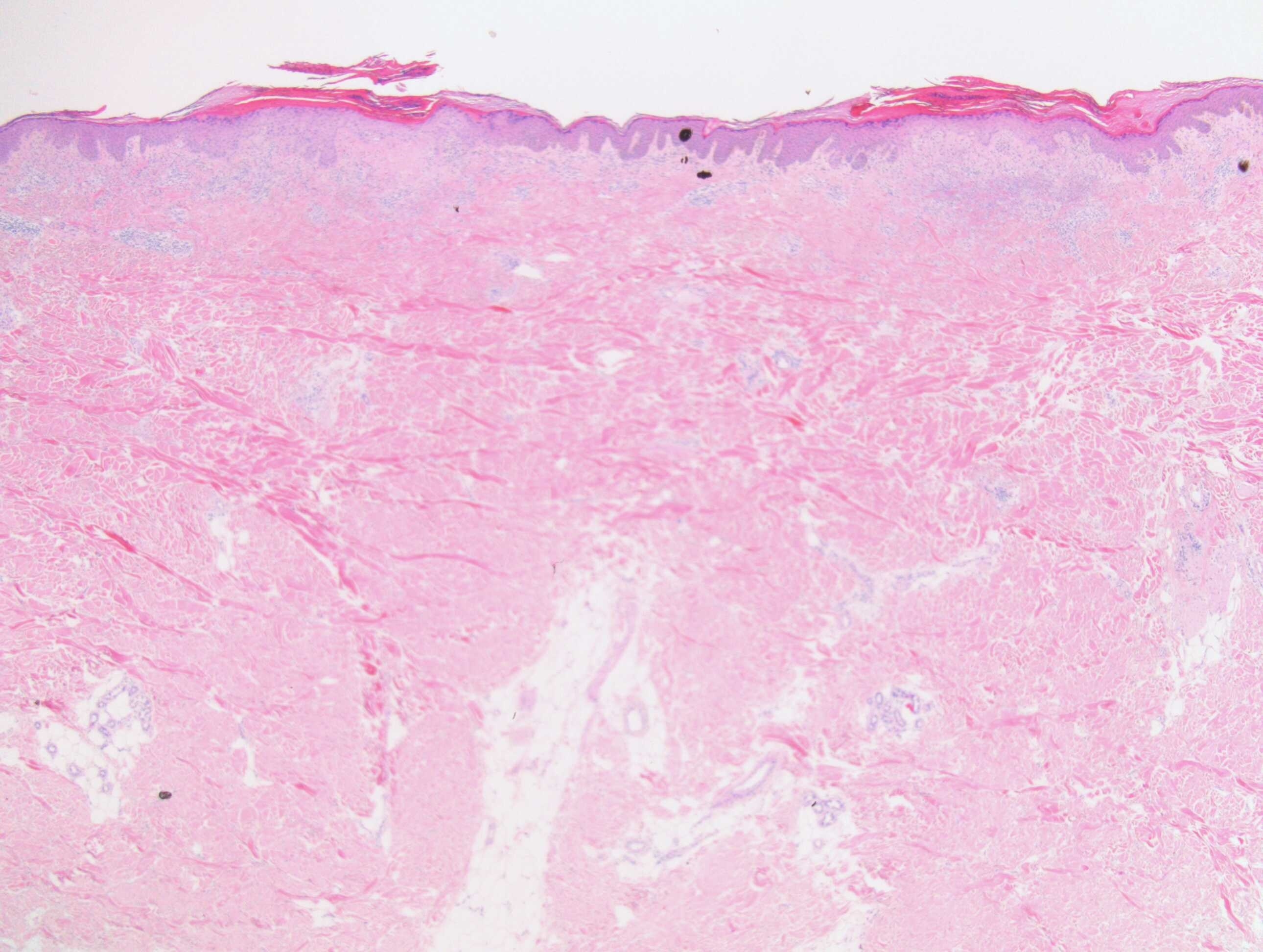
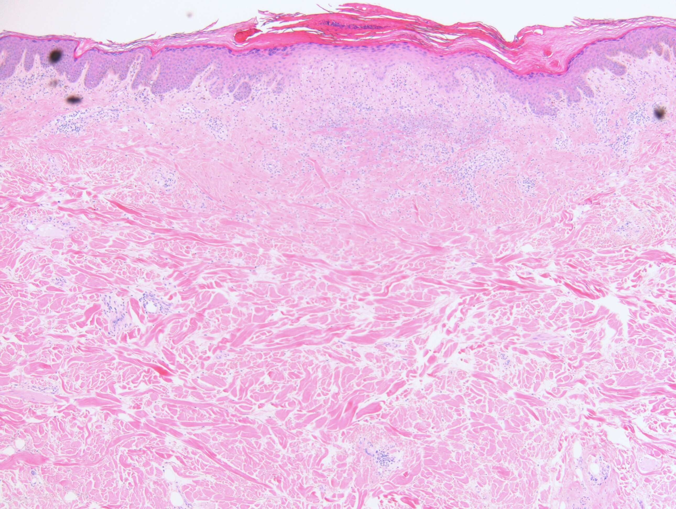
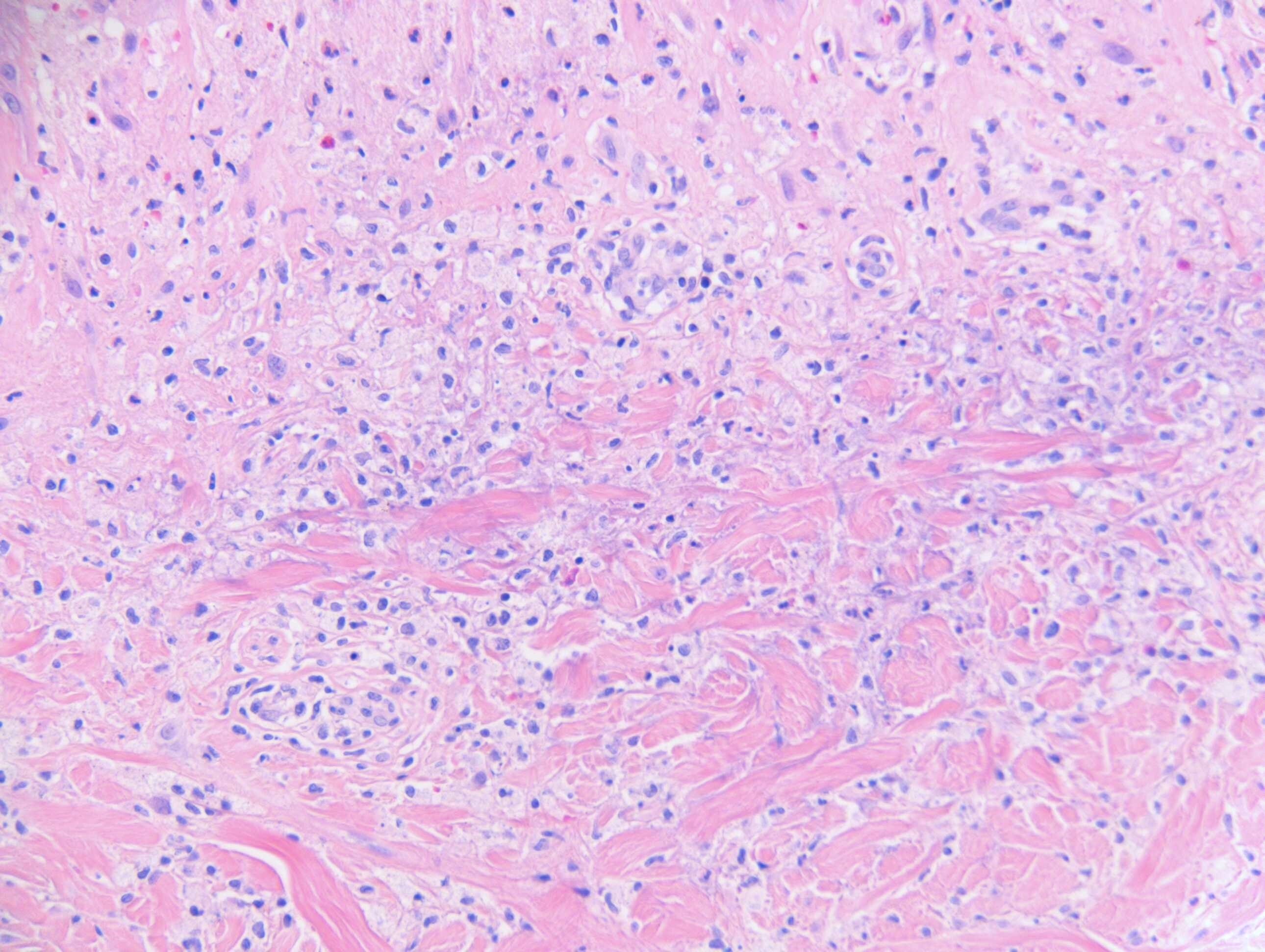
Itchy macular papular rash, prickling sensation since March 2019. Midline lower back,
back of shoulders & upper back, neck & scalp. ?lichen planus, ?lichen amyloidosis.
Skin type 4. Case c/o Dr Arti Bakshi

Edited by Admin_Dermpath


  Report Record

User Feedback


Maculopapular cutaneous mastocytosis (urticaria pigmentosa)?

Share this comment


Link to comment
share_externally

Eman El-Nabarawy

Posted (edited)

DH. Clinical clues; characteristic sites, itchy rash. Pathological clues, excoriations, edema, fibrin, acantholytic or shadow cells in the tips of dermal papillary, flame-like figures, few esinophils, neutrophils, dust.

Edited by Eman El-Nabarawy

Share this comment


Link to comment
share_externally

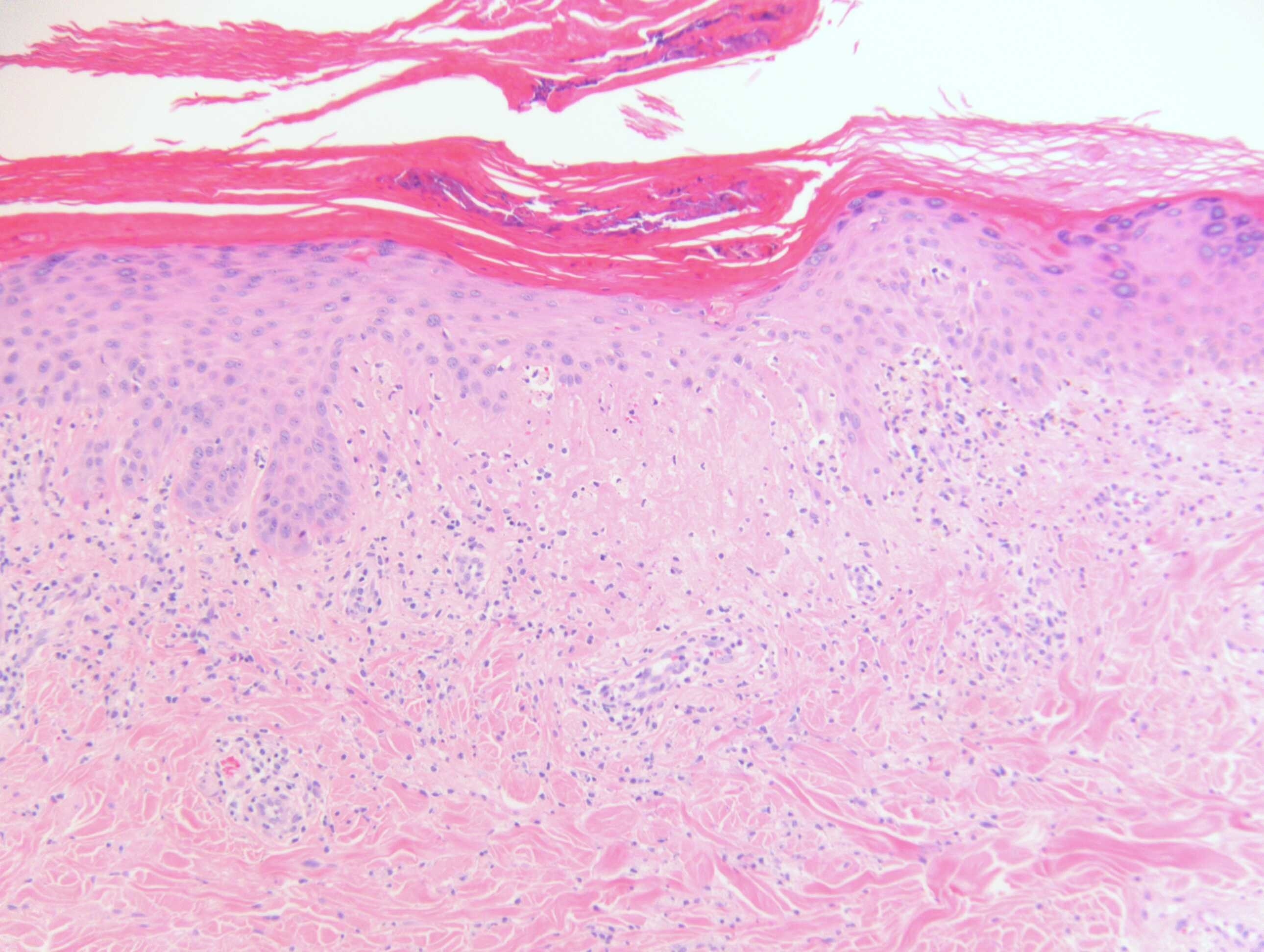
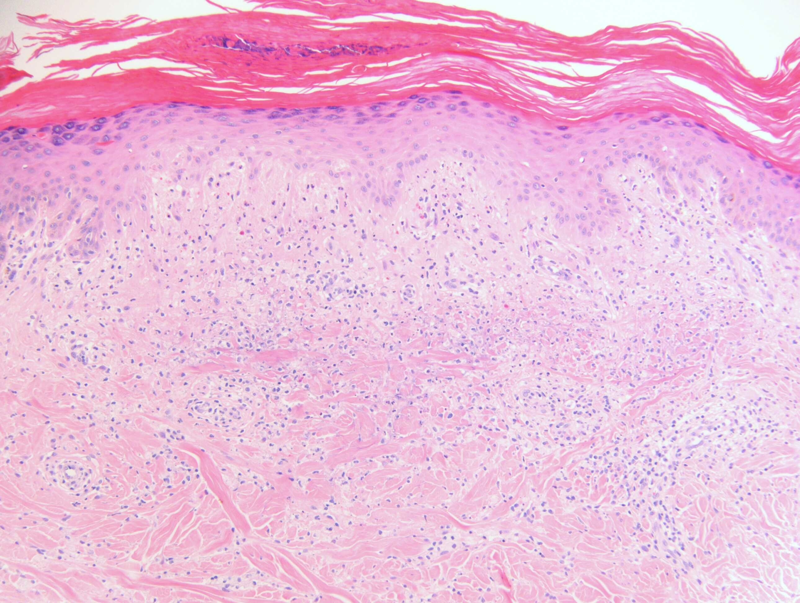
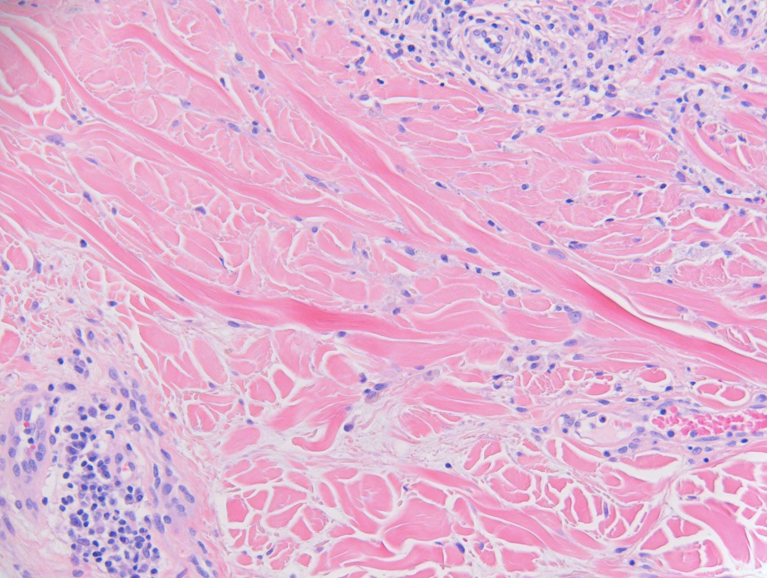
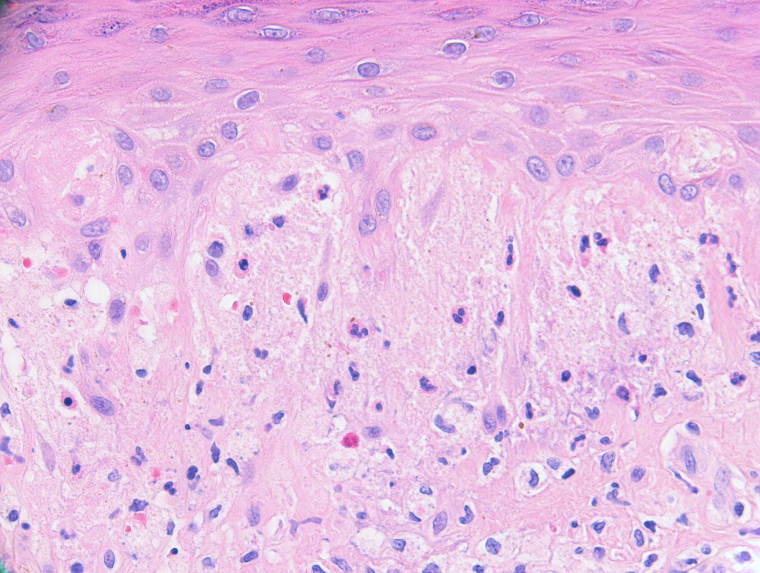
Hyperkeratosis with parakeratosis, fuzzy dermoepidermal junction, dermal mononuclear cell infiltrate and mucin deposits are the saliant features. I think it's LE or dermatomyositis (sine myositis if there is no myositis). 

Share this comment


Link to comment
share_externally

Dr. Mona Abdel-Halim

Posted

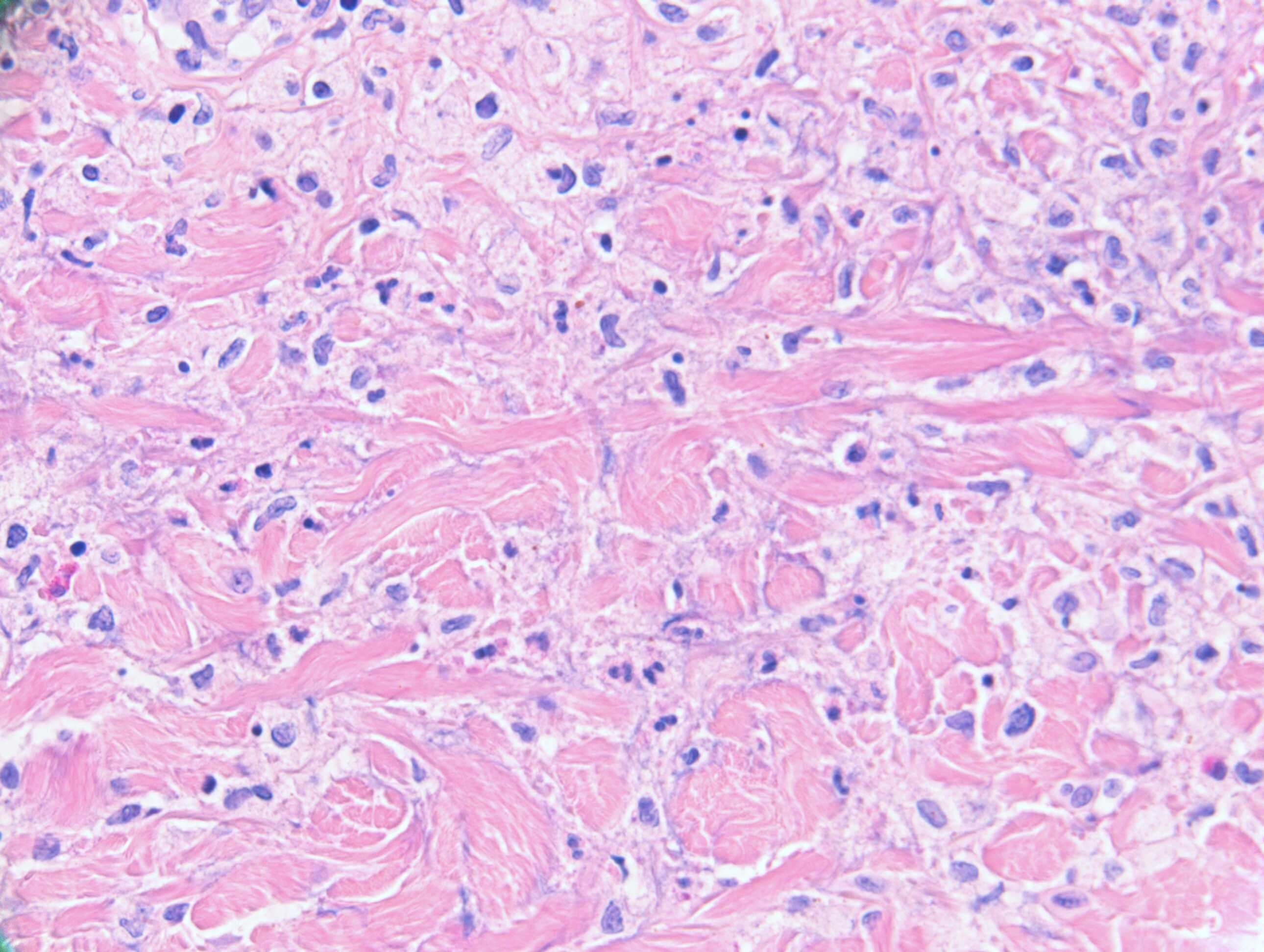
I think we are dealing with a connective tissue disease (LE or dermatmyositis) (epidermal changes fits with including interface changes and ? thick basement membrane) showing also features of interstitial granulomatous dermatitis (palisaded neutrophilic and granulomatous dermatitis).  So I am in simultaneous agreement with both Anil Patki and dermpath1! The distribution of the lesions including back of shoulders, upper back and neck makes me think of shawl sign of dermatomyositis. Interesting case! Looking forward to the correct answer. 

Share this comment


Link to comment
share_externally

Meenakshi Batrani

Posted

There is GA like pattern, significant eosinophils, neutrophils and plasma cells  along with focal epidermal necrosis with overlying parakeratotic scale.

In view of itchy maculo-papular rash, I would consider drug related interstitial granulomatous dermatitis (drug rash); there are even reports of generalized granuloma annulare associated with scabies and also secondary syphillis with IGD/GA like pattern. Although, IGD with connective tissue disease is a histological differential but in view of itchy maculopapular rash, it is less favoured.  

Share this comment


Link to comment
share_externally

Very difficult case. I think this is an intersticial granulomatous dermatosis, probably related to drug use. I do not think this is LE, despite it can simulate many dermatosis, because the lack of deep/periadnexial inflammation, the focal vacuolar alteration and the presence of eosinophils.

Share this comment


Link to comment
share_externally

Krishnakumar subramanian

Posted

dermatitis herpetiformis with GA like pattern

Share this comment


Link to comment
share_externally

After a closer look: palisaded neutrophilic and granulomatous dermatitis? 

Share this comment


Link to comment
share_externally

Dr. Richard Carr

Posted

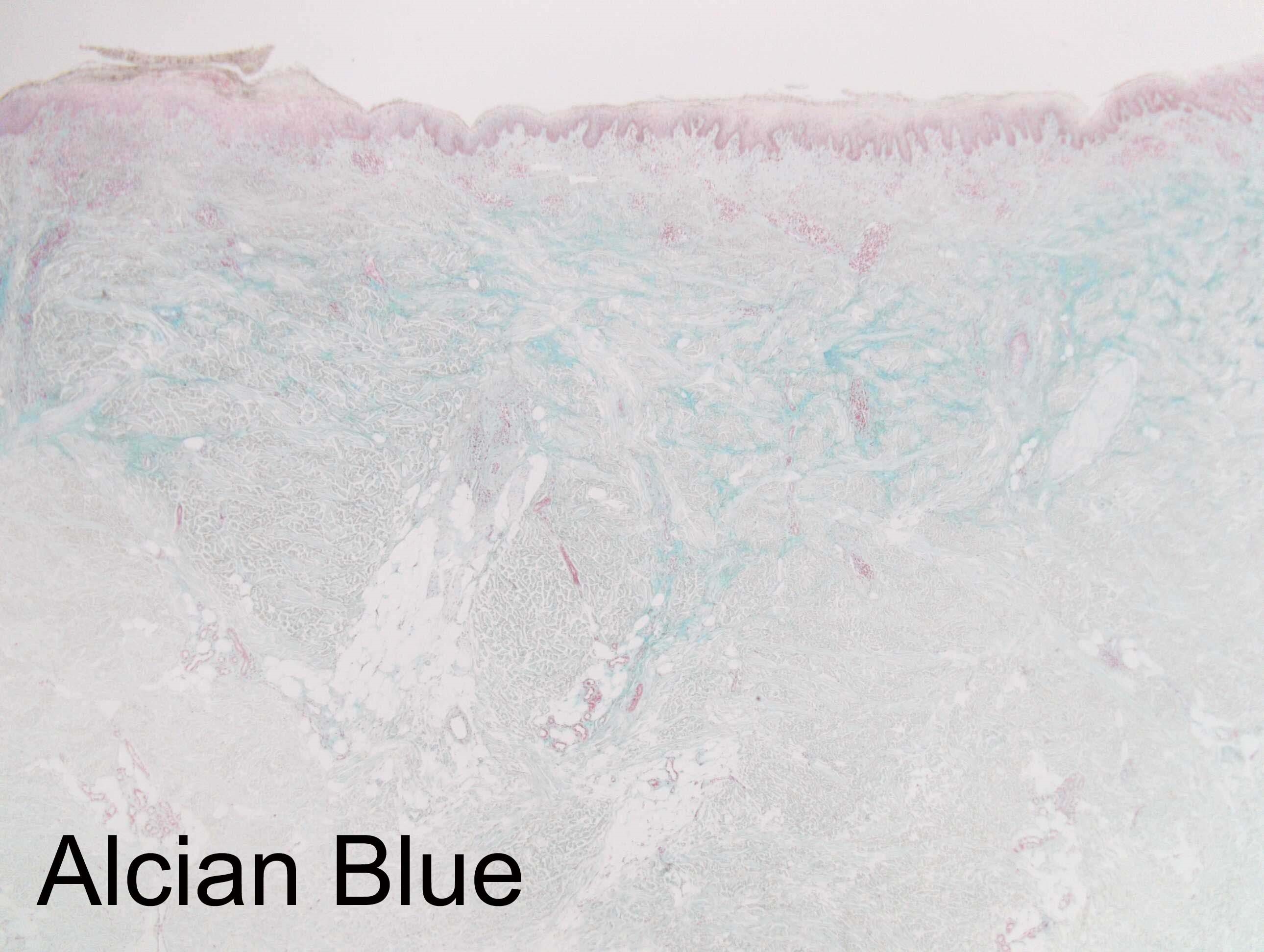
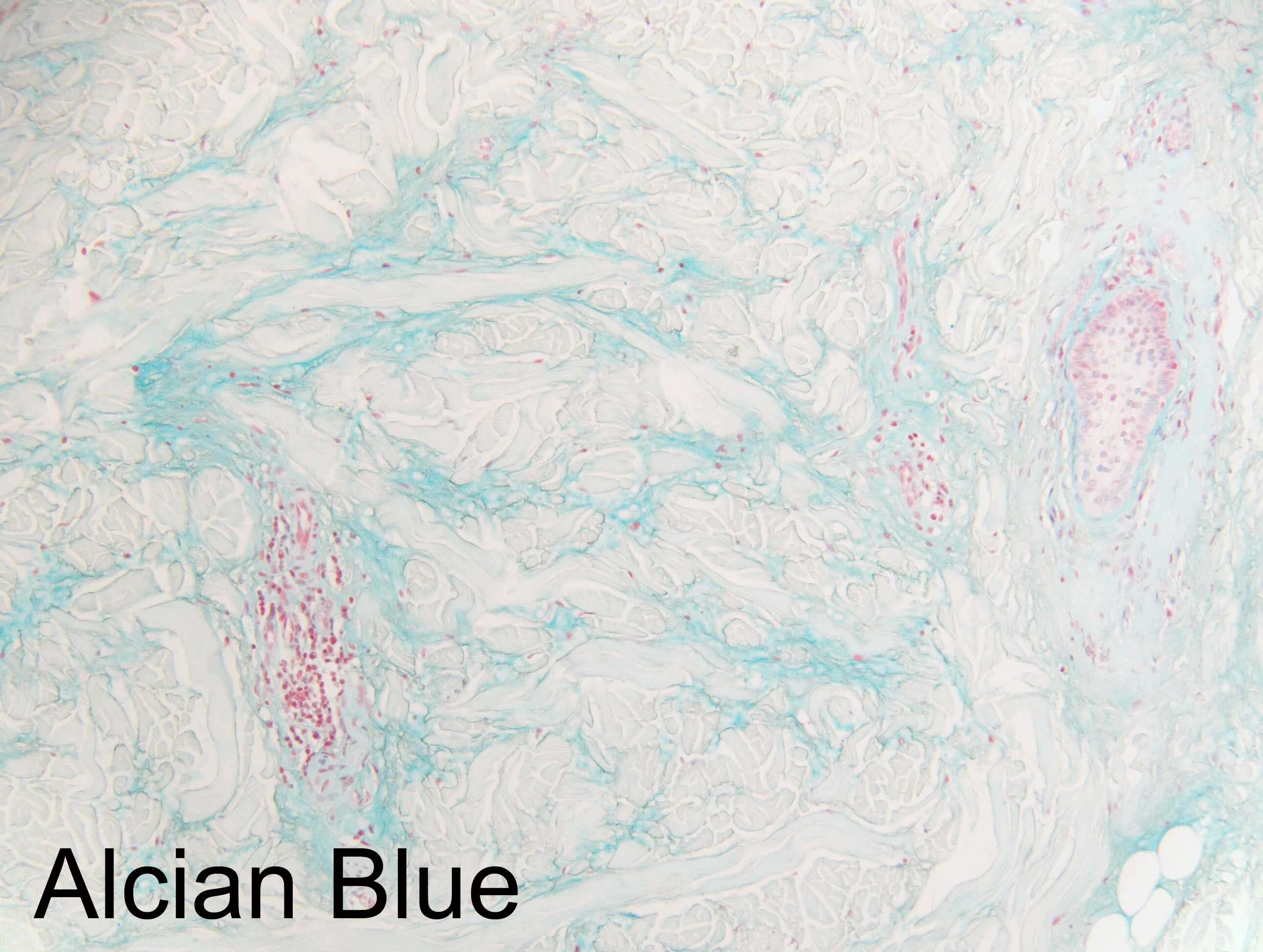
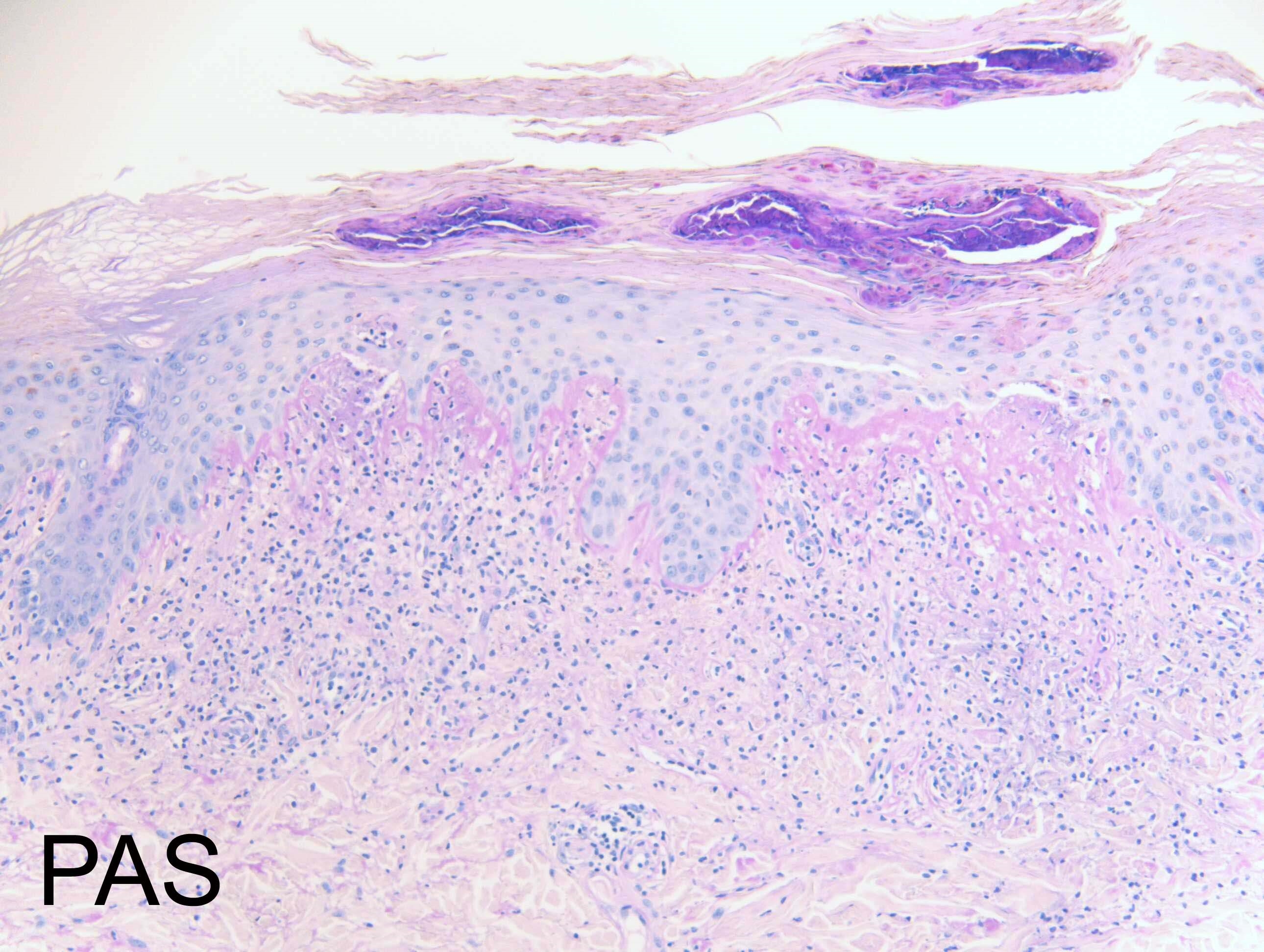
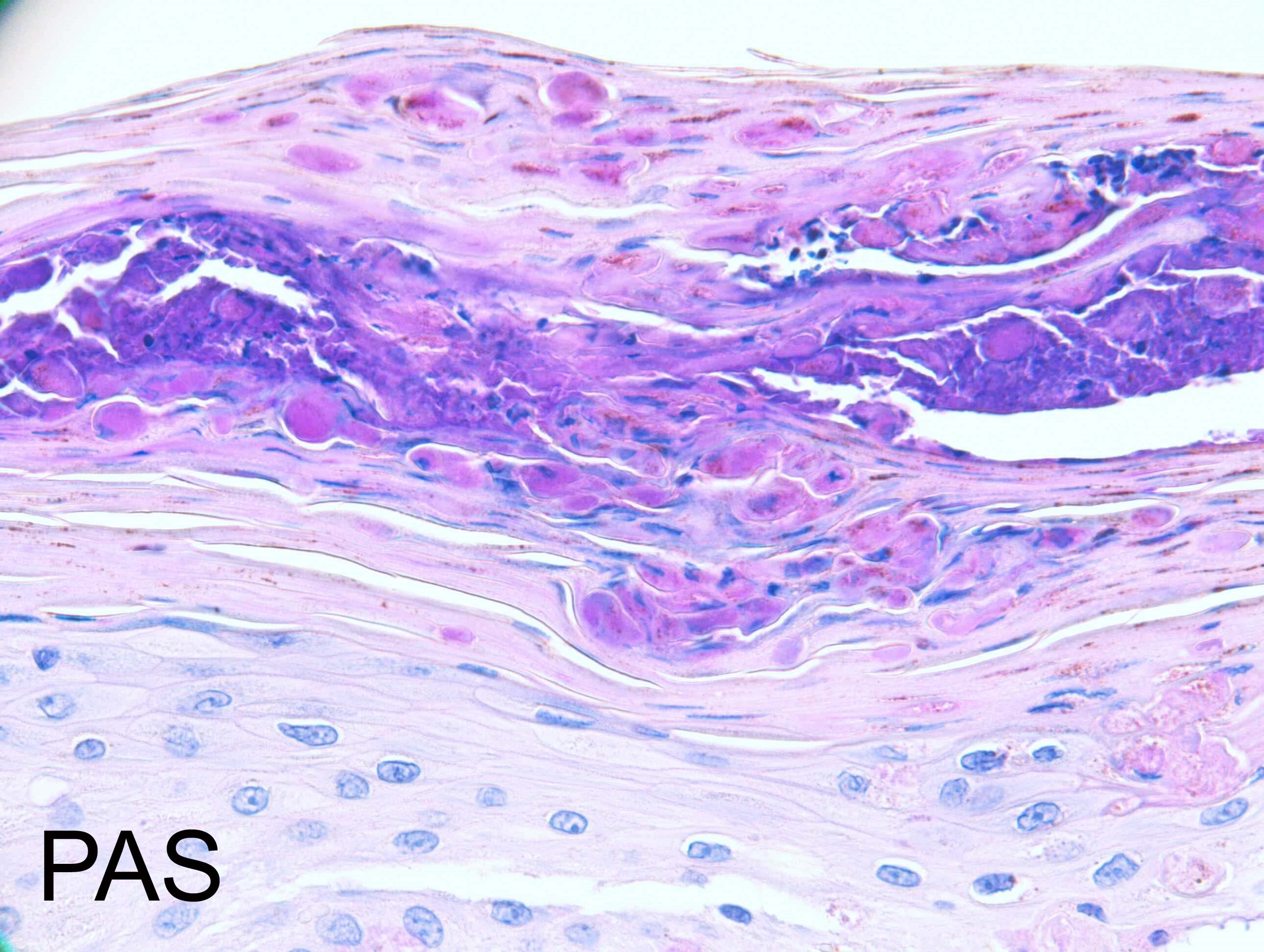
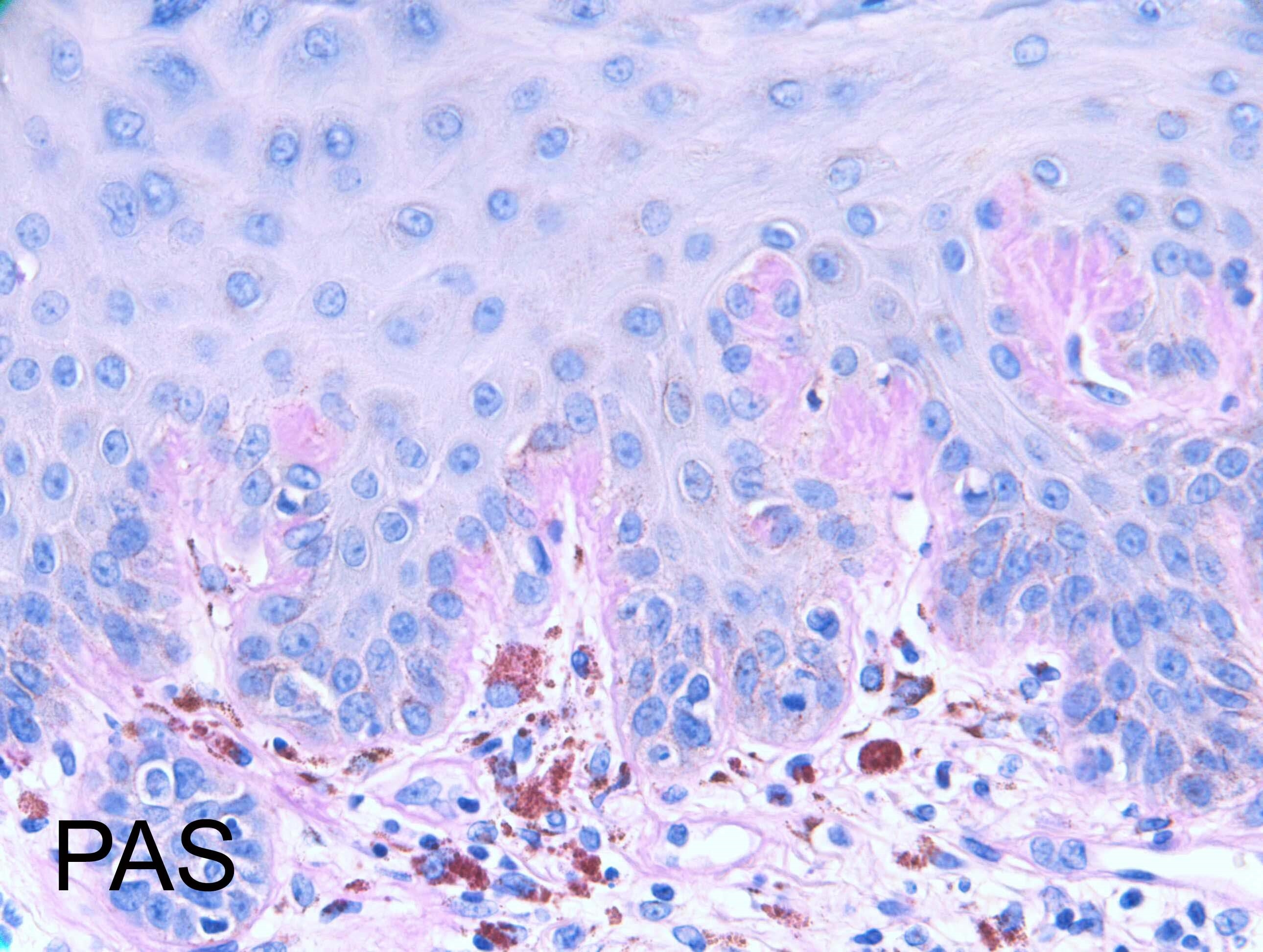
I've asked for some extra images to be posted of Alcian blue and PAS stain.

Share this comment


Link to comment
share_externally

Eman El-Nabarawy

Posted

Necrotic keratinocytes in the stratum corneum are clue to persistent lesions of adult-onset Still's disease (AOSD).

Share this comment


Link to comment
share_externally

Dr. Richard Carr

Posted

Nice discussion. This patient has SLE/Sjorgren's overlap. Recently started azothioprine & hydroxychloroquine. Arti and I both thought this was good for non-bullous neutrophilic deramtosis of autoimmune disease / SLE. 

I think we have a Churg-Strauss type granuloma here which can be a clue to lupus and rheumatoid. Nice increase mucin and focal basement membrane thickening on PAS. I agree old necrotic keratinocytes can be seen in the cornified layer probably when the process was more active.

Am J Dermatopathol. 2013 Aug;35(6):655-60. doi: 10.1097/DAD.0b013e3182840f7f.
Autoimmunity-related neutrophilic dermatosis: a newly described entity that is not exclusive of systemic lupus erythematosus.
Saeb-Lima M1, Charli-Joseph Y, Rodríguez-Acosta ED, Domínguez-Cherit J.
Author information
1 Pathology Department, National Institute of Medical Sciences and Nutrition, Salvador Zubiran, Mexico City, Mexico. Marcela.saeblima@gmail.com
Abstract
Neutrophilic dermatoses have long been known to be associated with autoinmune systemic diseases. Recently, a small number of cases of a disorder distinct from Sweet syndrome or bullous lupus erythematosus (LE) have been described as specifically related to systemic LE under diverse terms, including nonbullous neutrophilic dermatosis, nonbullous neutrophilic LE, and Sweet-like neutrophilic dermatosis. We describe 7 patients that developed urticarial lesions in the context of a known or concurrently diagnosed autoimmune connective tissue disease. Of a total of 7 patients, 6 were afflicted by systemic LE and 1 by rheumatoid arthritis and secondary Sjögren syndrome. Histological findings in all patients included an interstitial and perivascular neutrophilic infiltrate with leukocytoclasia, vacuolar alteration along the dermal-edidermal junction, and no vasculitis. Most patients had active systemic disease at the time of the cutaneous eruption. Skin lesions resolved rapidly after the administration of immunomodulating agents. In conclusion, we provide additional evidence of the existence of a recently defined nonbullous neutrophilic dermatosis in the context of autoimmune connective tissue diseases and propose the term autoimmunity-related neutrophilic dermatosis as an appropriate designation. Furthermore, we believe that this entity should prompt physicians to screen the presence of an active systemic disorder in afflicted patients.

Share this comment


Link to comment
share_externally


Join the conversation

You can post now and register later. If you have an account, sign in now to post with your account.

Guest
Add a comment...

×   Pasted as rich text.   Paste as plain text instead

  Only 75 emoji are allowed.

×   Your link has been automatically embedded.   Display as a link instead

×   Your previous content has been restored.   Clear editor

×   You cannot paste images directly. Upload or insert images from URL.

Loading...

×
×
  • Create New...