Jump to content
In this section we have spot diagnoses posted on a daily basis since June 2010, now over 4000! You can review the archived cases and read the suggested diagnoses by users and the final comment by the contributors.
Case are uploaded each week day by 10 am UK time with the correct diagnosis will generally be posted at 8 pm UK time. Why not view the most recent spot diagnosis and proffer a diagnosis?

Case Number : Case 2469 - 19 December 2019 Posted By: Saleem Taibjee

Please read the clinical history and view the images by clicking on them before you proffer your diagnosis.
Submitted Date :
   (0 reviews)

72 Male
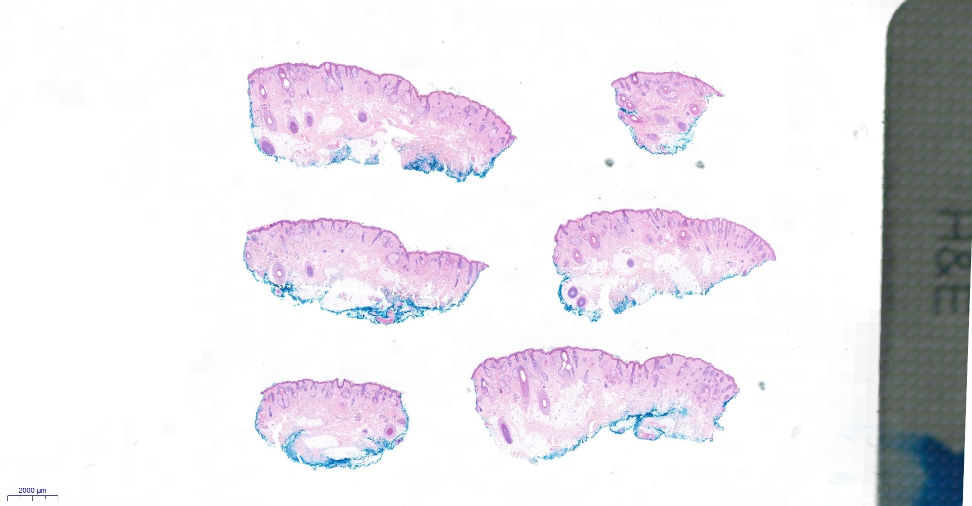
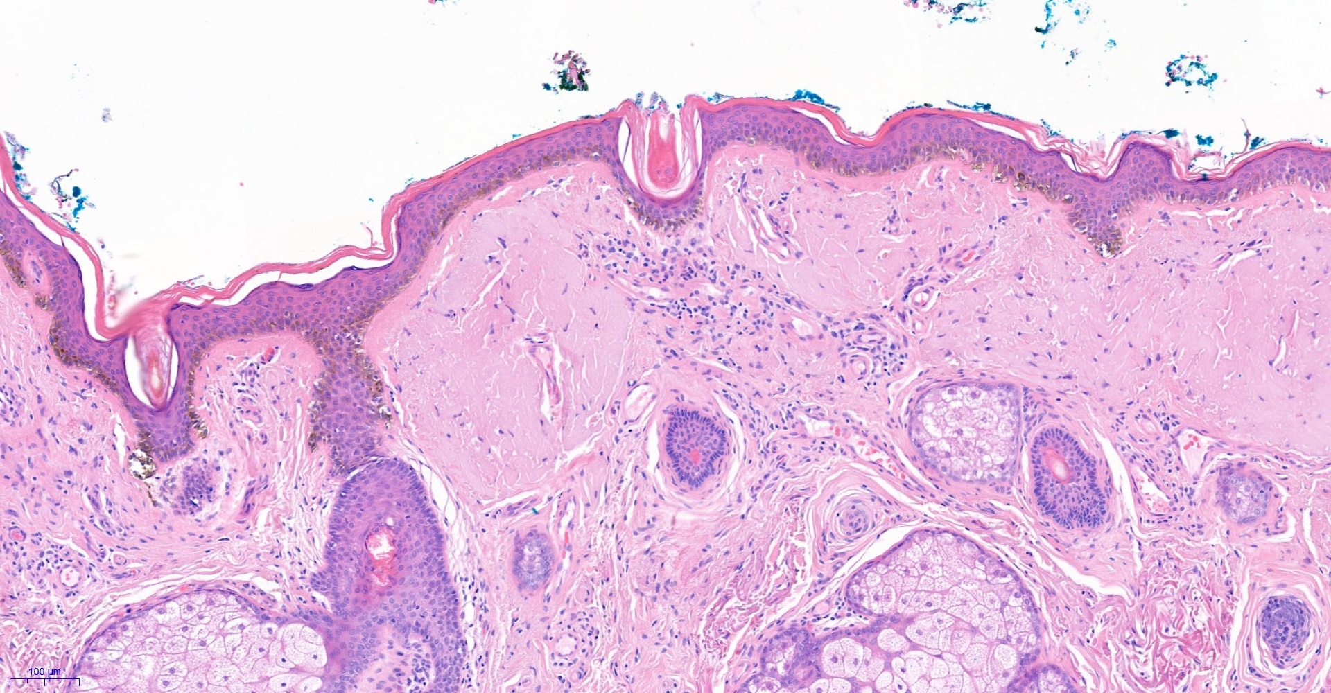
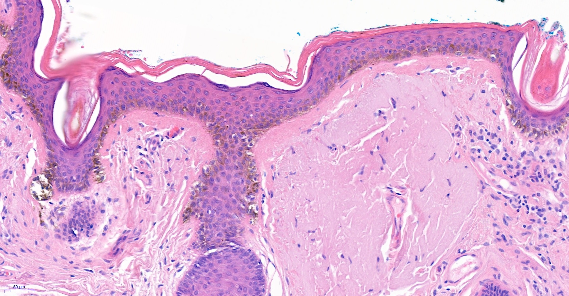
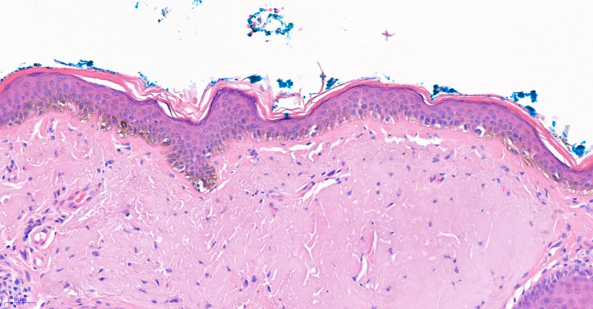
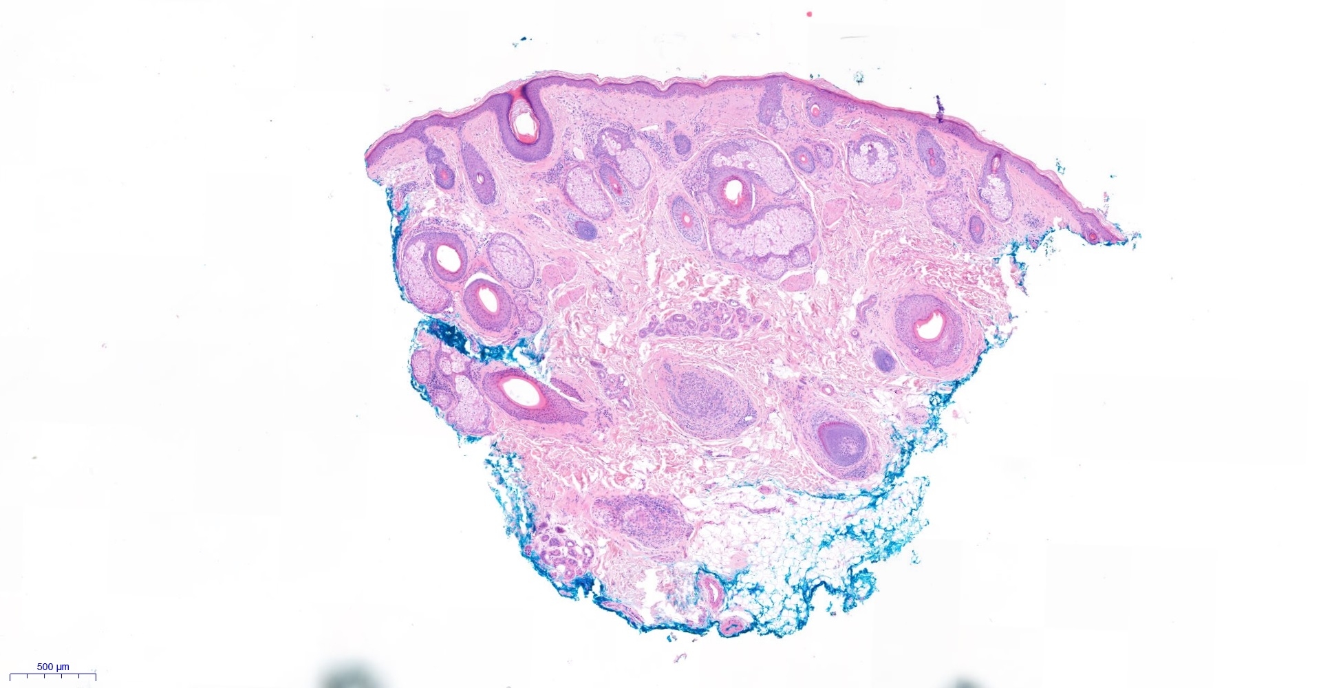
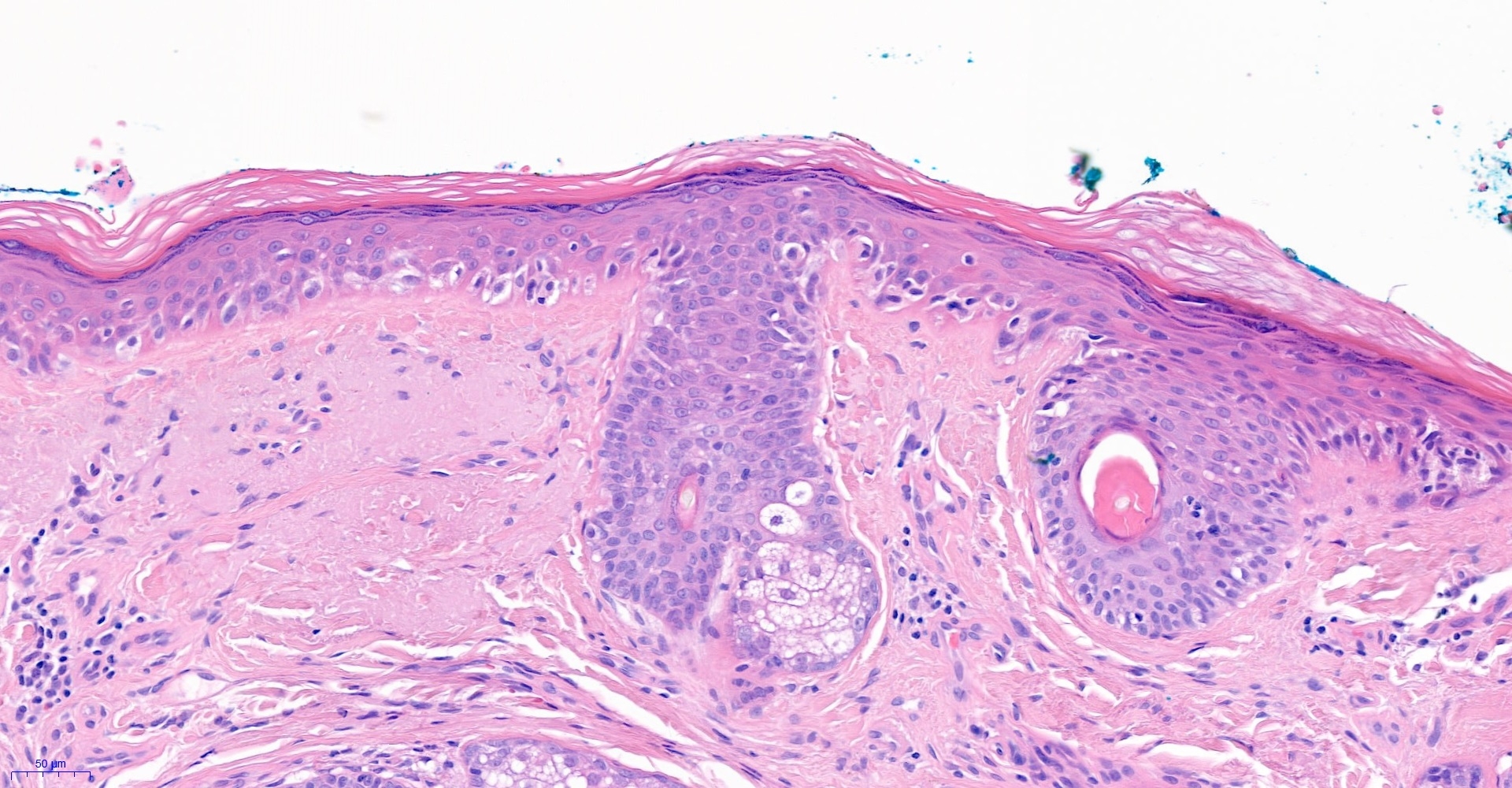
pigmented lesion right cheek ?recurrence of lentigo maligna.

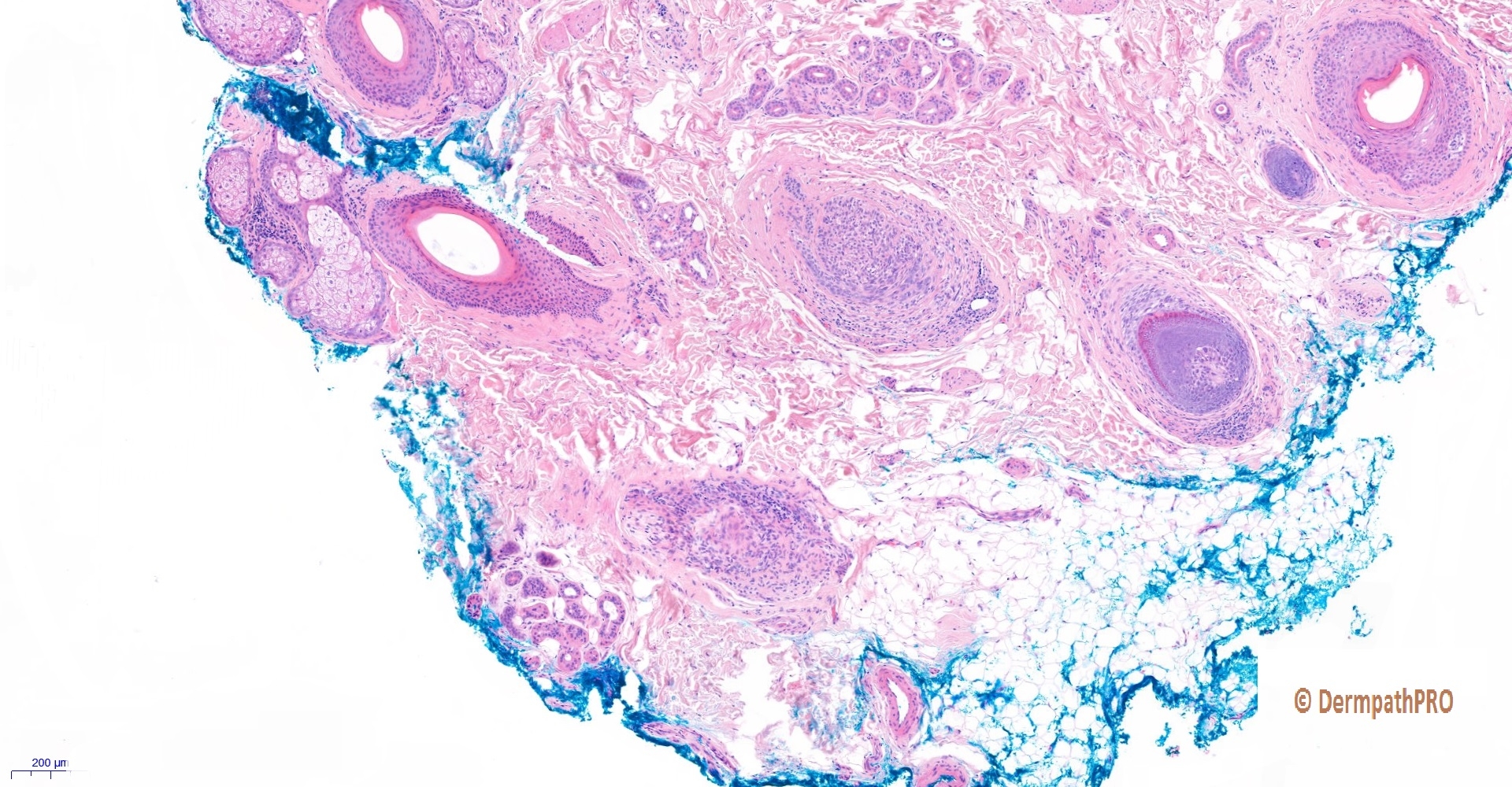
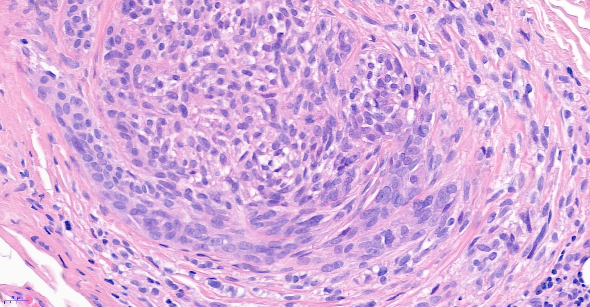
First 4 images are from central transverse section. Last 4 images are from the tip.


  • Like 1
  • Thanks 1
  Report Record

User Feedback


Dr. Mona Abdel-Halim

Posted

I think we r dealing with a recurrence yes, involving the surface epidermis but predominantly involving the follicular epithelium what is called: follicular malignant melanoma (in this case, it will be MIS with extensive follicular involvement). Waiting for beautiful IHC images! 

Share this comment


Link to comment
share_externally

Difficult!  It could be an atypical actinic melanocytic hyperplasia...

Share this comment


Link to comment
share_externally

Recurrente of LM with deep extension trought the follicular epithelium. Agree with Mona.

Share this comment


Link to comment
share_externally

Eman El-Nabarawy

Posted

I would like to see SOX10 or MelanA to verify follicular involvement.

Share this comment


Link to comment
share_externally

Saleem Taibjee

Posted

Yes, this is recurrent lentigo maligna, it is quite possible that the recurrence has arisen from persistent deeper follicular involvement. There also appears to be micro-invasion within the follicular involvement. My only hesitation in this case was the relatively bland cytology. However, the finding of PRAME positivity (overlying epidermis with positive control of sebaceous glands, then separate image of follicle) also supports melanoma. Melan-A and PRAME are shown below.

To be honest, I nearly missed the follicular involvement - it was only present in the tip of the specimen.

Mona, thanks for the useful reference which I was not aware of.

Festive greetings to everyone, back in touch after the New Year!

Saleem

10670_15.0x Melan-A labelled.jpg51246_10.0x PRAME labelled.jpg51246_10.0x PRAMEb labelled.jpgadditionally

Share this comment


Link to comment
share_externally

Dr. Richard Carr

Posted

Mona got the beautiful images. Nice case Saleem.

Share this comment


Link to comment
share_externally


Join the conversation

You can post now and register later. If you have an account, sign in now to post with your account.

Guest
Add a comment...

×   Pasted as rich text.   Paste as plain text instead

  Only 75 emoji are allowed.

×   Your link has been automatically embedded.   Display as a link instead

×   Your previous content has been restored.   Clear editor

×   You cannot paste images directly. Upload or insert images from URL.

Loading...

×
×
  • Create New...