Jump to content
In this section we have spot diagnoses posted on a daily basis since June 2010, now over 4000! You can review the archived cases and read the suggested diagnoses by users and the final comment by the contributors.
Case are uploaded each week day by 10 am UK time with the correct diagnosis will generally be posted at 8 pm UK time. Why not view the most recent spot diagnosis and proffer a diagnosis?

Case Number : Case 2474 - 27 December 2019 Posted By: Dr. Richard Carr

Please read the clinical history and view the images by clicking on them before you proffer your diagnosis.
Submitted Date :
   (0 reviews)

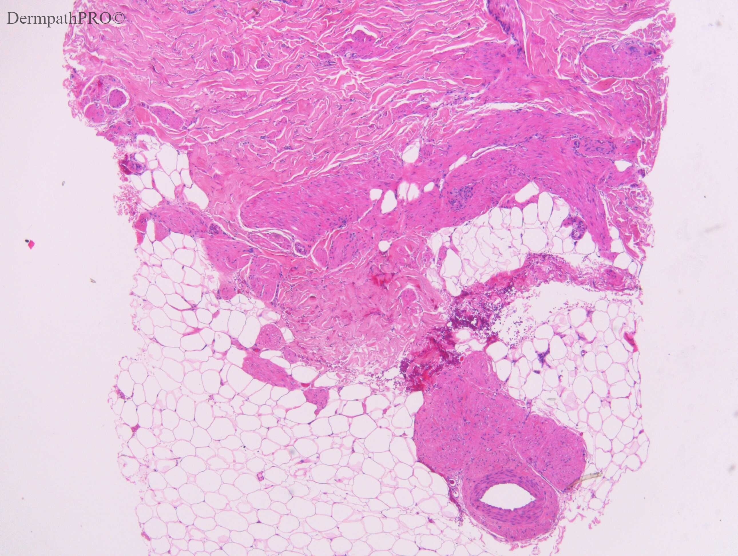
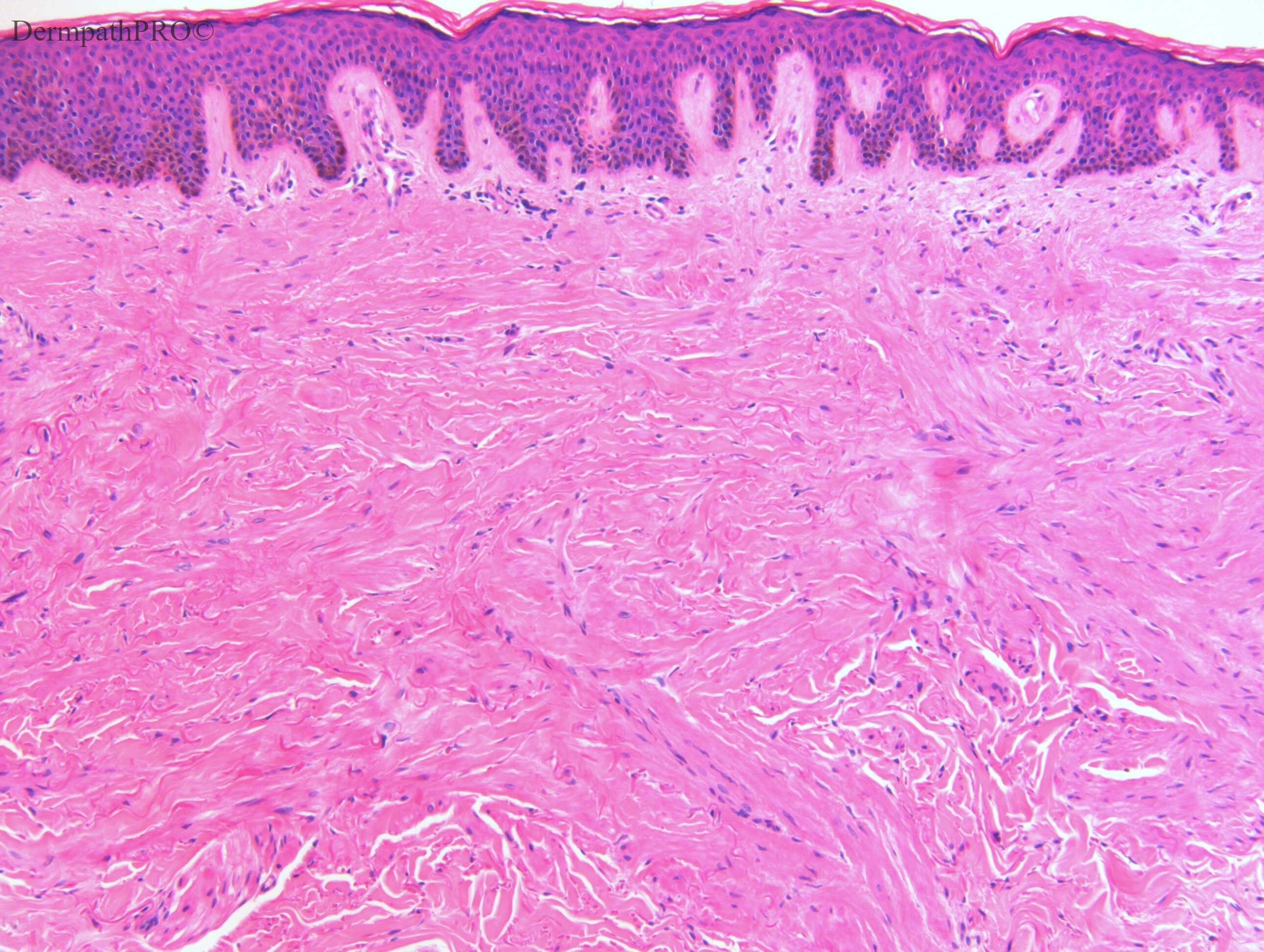
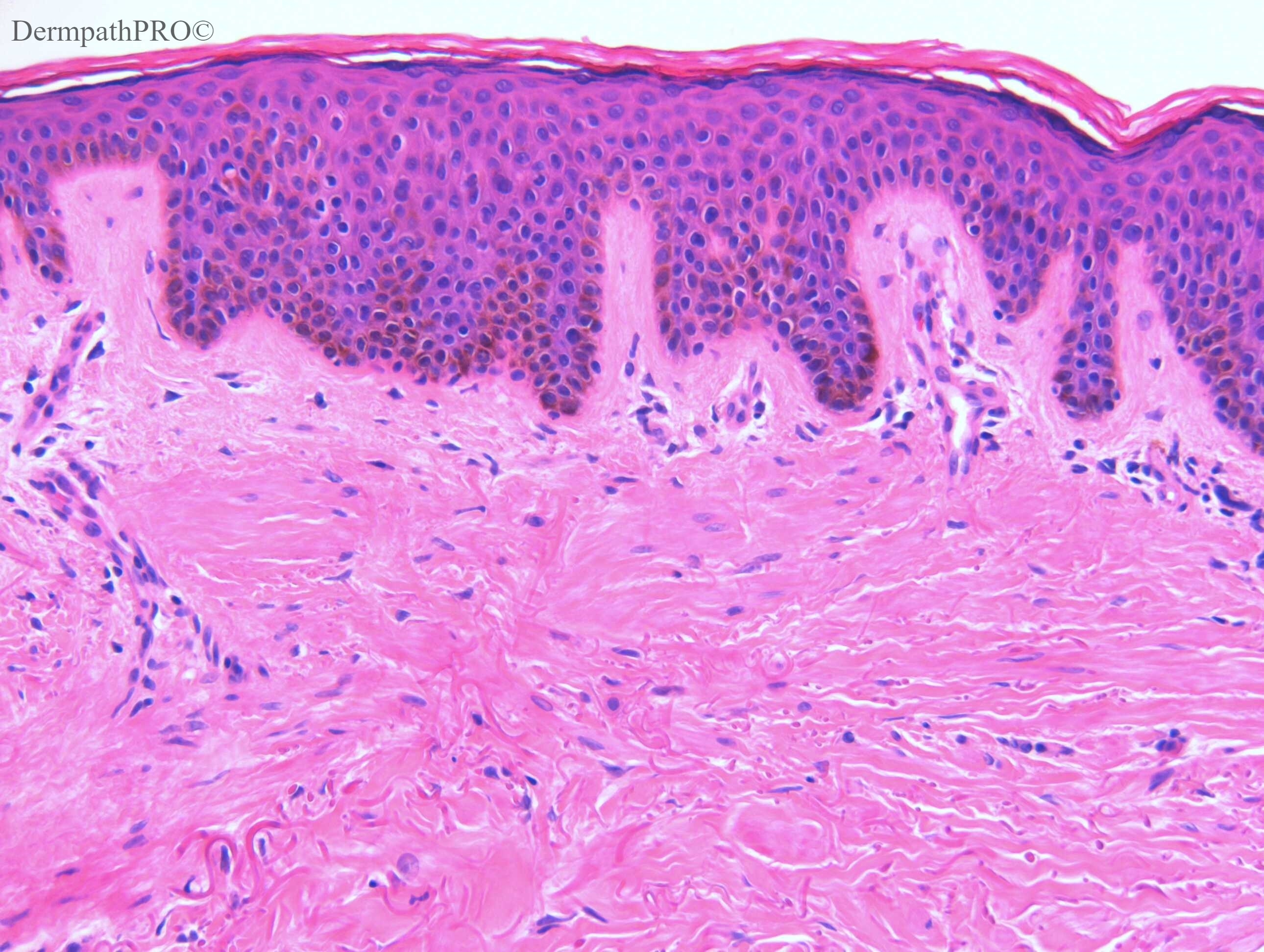
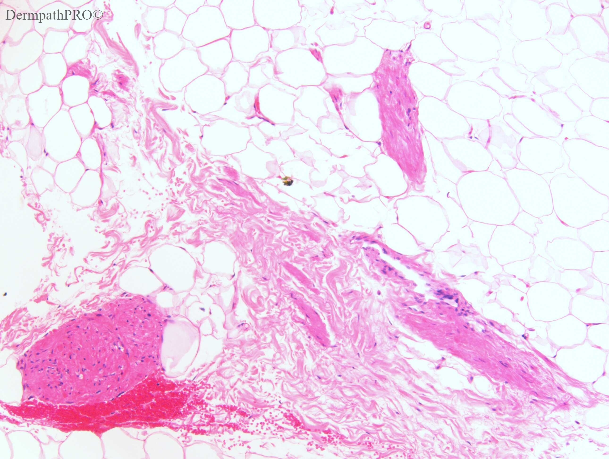
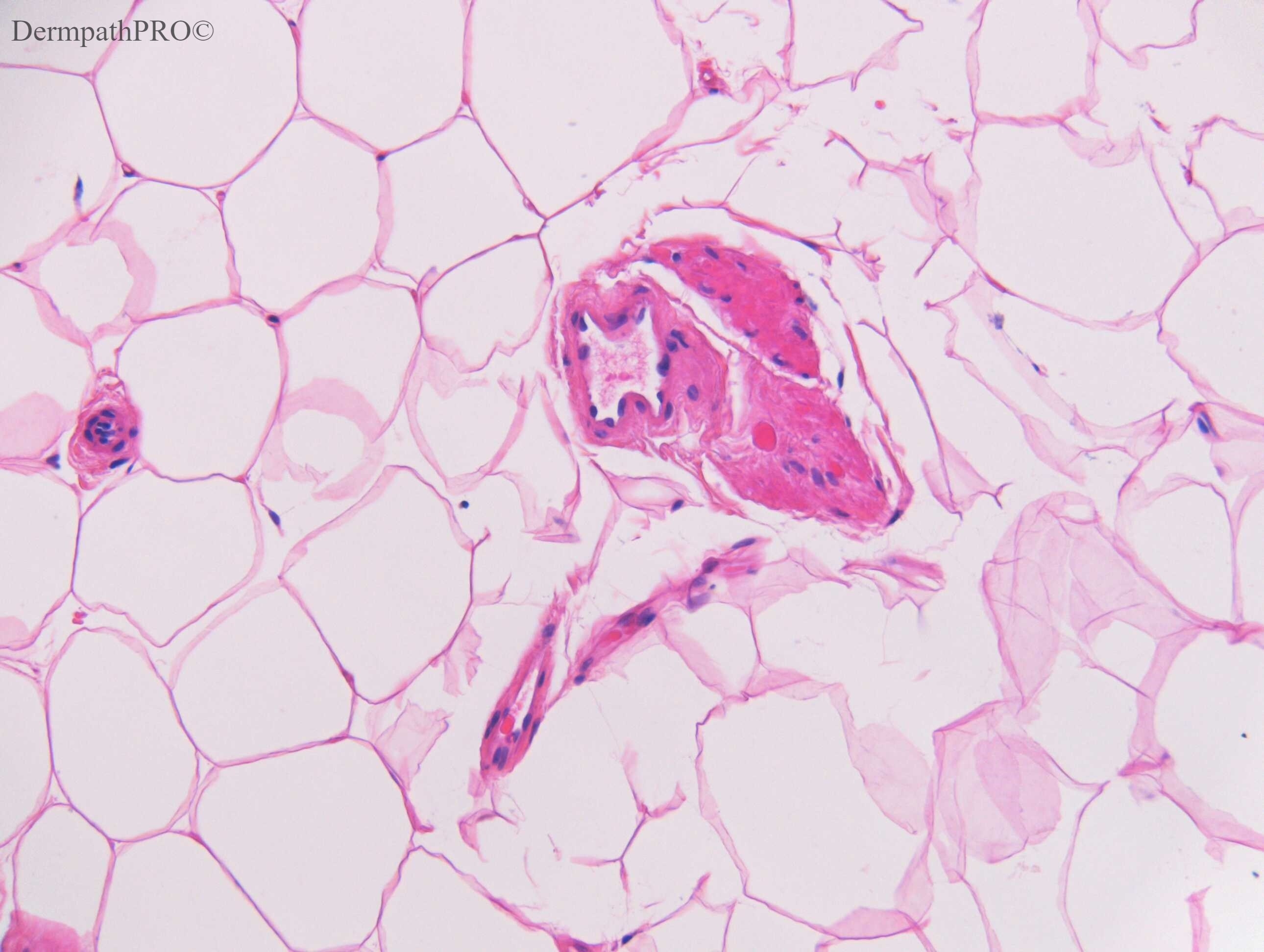
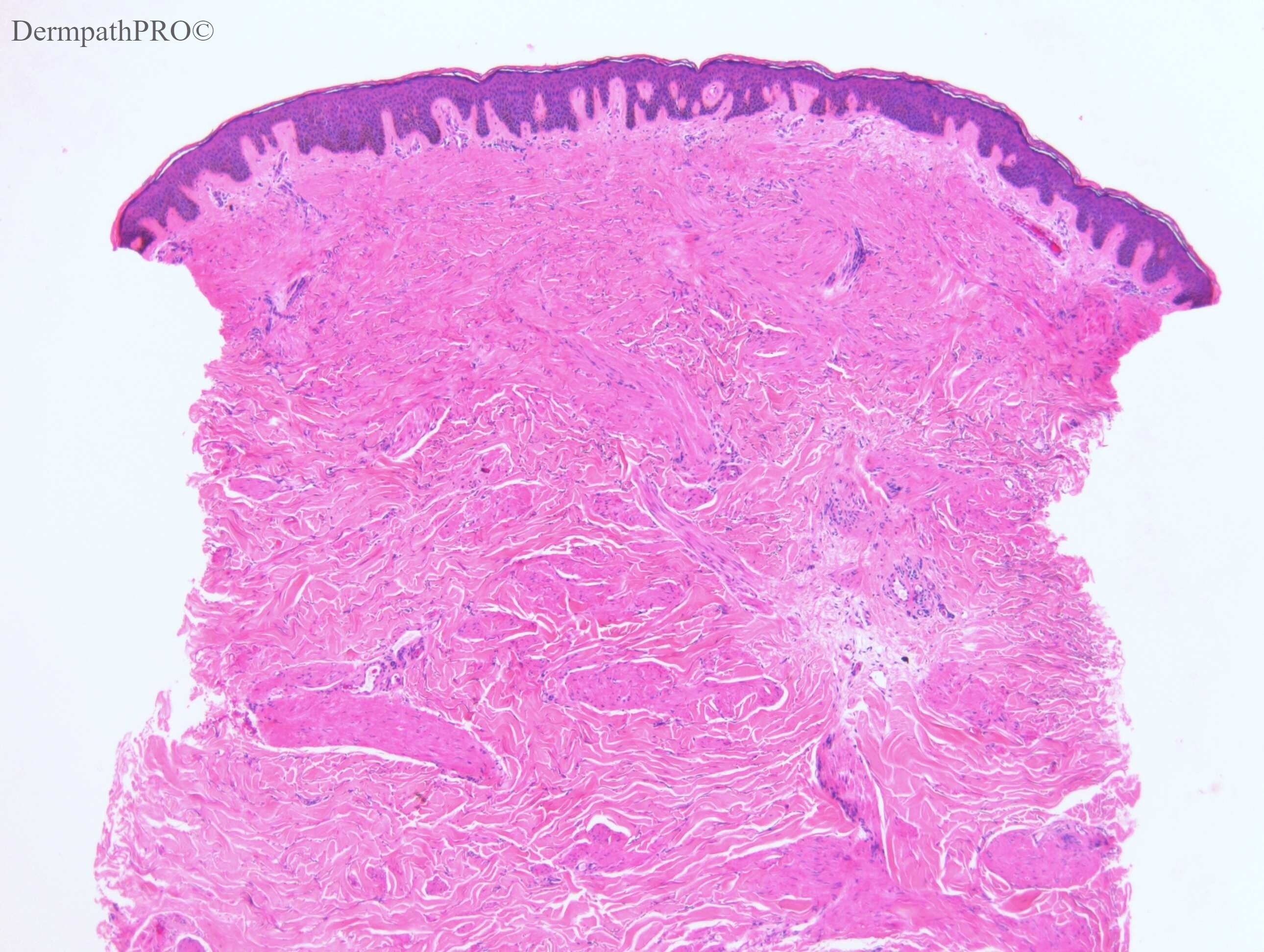
F60. Left arm. 3 x 5cm raised plaque, irregular surface, slightly rippled pattern, normal colour, ?lichen amyloidosis, ?neurofibroma

Edited by Admin_Dermpath


  Report Record

User Feedback


Dr. Mona Abdel-Halim

Posted

Dermatomyofibroma? SMA and Muscle specific actin? CD34?

Share this comment


Link to comment
share_externally

Krishnakumar subramanian

Posted

epidermal reaction with a green zone and somewhat spindle cell tumor is seen

No invading the sub cutaneous fat

I agree with dermatomyofibroma

Share this comment


Link to comment
share_externally

Eman El-Nabarawy

Posted

I saw very thick elastic fibers with fragmented some. ? Verheoff van Gieson stain.So I would like to add elastofibroma.

Share this comment


Link to comment
share_externally

Dr. Mona Abdel-Halim

Posted

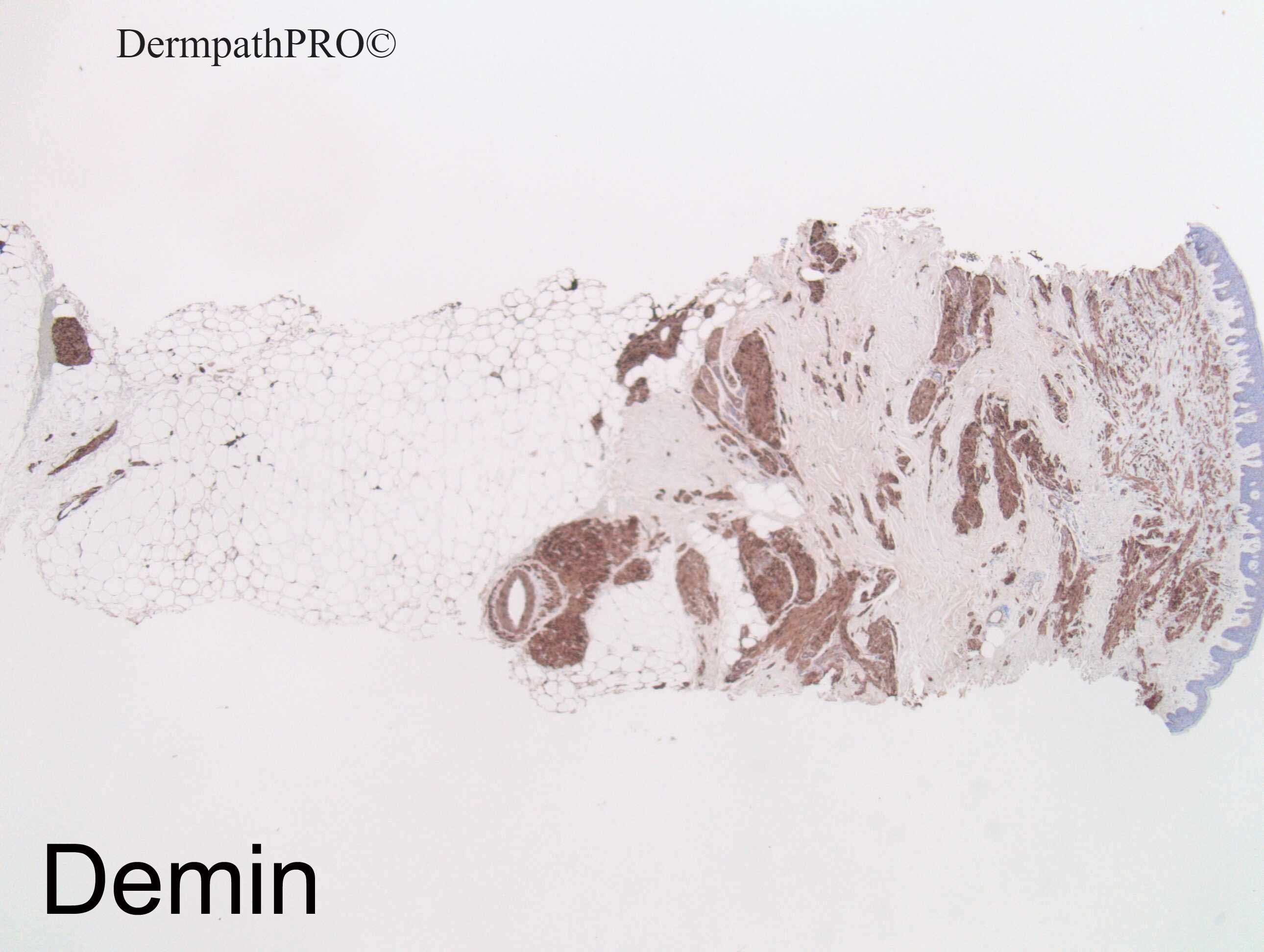
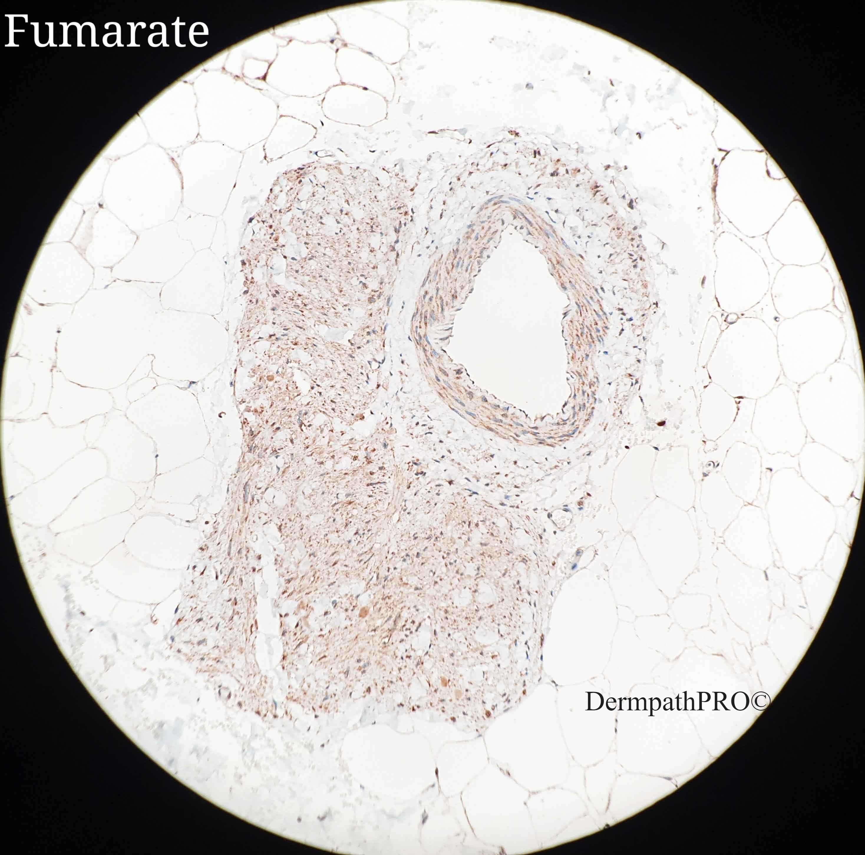
Desmin positivity will make it leiomyoma not dermatomyofibroma  (I think this is the plaque variant of leiomyoma from the clinical description). I was not aware with Fumarate IHC (Fumarate hydratase) in leiomyomas so I had to read about it. It appeared that its loss in cutaneous leiomyomas is a sensitive and specific marker for detection of Hereditary Leiomyomatosis and Associated Renal Cell Carcinoma with an overall sensitivity and specificity of 70% and 97% respectively according to this article: 

https://journals.lww.com/ajsp/pages/articleviewer.aspx?year=2017&issue=06000&article=00010&type=Abstract

Share this comment


Link to comment
share_externally

Dr. Richard Carr

Posted

Thanks Mona that was the reference I wanted to highlight.

In this case Raul Perret kindly reviewed the case and undertook the staining.

My differential included plaque like leiomyoma and a smooth muscle hamartoma / Becker's. Clinically is was not suggestive of a Becker. She did have a history of fibroids so we wanted to r/o Reed's syndrome. Raul states: Fumarate expression was conserved (see attached image). Based on this phenotype, the possibility of Reed sybdrome is low.

Share this comment


Link to comment
share_externally


Join the conversation

You can post now and register later. If you have an account, sign in now to post with your account.

Guest
Add a comment...

×   Pasted as rich text.   Paste as plain text instead

  Only 75 emoji are allowed.

×   Your link has been automatically embedded.   Display as a link instead

×   Your previous content has been restored.   Clear editor

×   You cannot paste images directly. Upload or insert images from URL.

Loading...

×
×
  • Create New...