Jump to content
In this section we have spot diagnoses posted on a daily basis since June 2010, now over 4000! You can review the archived cases and read the suggested diagnoses by users and the final comment by the contributors.
Case are uploaded each week day by 10 am UK time with the correct diagnosis will generally be posted at 8 pm UK time. Why not view the most recent spot diagnosis and proffer a diagnosis?

Case Number : Case 2338 - 3 June 2019 Posted By: Limin Yu

Please read the clinical history and view the images by clicking on them before you proffer your diagnosis.
Submitted Date :
   (0 reviews)

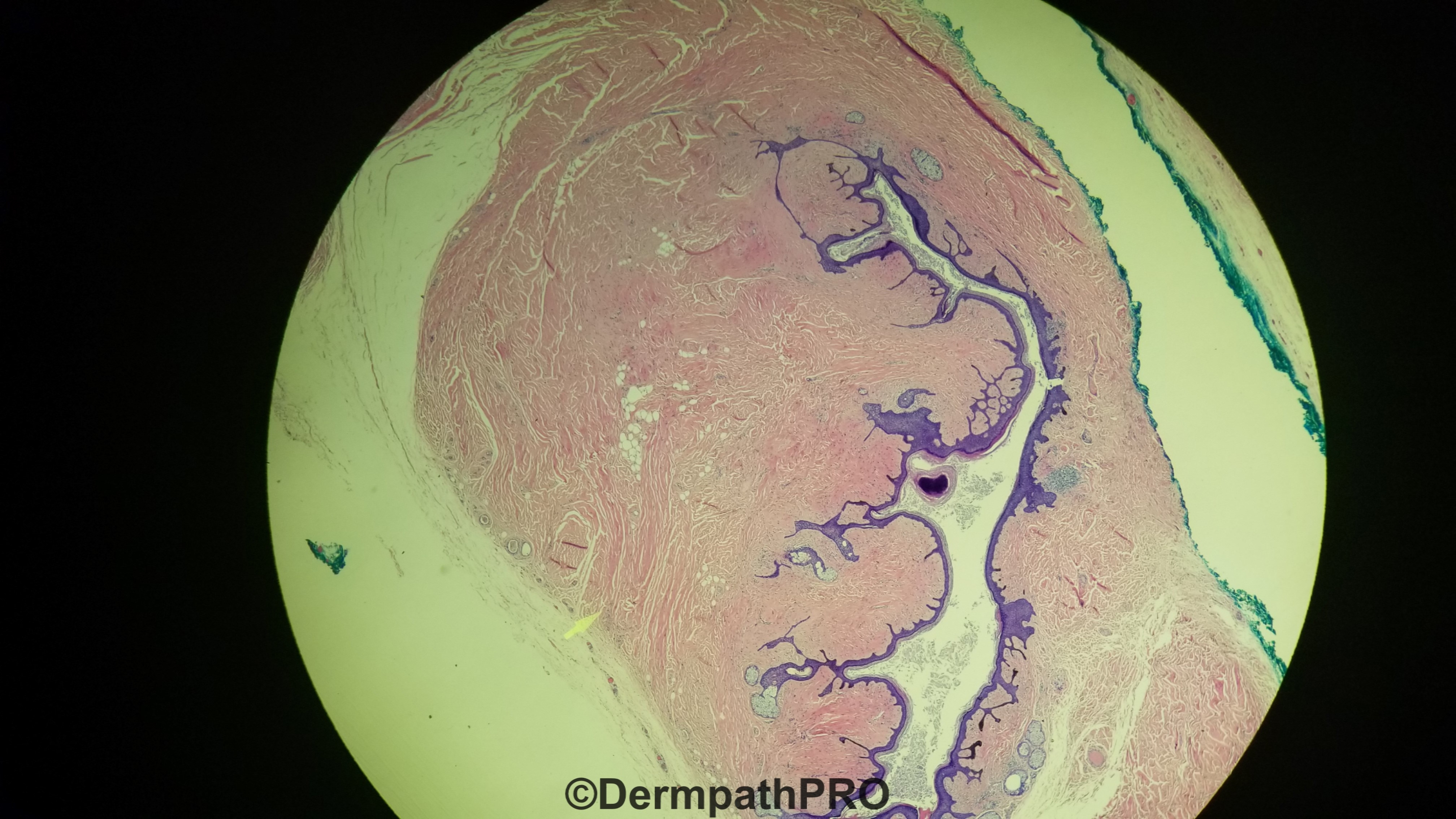
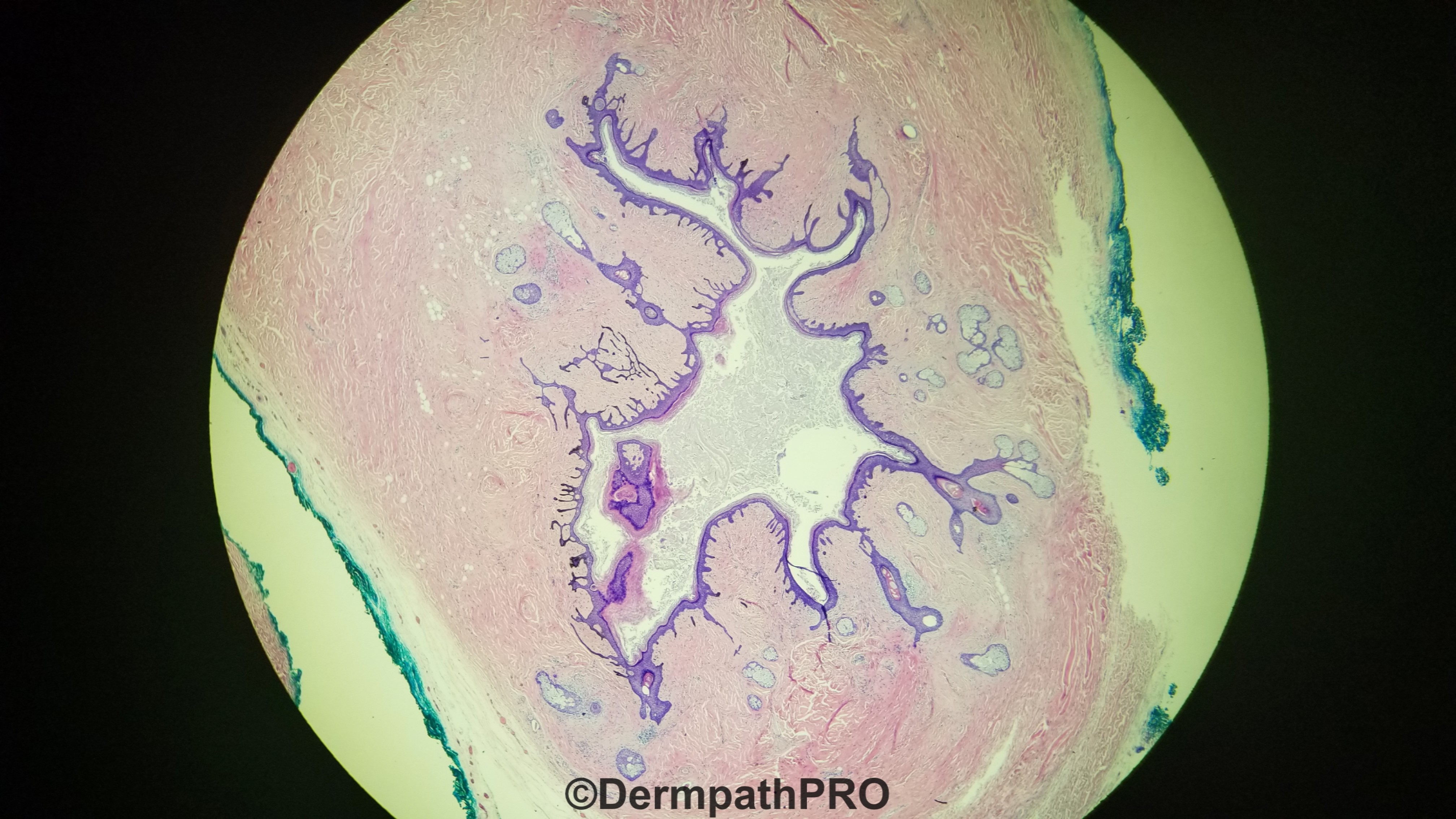
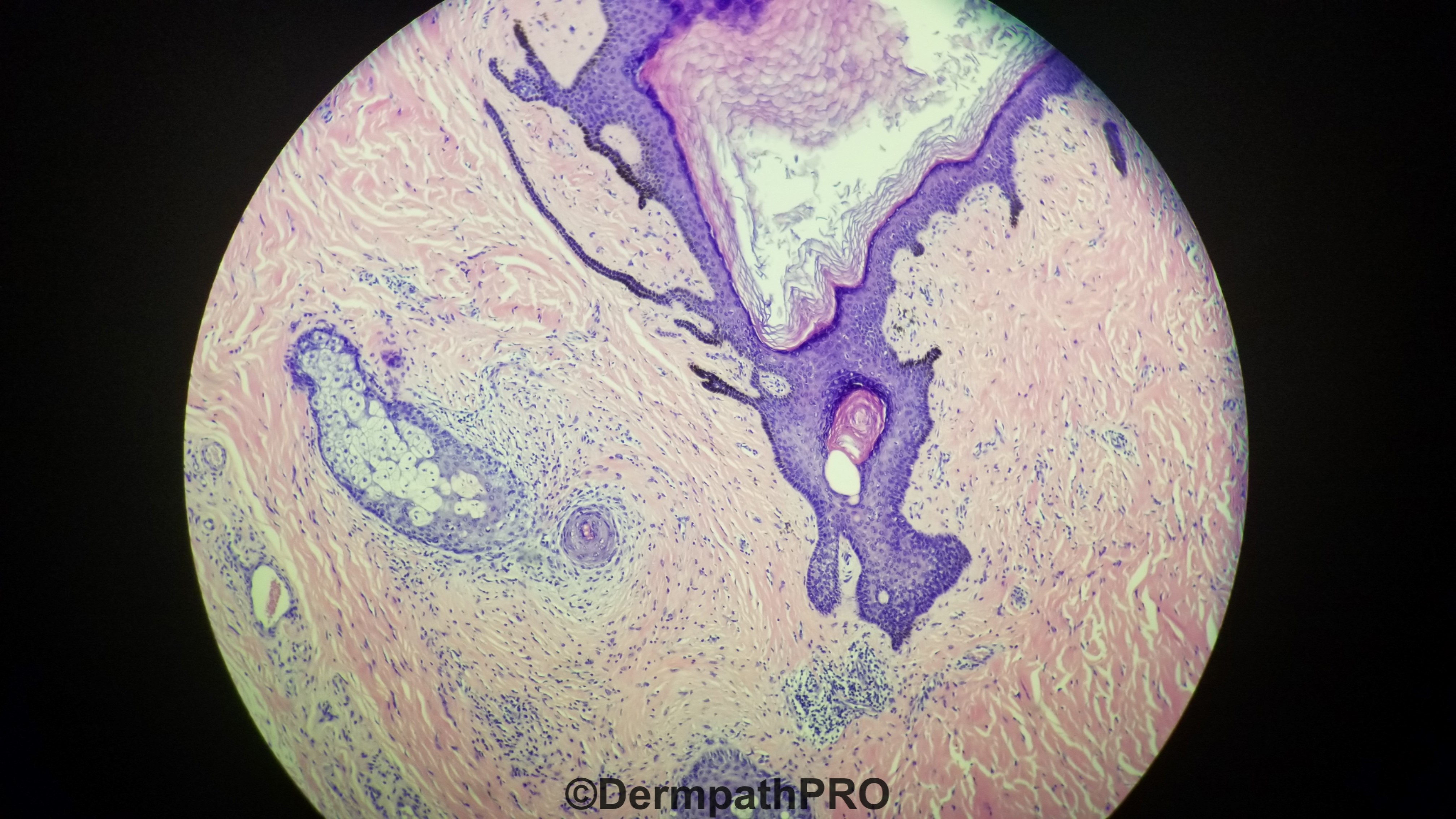
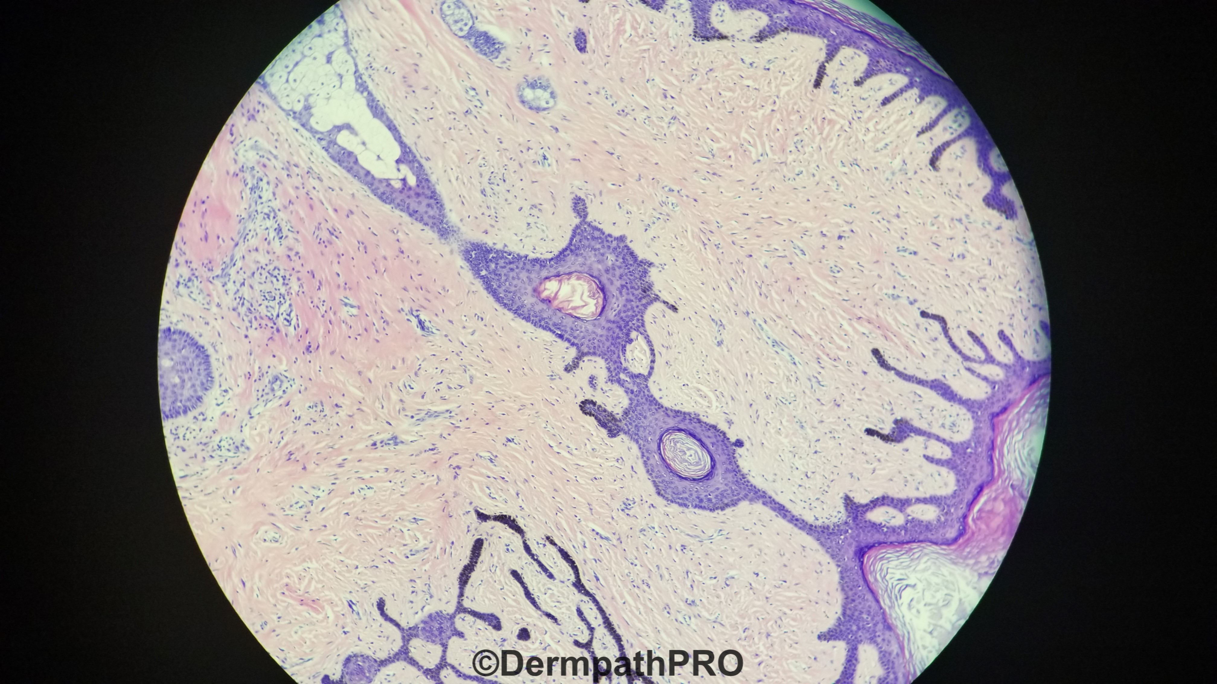
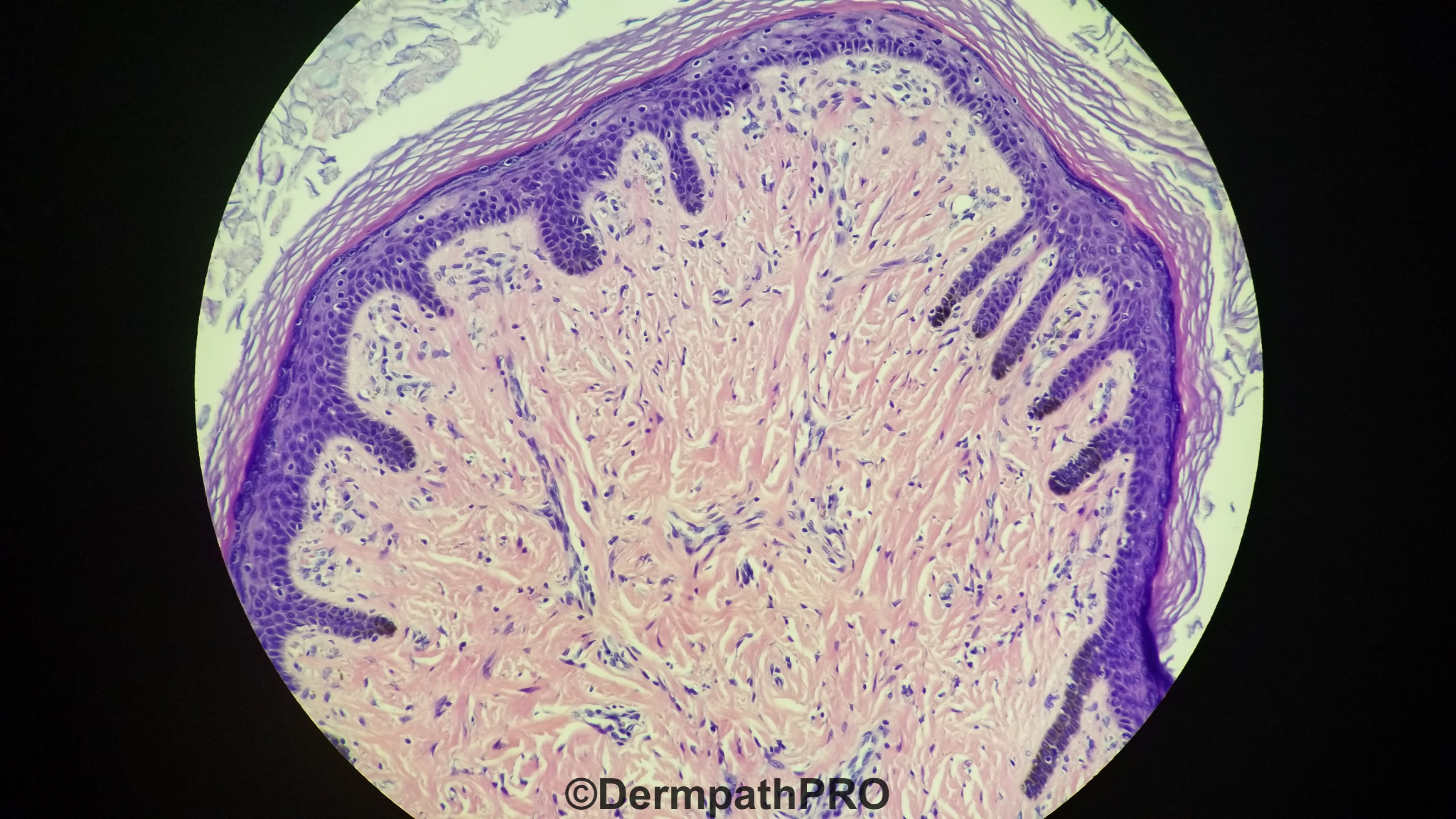
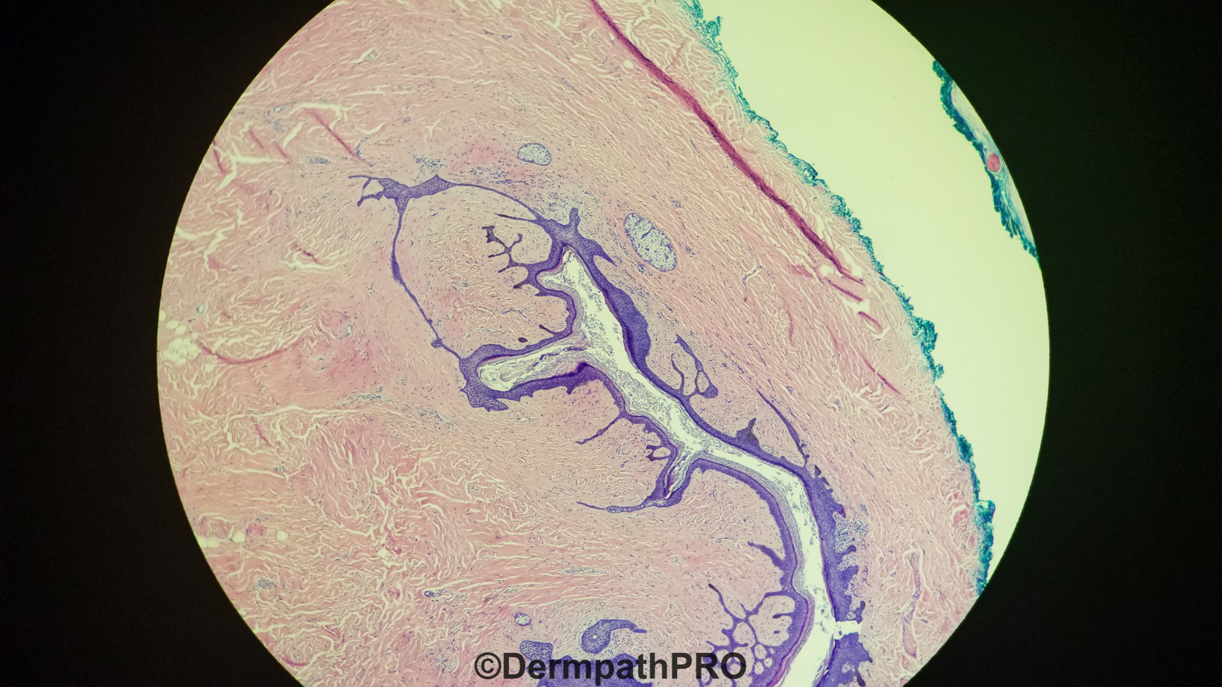
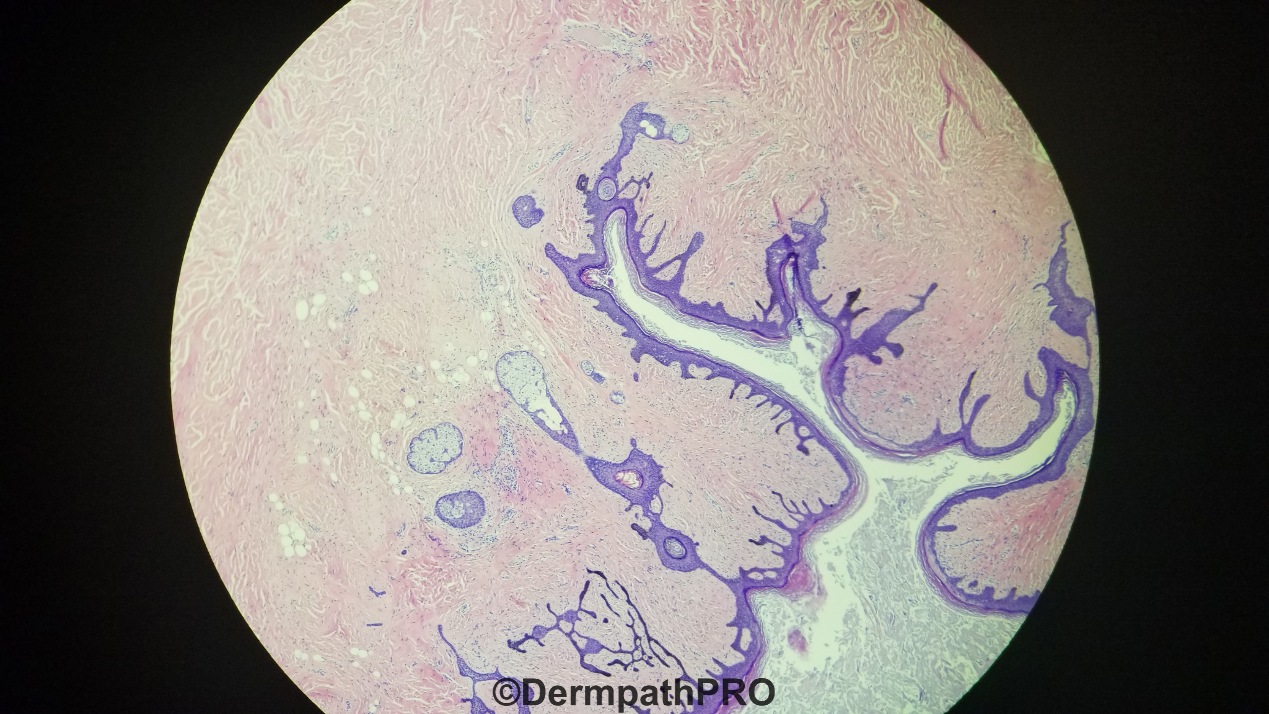
76 year old with lesion on the nose

  Report Record

User Feedback


Benign hamartomatous lesion, most likely fibrofolliculoma/trichodiscoma type lesions.

Share this comment


Link to comment
share_externally

John Zhang

Posted

3 hours ago, HENRY said:

Benign hamartomatous lesion, most likely fibrofolliculoma/trichodiscoma type lesions.

Great point Henry, Agree!

At first glance I was debating if this should be classified as a fibrofolliculoma, or a trichofolliculoma. I looked at the photos hard, and couldn't see any hair bulbs (follicular germ). I think if there is no follicular germ, a lesion should be classified as fibrofolliculoma rather than trichofolliculoma.

Share this comment


Link to comment
share_externally

Meenakshi Batrani

Posted

Folliculosebaceous cystic hamartoma

Share this comment


Link to comment
share_externally

Alex-Ventura-Leon

Posted

Trichofolliculoma vs Fibrofolliculoma.

Agree with the observation of Henry and Zhang. This lesion fit more with Fibrofolliculoma

Share this comment


Link to comment
share_externally

Anil Patki

Posted

Dilated cystic structure in the centre along with follicular, sebaceous and mesenchymal elements together suggest folliculosebaceous cystic hamartoma.

Share this comment


Link to comment
share_externally

We all agree that this is a form of hamatomatous lesion with both epithelial and mesenchymal components. There are a lot of overlapping histologic features between follicullosebaceous cystic hematoma (FSCH )and Fibrofolliculoma / trichodiscoma. I tried one time to read about these entities and I came to my own conclusion (right or wrong) that FCH  usually has a centrally dilated cystic structure in the center with infundibulocystic and sebaceous differentiation surrounded by mesenchymal storma. The epithelial component is located only in the dermis and not connected to the epidermis.


Fibrofolliculoma /trichodiscoma are also hamartomatous lesions but the epithelial components can be connected to the overlying epidermis and usually not strictly located in the center of the lesion. In fact, the mesenchymal component is more likely located in the center, surrounded by the epithelial component. I was told that the epithelial component in trichodiscoma is more mature then fibrofolliculoma, to wit, has sebaceous differentiation. So if I see sebaceous differentiation, I would classify the lesion as trichodiscoma instead of fibrofolliculoma.


So if I apply my reading to this lesion, I would say that this lesion is completely located in the dermis and the epithelial component in this lesion is in the center surrounded by a mesenchymal storma. There are no epithelial strands attached to the overlying epidermis that I could see. So I would classify it as FSCH.


If someone wants to place this lesion in the Fibrofolliculoma / trichodiscoma group, then it should be a trichodiscoma based on the presence of the sebaceous glands (fig 5).


But that's my own practical approach to this group of confusing hamartomatous lesions. Any suggestions would be greatly appreciated so that I can improve my criteria.

Share this comment


Link to comment
share_externally

vincenzo

Posted

Having learnt the adnexal neoplasm by Requena-Sangueza, I'd rather define this tumor as trichofolliculoma ( sebaceous t. aka FSCH of course ), but I'm learning a lot by Colleagues, so thanks very much.

Share this comment


Link to comment
share_externally

Dr. Mona Abdel-Halim

Posted

I believe it is fibrofolliculoma/trichodiscoma

Share this comment


Link to comment
share_externally


Join the conversation

You can post now and register later. If you have an account, sign in now to post with your account.

Guest
Add a comment...

×   Pasted as rich text.   Paste as plain text instead

  Only 75 emoji are allowed.

×   Your link has been automatically embedded.   Display as a link instead

×   Your previous content has been restored.   Clear editor

×   You cannot paste images directly. Upload or insert images from URL.

Loading...

×
×
  • Create New...