Jump to content
In this section we have spot diagnoses posted on a daily basis since June 2010, now over 4000! You can review the archived cases and read the suggested diagnoses by users and the final comment by the contributors.
Case are uploaded each week day by 10 am UK time with the correct diagnosis will generally be posted at 8 pm UK time. Why not view the most recent spot diagnosis and proffer a diagnosis?

Case Number : Case 2347 - 14 June 2019 Posted By: Dr. Richard Carr

Please read the clinical history and view the images by clicking on them before you proffer your diagnosis.
Submitted Date :
   (0 reviews)

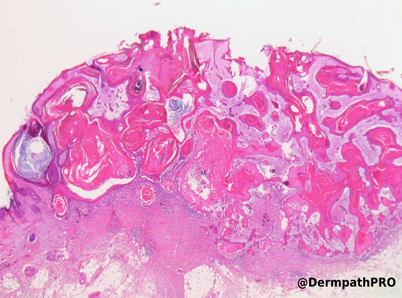
Female, age 85. >9 months h/o lesion left cheek. Biopsied at 6 months as suspected BCC and reported as well differentiated SCC


  • Like 1
  Report Record

User Feedback


Krishnakumar subramanian

Posted

is it proliferating trichelemmal cyst mimicking SCC

Share this comment


Link to comment
share_externally

Why not a SCC-follicular type? There is a "spontaneous" acantholysis with mucinous pools.

Share this comment


Link to comment
share_externally

Alex-Ventura-Leon

Posted

Same impression as Vincenzo.

Follicular SCC, pushing borders.

Share this comment


Link to comment
share_externally

Anil Patki

Posted

KA-like SCC showing acantholysis

Share this comment


Link to comment
share_externally

Dr. Richard Carr

Posted

Well done Vincenzo - spot on. I don't allow obvious acantholysis and folliuclar mucin in KA so at least this case must be signed out cautiously. As it is well differentiated and relatively pushing of margin I suspect it's a low risk lesion and probably would not require follow-up but I also suspect if left it would have grown relentlessly. Another small print aspect is the tumour show calcification - not seen that in KA (which grow quickly then involute) common in other follicular tumours (BCC, pilar tumours, pilomatrixoma etc). There is one subtype of KA, centrifugum marginatum, that may show persisent growth but also has regression centrally. The lesion ilustrated show some regressive features e.g. keratin granulomas but also looks rather indolent. Slight asymetry, not so good development of lips, no elastic or collagen trapping (a major helpful feature of KA), not much intra-epithelial neutrophil abscess formation (a good feature for typical KA), relatively slight lichenoid reaction/colloid bodies (as opposed in to brisk in KA), some stromal desmoplasia (not a feature of proliferative KA), 

I've been working on my criteria for KA v's follicular (infundibular-tricholemmal) SCC to refine the diagnostic challenge in most cases. 

This case would actually score in the "Don't know" group but it certainly is not a typical KA. Remember typical proliferative KA look highly malignant and infiltrative at the peripheries of the invasive front but should show central maturation in all areas. Signs of brisk inflammatory reaction and regression are always nice to see. In the biopsy which I also reported the lesion was involving fat (again I don't like to see that in KA). It did not have much of an inflammatory reaction and had acantholysis although the mucin was less well developed. The borders were just not "malignant" looking enough.

I suggest you check out my you tube again and pay attention to all the small print. I hope to see many of you at the Lisbon ISDP meeting in the autumin. I've been give a slot to talk about follicular SCC and KA. Hopeful the paradigm shifting work will get greater appreciated and the tumour will be listed in the next WHO classfication!  One caveat is that KA is not a safe diagnosis unless you've spent a lot of time working out all the nuances and you have to work within the setting of a multidisciplinary team. If every one understands it's challenging and it's a risk balance patients will get the most appropriate label and better management. Many circumscript fSCC don't need to be followed up in my opinion and we need some large research studies to confirm this. A lot more research is required on KA obviously.


Share this comment


Link to comment
share_externally

Krishnakumar subramanian

Posted

sir wonderful lesson on follicular SCC and the point on inverted follicular keratosis with mucin

great


Share this comment


Link to comment
share_externally


Join the conversation

You can post now and register later. If you have an account, sign in now to post with your account.

Guest
Add a comment...

×   Pasted as rich text.   Paste as plain text instead

  Only 75 emoji are allowed.

×   Your link has been automatically embedded.   Display as a link instead

×   Your previous content has been restored.   Clear editor

×   You cannot paste images directly. Upload or insert images from URL.

Loading...

×
×
  • Create New...