Jump to content
In this section we have spot diagnoses posted on a daily basis since June 2010, now over 4000! You can review the archived cases and read the suggested diagnoses by users and the final comment by the contributors.
Case are uploaded each week day by 10 am UK time with the correct diagnosis will generally be posted at 8 pm UK time. Why not view the most recent spot diagnosis and proffer a diagnosis?

Case Number : Case 2351 - 20 June 2019 Posted By: Dr. Richard Carr

Please read the clinical history and view the images by clicking on them before you proffer your diagnosis.
Submitted Date :
   (0 reviews)

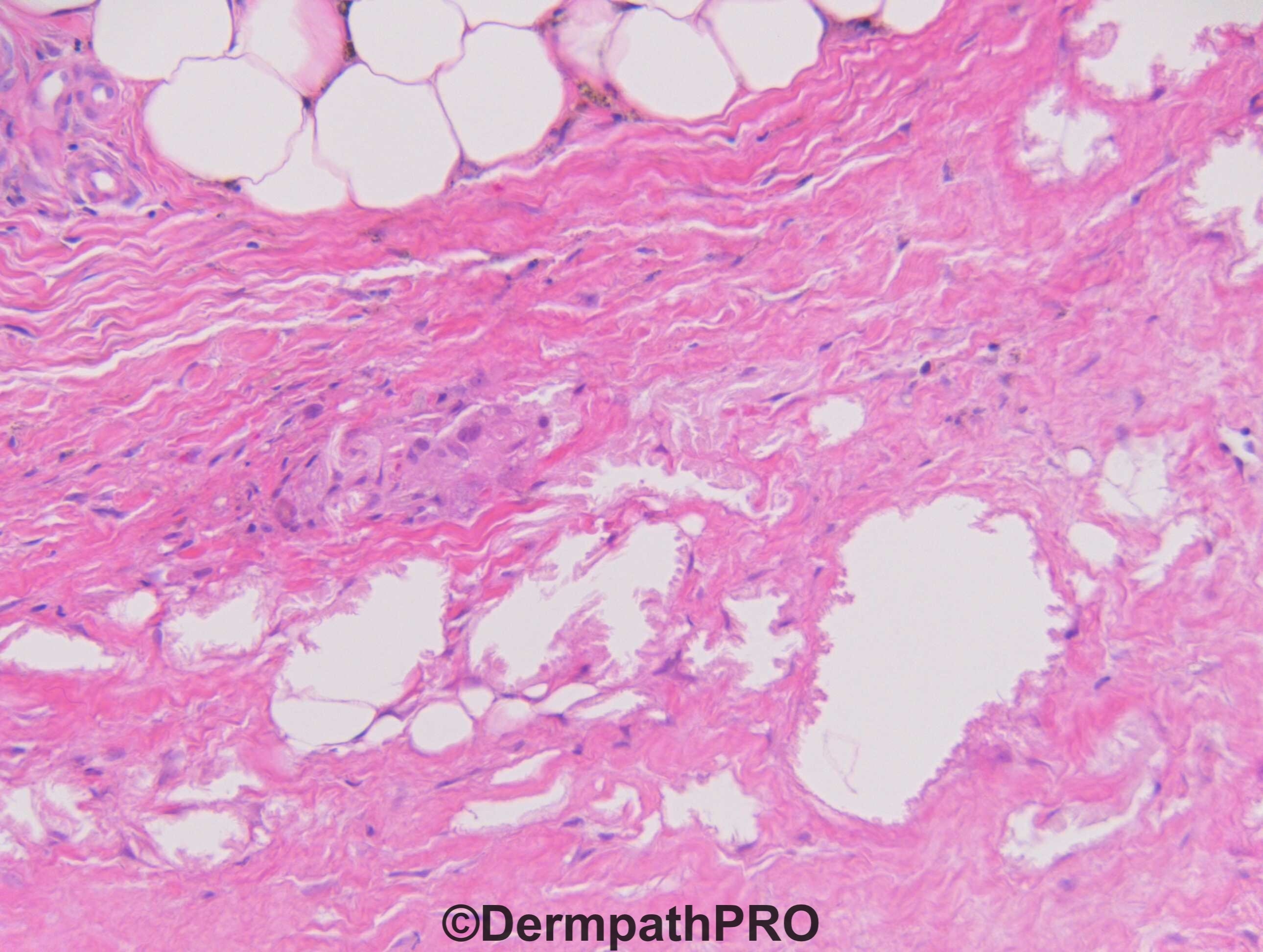
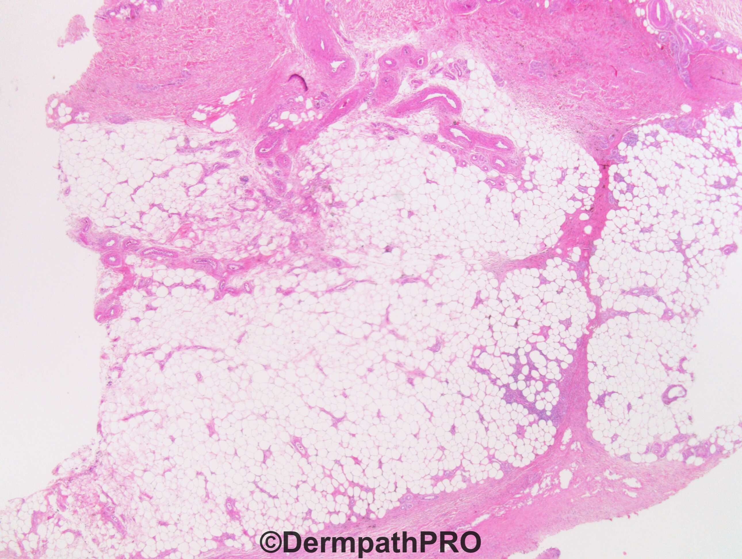
M55. Sri Lankan. >1 year asymptomatic, non-tender, firm, dark discolouration to both lower legs. Rt > Lt.
?Erythema nodosum clinically. No resolution over 9/12. Denies joint pains, swellings, other rashes, weight loss. Dermatologist queries pretibial myxoedema ?scleroderma, ?deep lupus / panniculitis

Edited by Admin_Dermpath


  Report Record

User Feedback


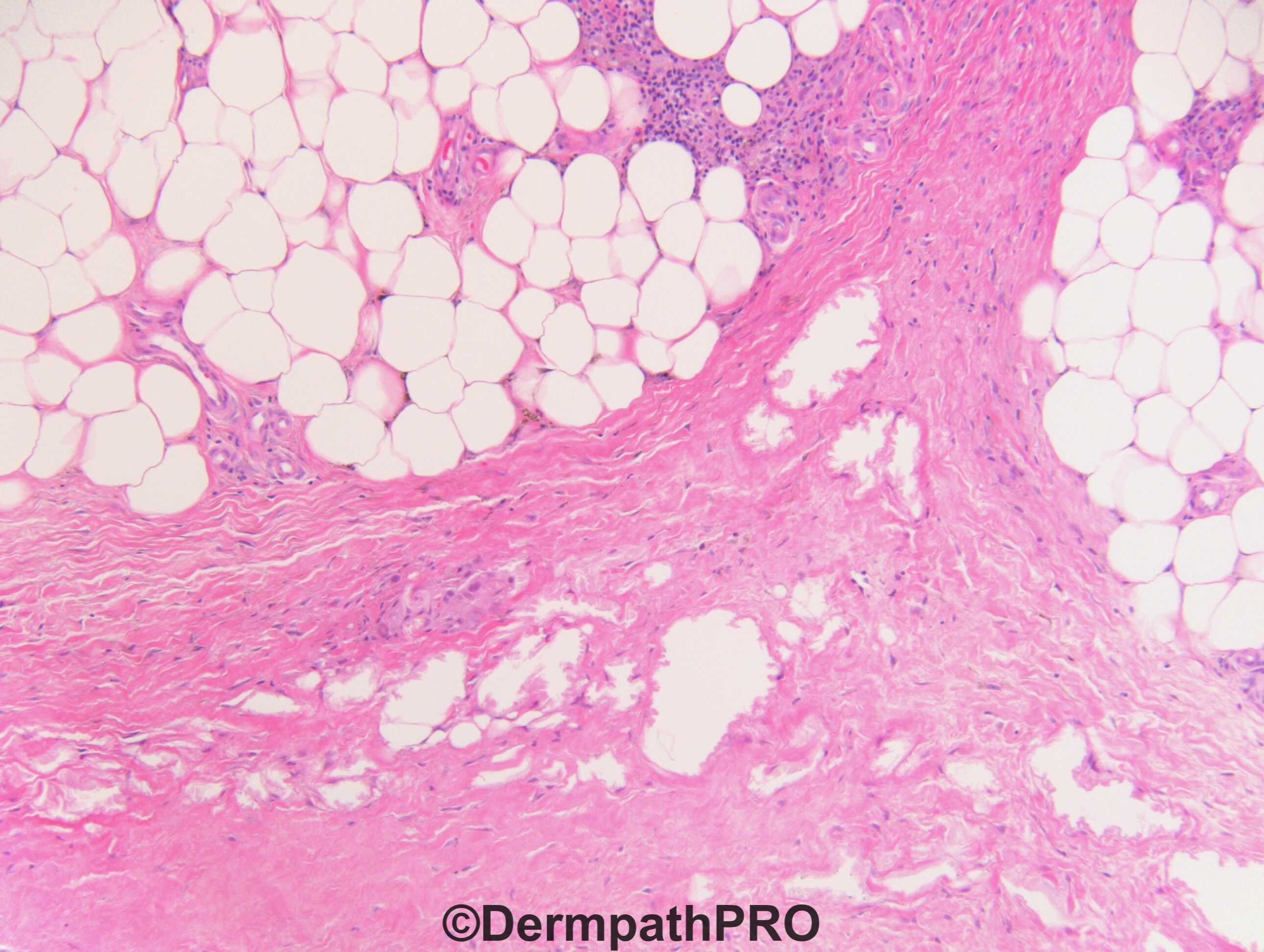
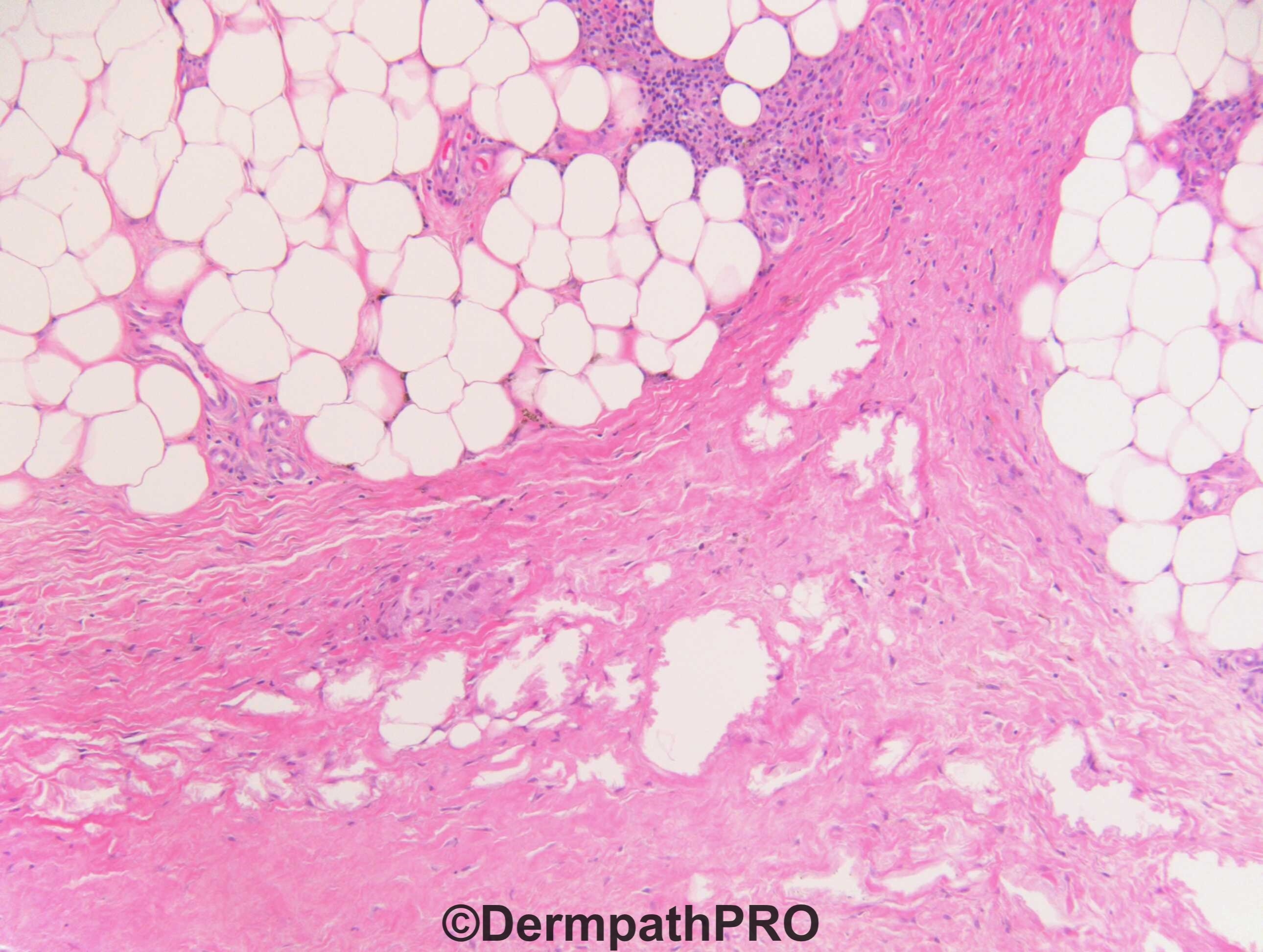
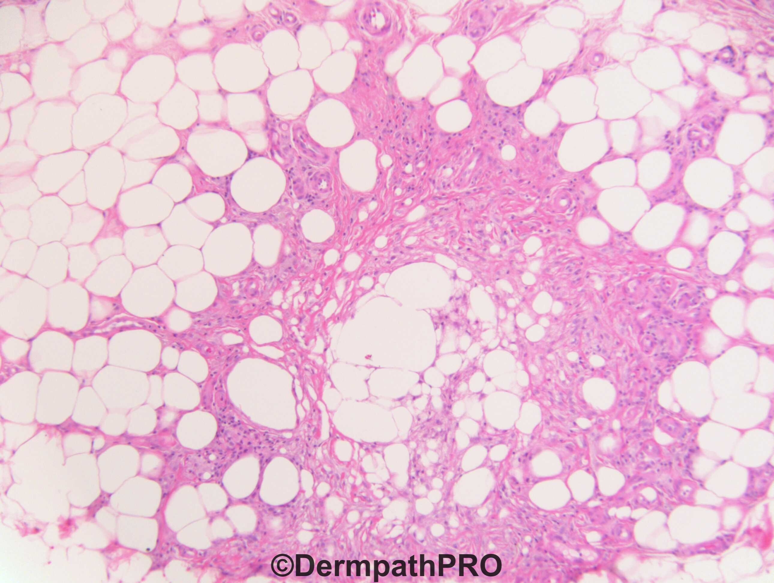
Hemorragy is a common event in Erythema Nodosum, as the lipomembranous fat necrosis. So my provisional spot is EN.

Share this comment


Link to comment
share_externally

Krishnakumar subramanian

Posted

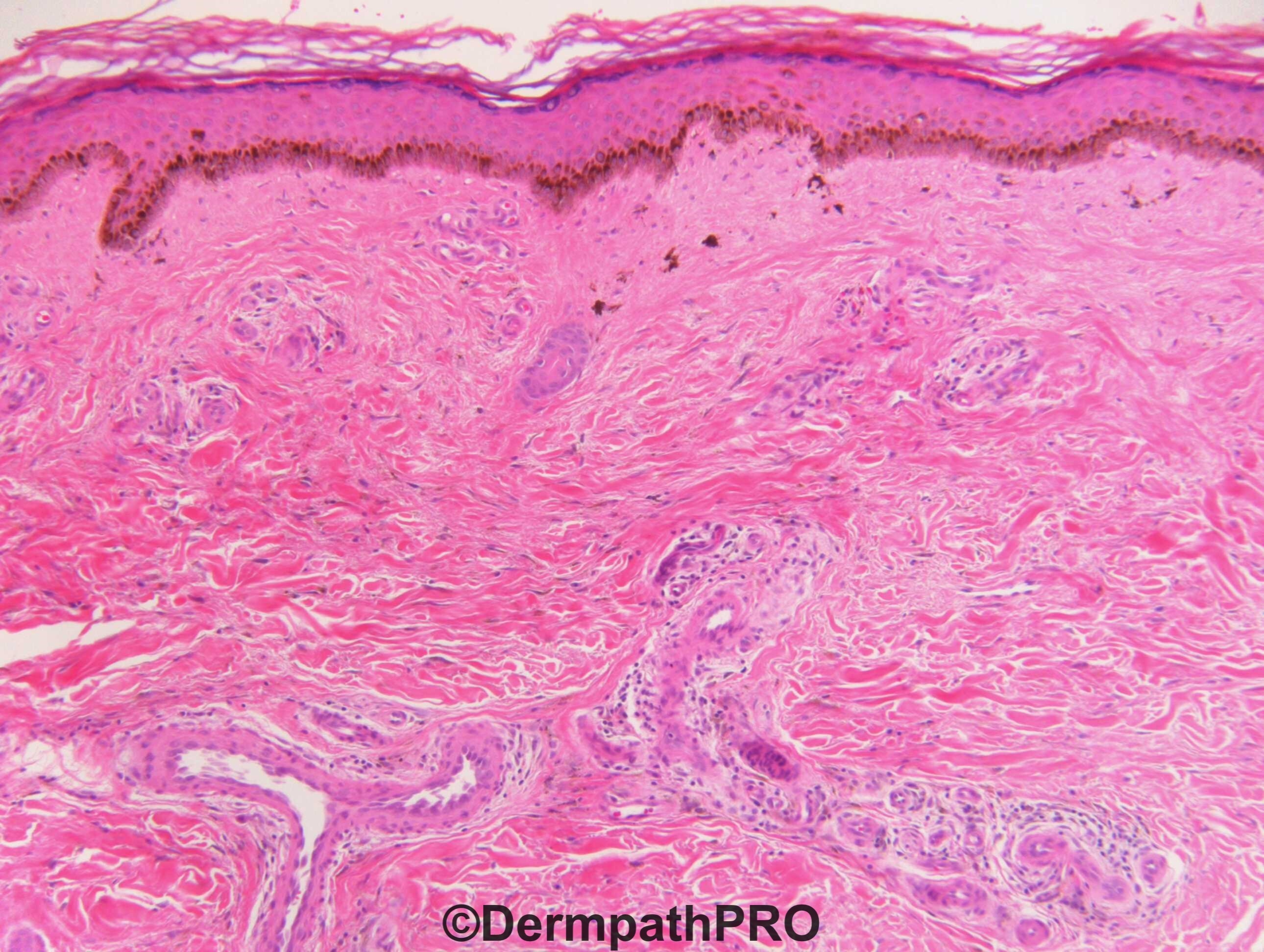
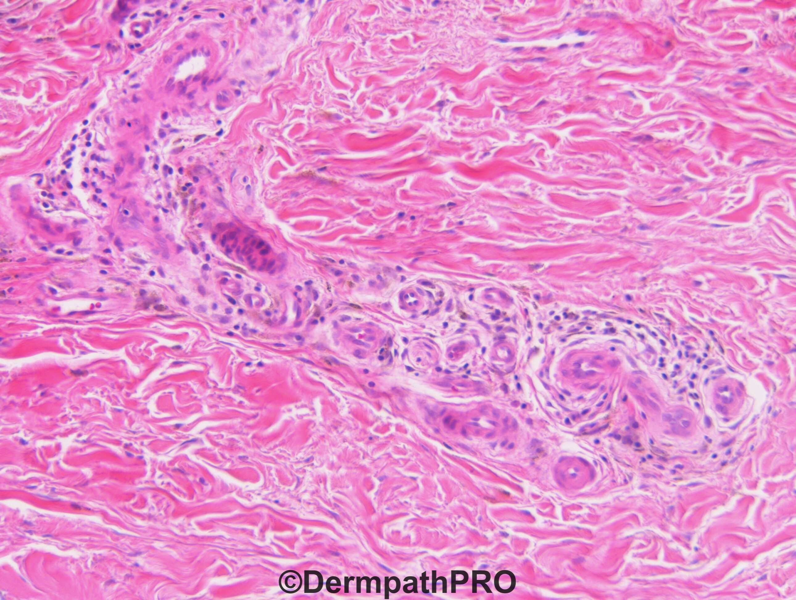
stasis with septal panniculitis and fibrosis

something like lipodermatofibrosis

Share this comment


Link to comment
share_externally

John Zhang

Posted

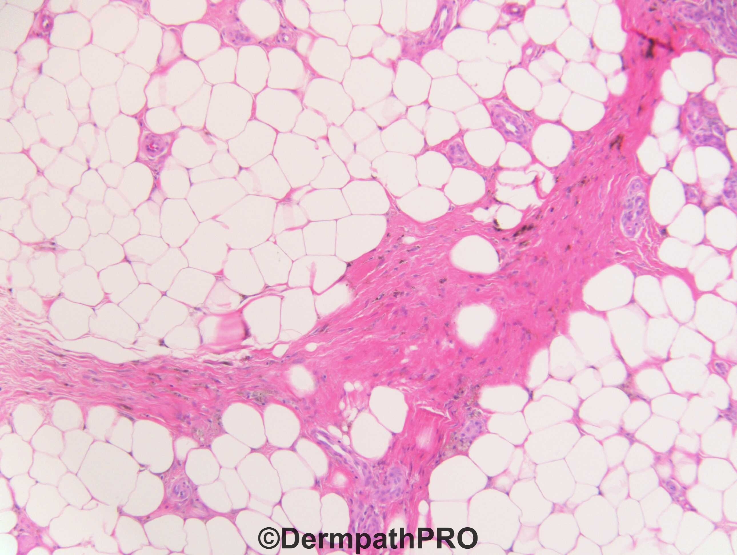
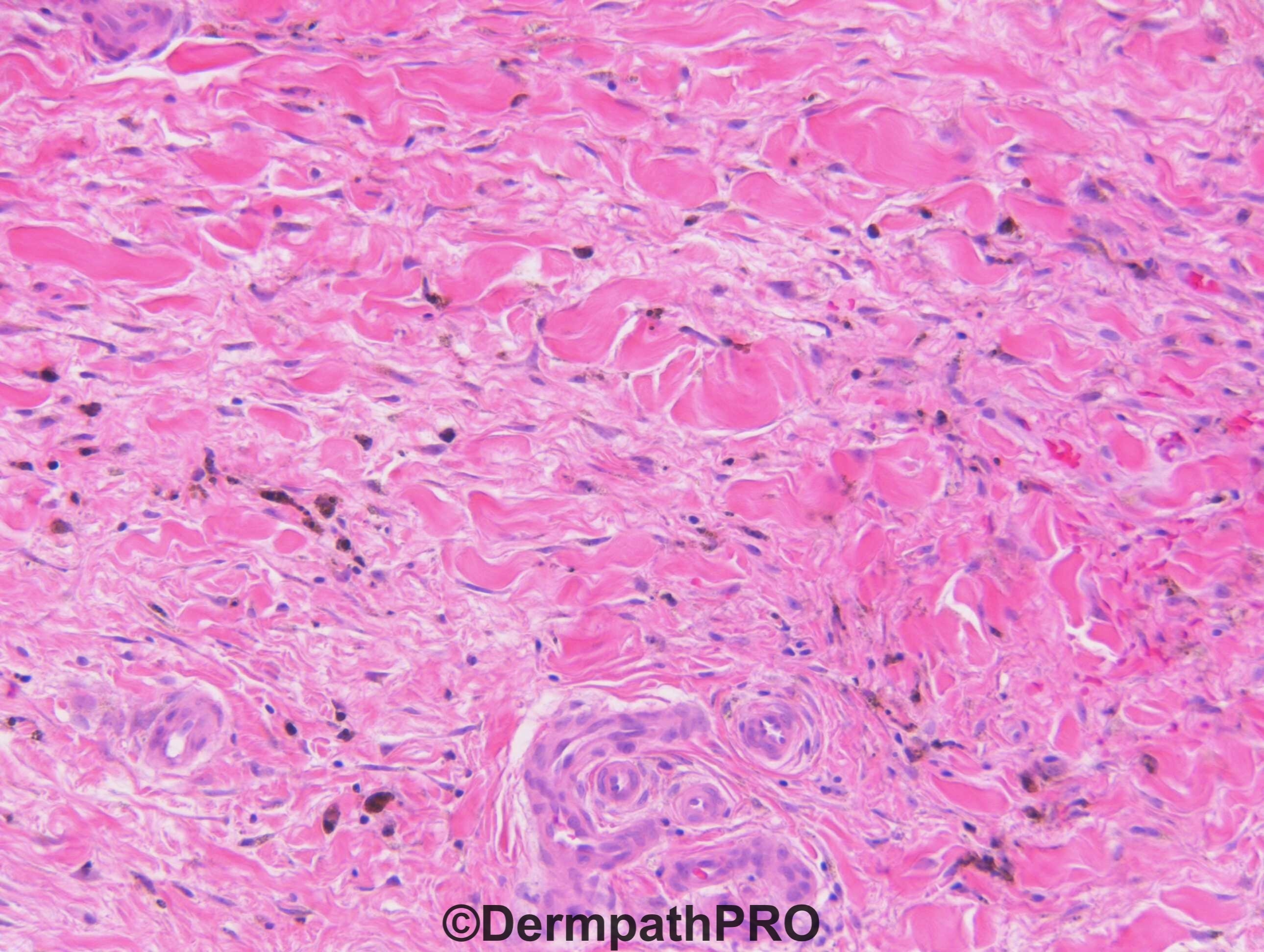
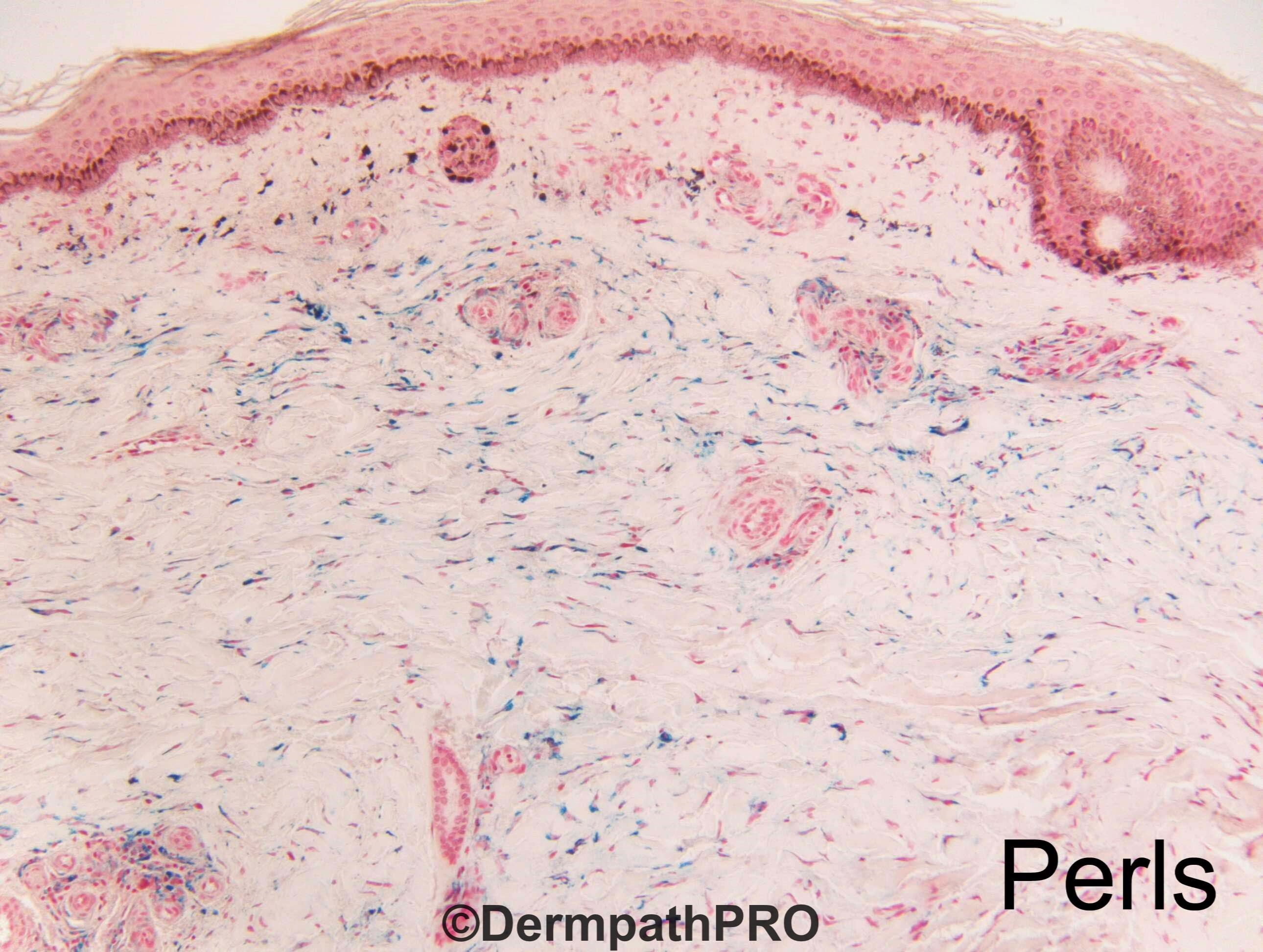
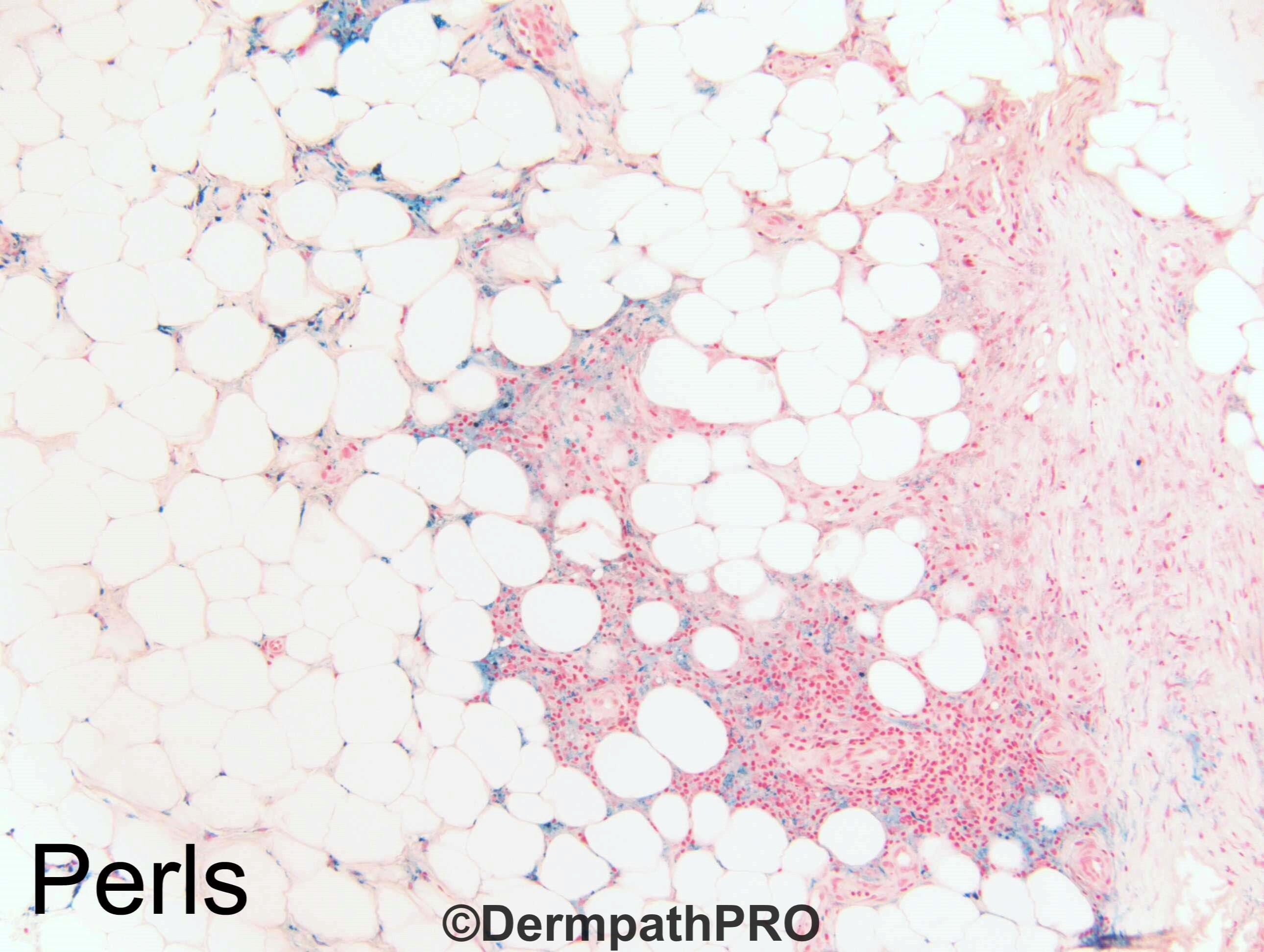
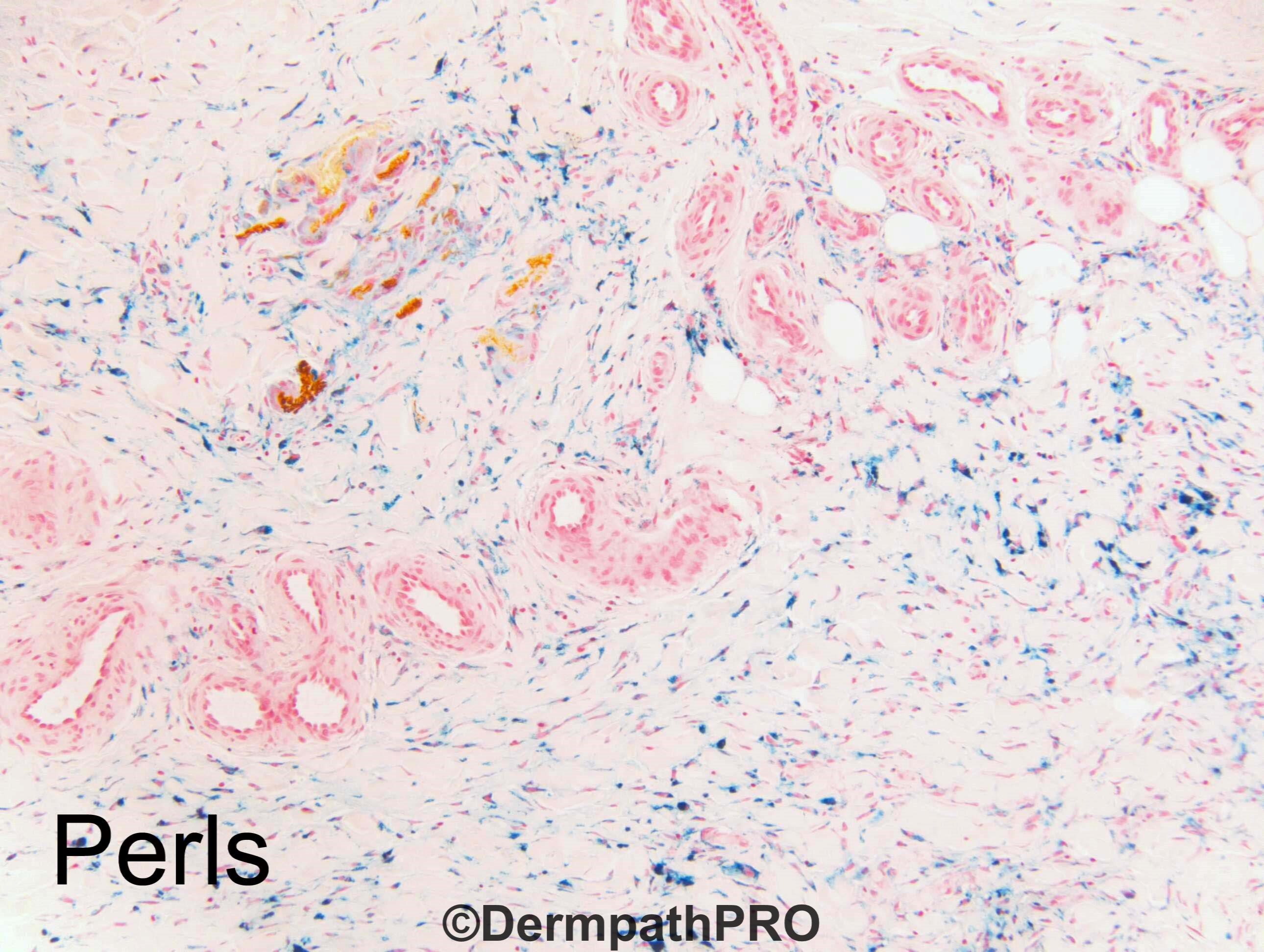
My first impression was lipodermatosclerosis. The hemosiderin in fig 3 seems too superficial for EN.

Share this comment


Link to comment
share_externally

Dr. Richard Carr

Posted

Think we're missing some images. I'll enquire.

Share this comment


Link to comment
share_externally

On 19/06/2019 at 21:08, Krishnakumar subramanian said:

stasis with septal panniculitis and fibrosis

something like lipodermatofibrosis

Agree. I would like to see more lobular inflammation/adipocytic necrosis, but I think this is the best diagnosis.

Share this comment


Link to comment
share_externally

Erythema nodosum is usually tender or painful, so may be I'm wrong...

Share this comment


Link to comment
share_externally

Neil Catterall

Posted

Lipodermatosclerosis with membranocystic change 


Share this comment


Link to comment
share_externally

Lipodermatosclerosis and venous insufficiency 

Share this comment


Link to comment
share_externally

Dr. Richard Carr

Posted

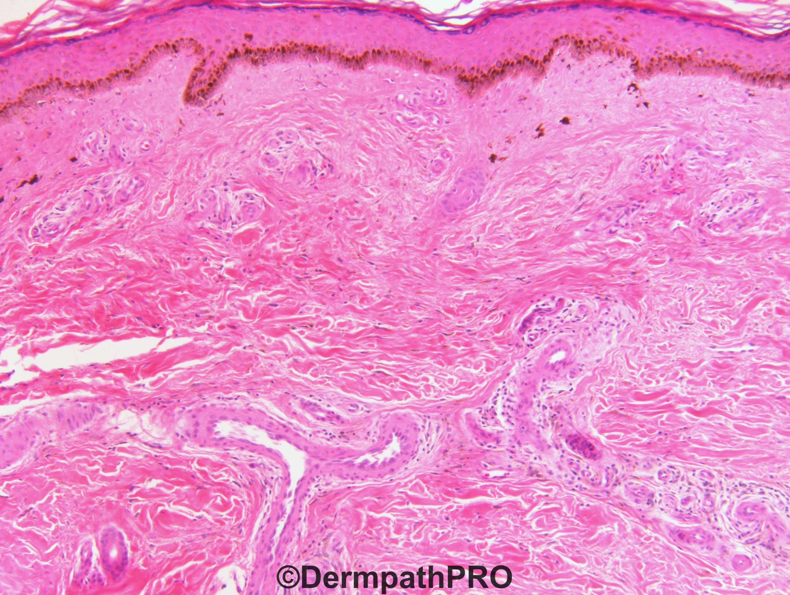
Yes I agree with prominent haemosiderin deposition, minimal inflammatory changes, focal lipomembranous fat necrosis in keeping with venous insufficiency and lipodermatosclerosis. I suspect increased basal melanin pigmentation may also be contributing to the clinical appearances.

Share this comment


Link to comment
share_externally


Join the conversation

You can post now and register later. If you have an account, sign in now to post with your account.

Guest
Add a comment...

×   Pasted as rich text.   Paste as plain text instead

  Only 75 emoji are allowed.

×   Your link has been automatically embedded.   Display as a link instead

×   Your previous content has been restored.   Clear editor

×   You cannot paste images directly. Upload or insert images from URL.

Loading...

×
×
  • Create New...