Jump to content
In this section we have spot diagnoses posted on a daily basis since June 2010, now over 4000! You can review the archived cases and read the suggested diagnoses by users and the final comment by the contributors.
Case are uploaded each week day by 10 am UK time with the correct diagnosis will generally be posted at 8 pm UK time. Why not view the most recent spot diagnosis and proffer a diagnosis?

Case Number : Case 2316 - 2 May 2019 Posted By: Raul Perret

Please read the clinical history and view the images by clicking on them before you proffer your diagnosis.
Submitted Date :
   (0 reviews)

Spot Diagnosis


  Report Record

User Feedback


msofopoulos

Posted

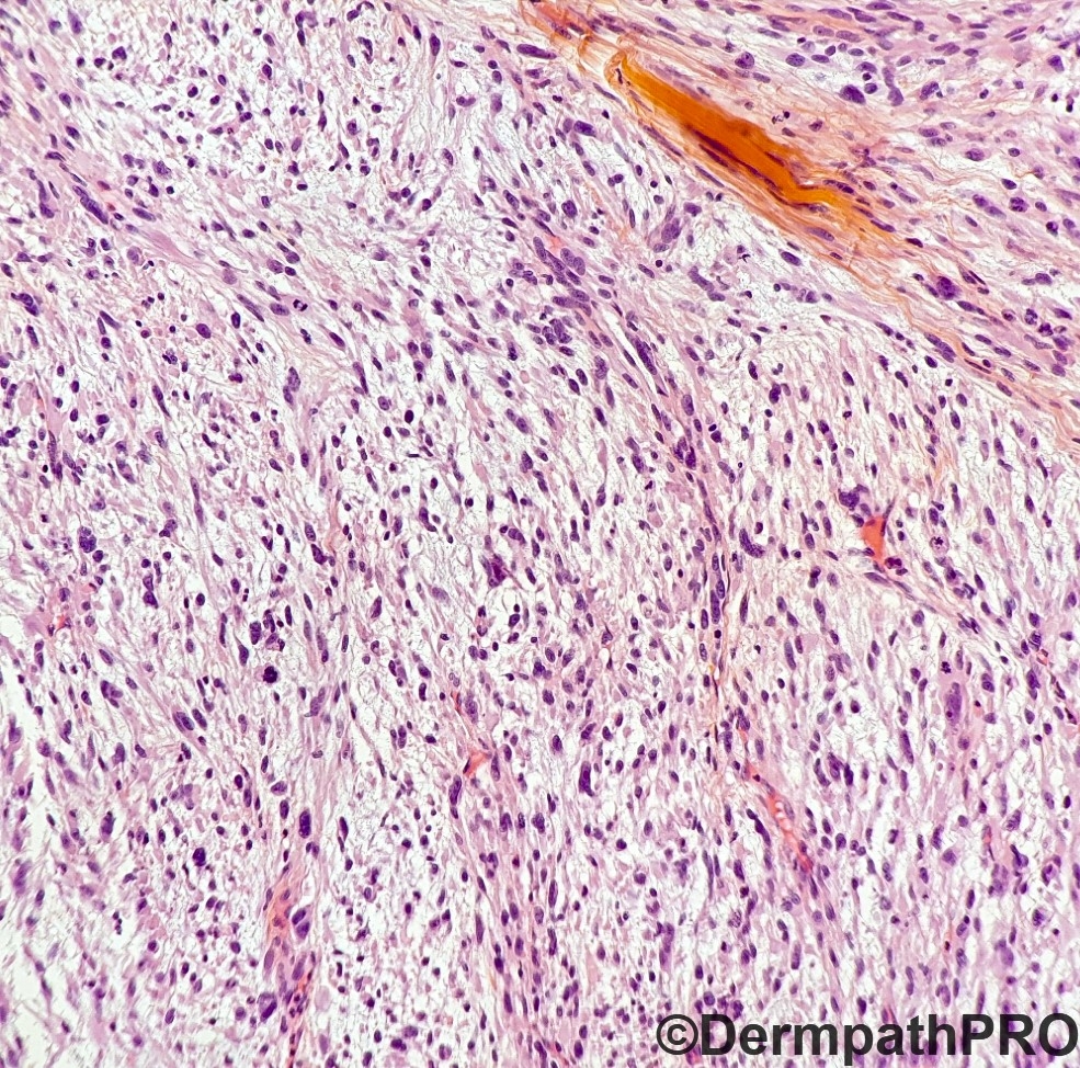
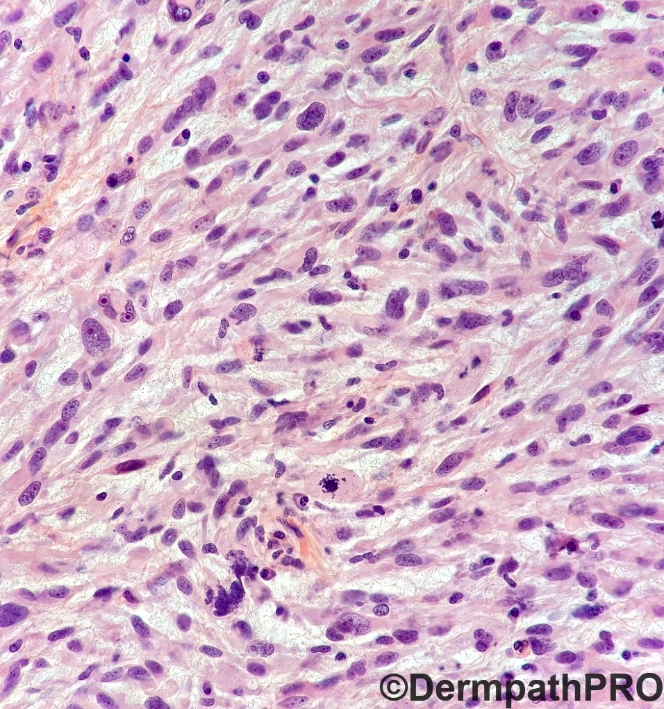
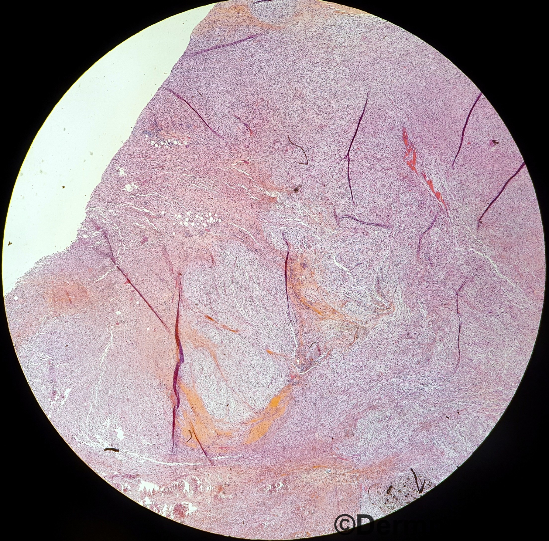
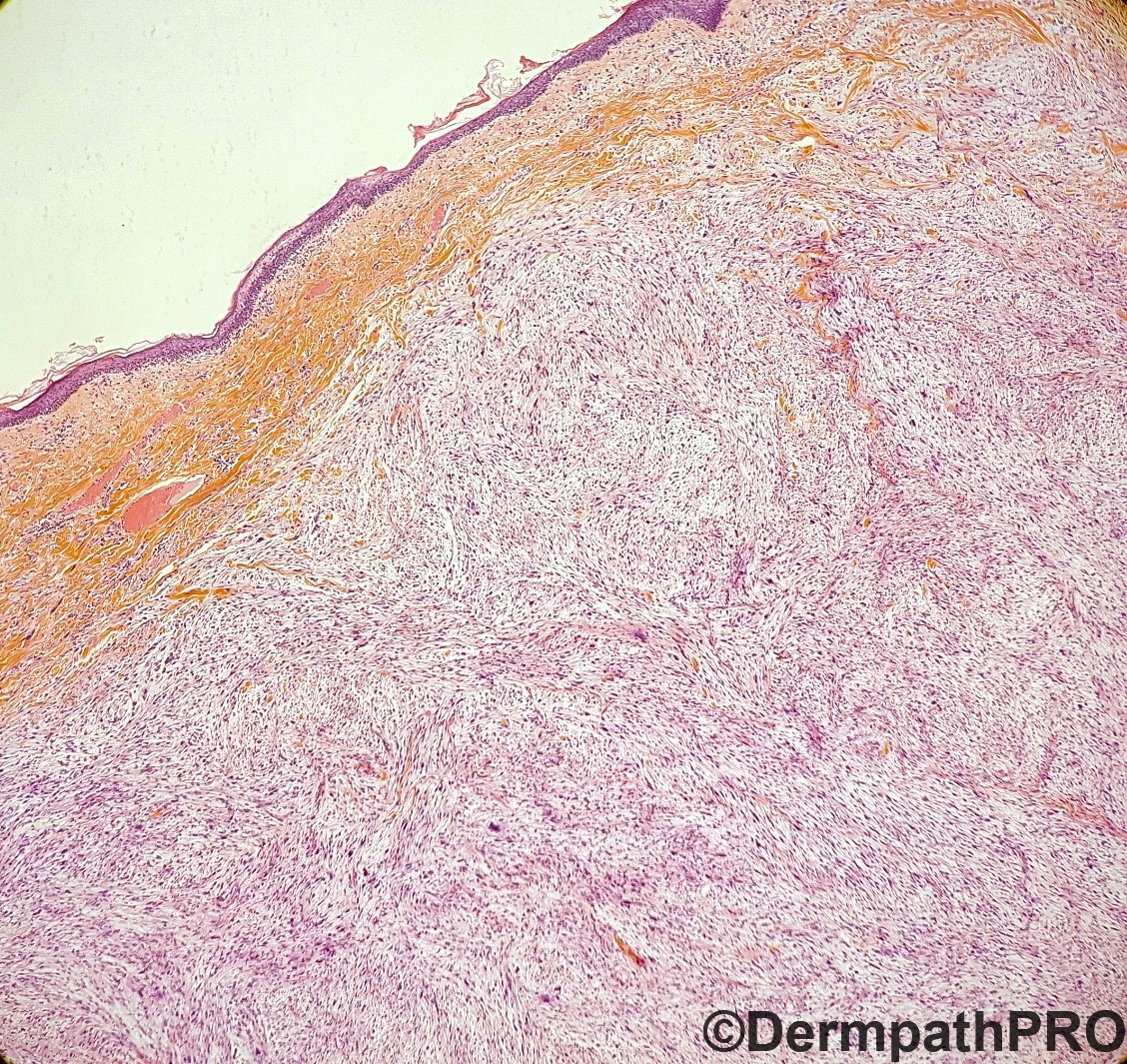
Looks like nodular Fasciitis. How big is it? Is it well circumscribed? Rapid growth? I cant rule out pleomorphic dermal sarcoma (although they usually uglier)

Share this comment


Link to comment
share_externally

John Zhang

Posted

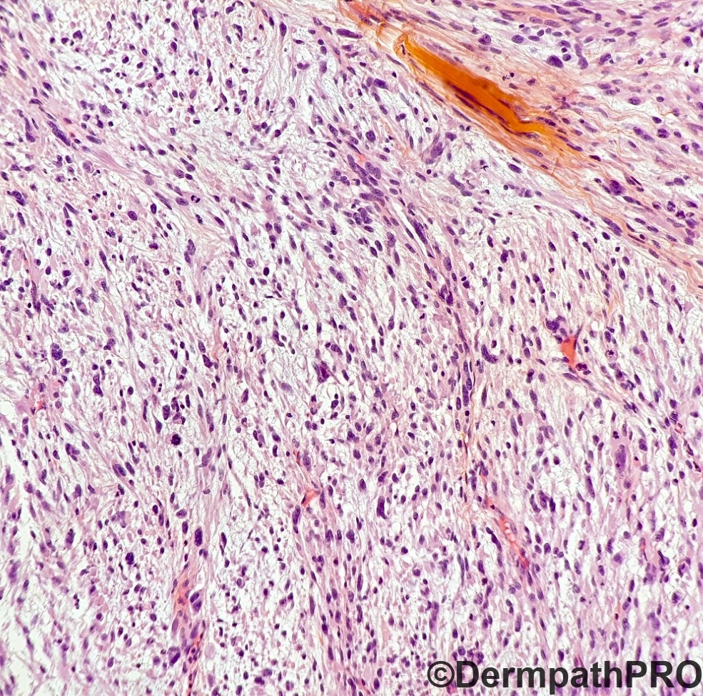
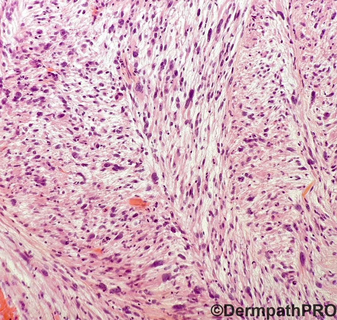
Agree with Anh, My first thought was Myxofibrosarcoma: pleomorphic, myxoid, curvy linear vessels. Interestingly focally there is almost a hint of herring bone pattern. I don't favor nodular fasciitis but will probably need clinical info (age of patient, how fast it grew) to rule it out.

Share this comment


Link to comment
share_externally

Alex-Ventura-Leon

Posted

Myxofibrosarcoma is my first thought.

Share this comment


Link to comment
share_externally

Krishnakumar subramanian

Posted

myxoid dermatofibrosarcoma

Share this comment


Link to comment
share_externally

Meenakshi Batrani

Posted

Myxofibrosacroma primary or DFSP with transformation to myxofibrosarcoma (considering the upper dermal component).

Share this comment


Link to comment
share_externally

vincenzo

Posted

9 hours ago, John Zhang said:

Agree with Anh, My first thought was Myxofibrosarcoma: pleomorphic, myxoid, curvy linear vessels. Interestingly focally there is almost a hint of herring bone pattern. I don't favor nodular fasciitis but will probably need clinical info (age of patient, how fast it grew) to rule it out.


Agree. AFX could be a diff, but it seems a large mass coming from the deep soft parts.

Share this comment


Link to comment
share_externally

Dr. Mona Abdel-Halim

Posted

Myxoinflammatory fibroblastic sarcoma

Share this comment


Link to comment
share_externally

vincenzo

Posted (edited)

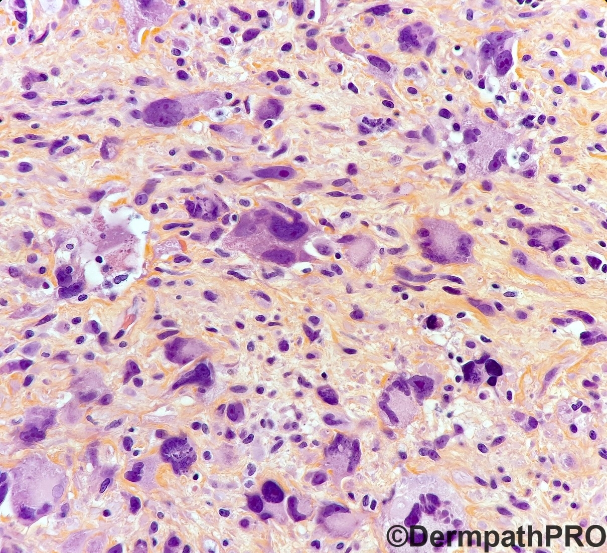
Good diff, Mona. A poorly inflammatory MIFS could be a possibility, because we can see ganglion like atypical cells and some sternbergoid cell(fig3). But it don't seems me at a glance. Waiting for the diagnosis. 

Edited by vincenzo

Share this comment


Link to comment
share_externally

Raul Perret

Posted

Good differential. This was a grade 2 myxofibrosarcoma. Note the multinodular architecture and the curvilinear blood vessels that are a key finding for this diagnosis. There are not virocyte-like cells in this case but rather multinucleated "myoid" cells which can be sometimes seen in Mixofibrosarcoma. Thanks for participating 

PSX_20190409_090009.jpg

Share this comment


Link to comment
share_externally


Join the conversation

You can post now and register later. If you have an account, sign in now to post with your account.

Guest
Add a comment...

×   Pasted as rich text.   Paste as plain text instead

  Only 75 emoji are allowed.

×   Your link has been automatically embedded.   Display as a link instead

×   Your previous content has been restored.   Clear editor

×   You cannot paste images directly. Upload or insert images from URL.

Loading...

×
×
  • Create New...