Jump to content
In this section we have spot diagnoses posted on a daily basis since June 2010, now over 4000! You can review the archived cases and read the suggested diagnoses by users and the final comment by the contributors.
Case are uploaded each week day by 10 am UK time with the correct diagnosis will generally be posted at 8 pm UK time. Why not view the most recent spot diagnosis and proffer a diagnosis?

Case Number : Case 2317 - 3 May 2019 Posted By: Dr. Richard Carr

Please read the clinical history and view the images by clicking on them before you proffer your diagnosis.
Submitted Date :
   (0 reviews)

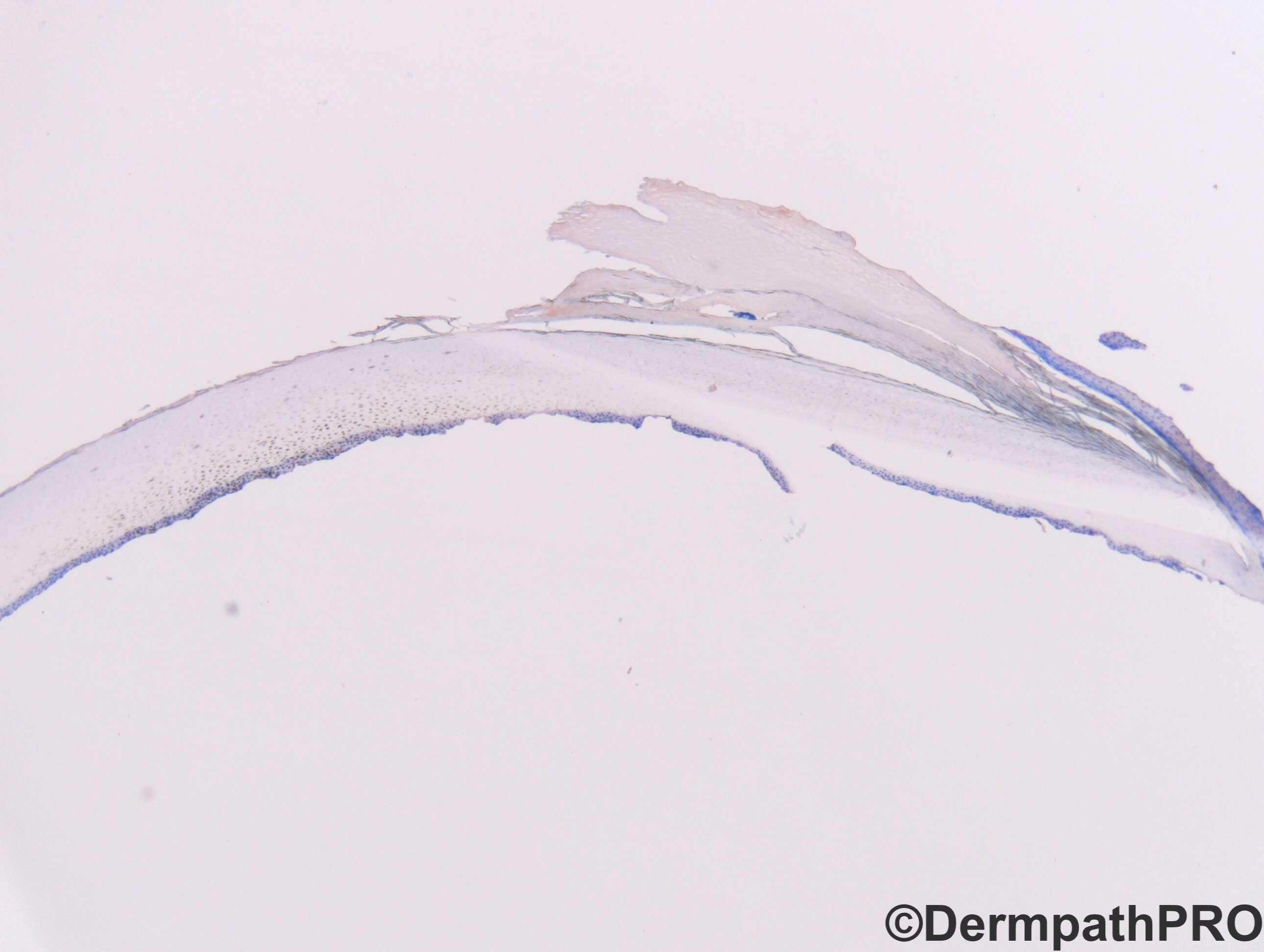
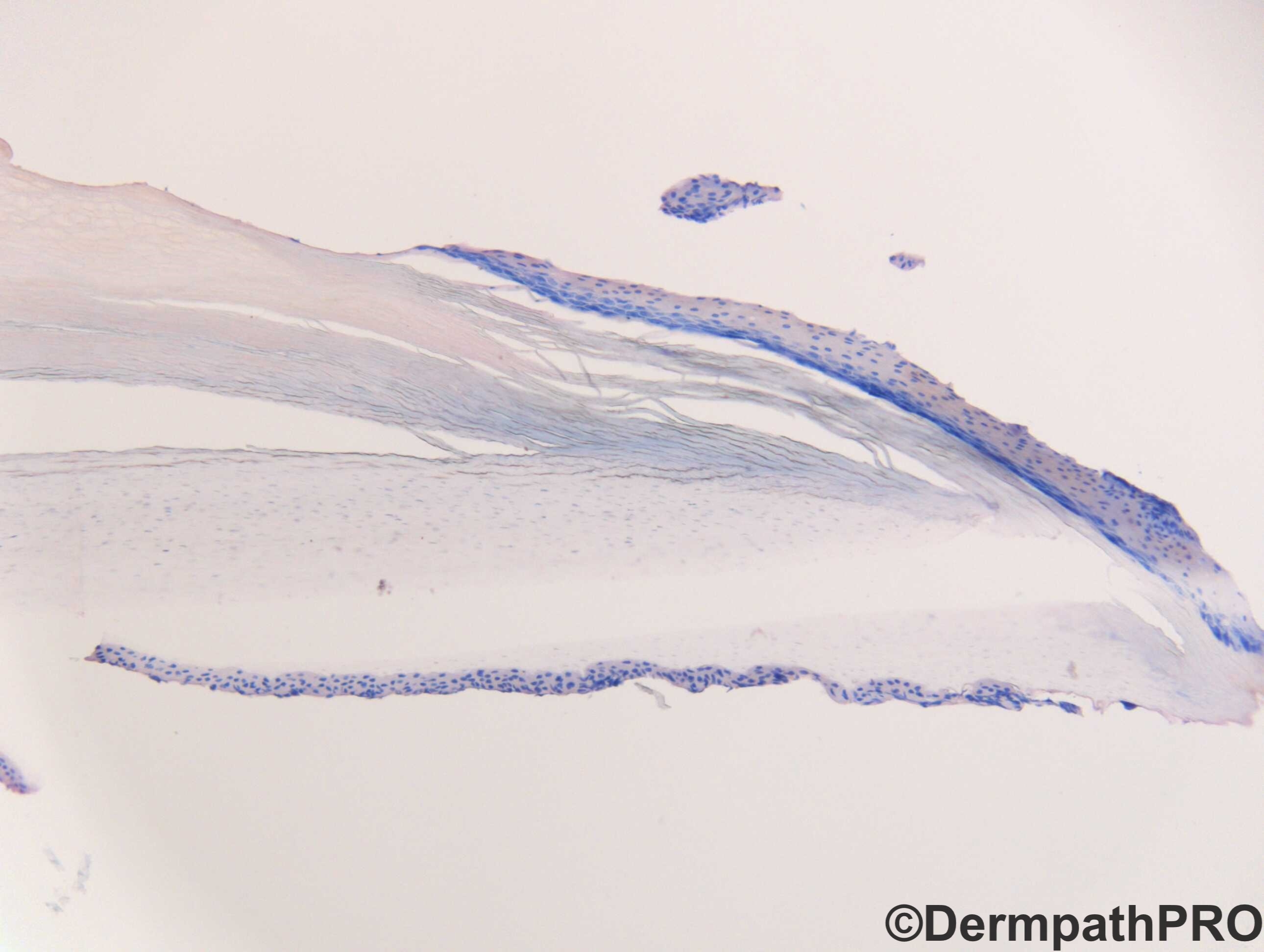
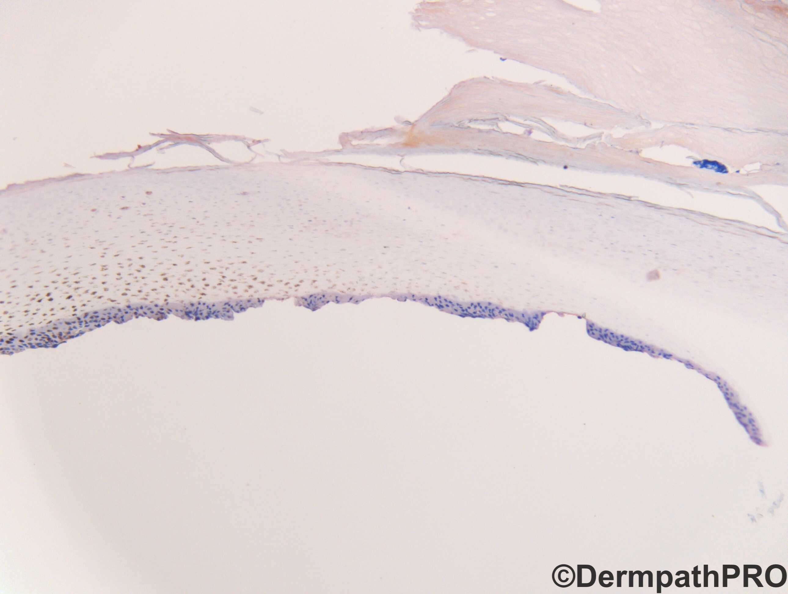
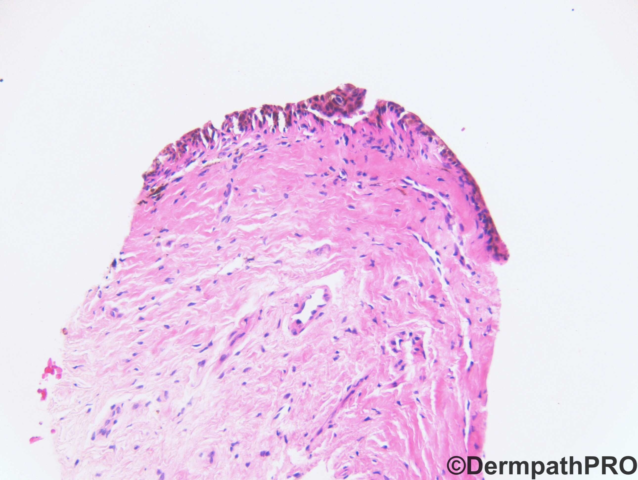
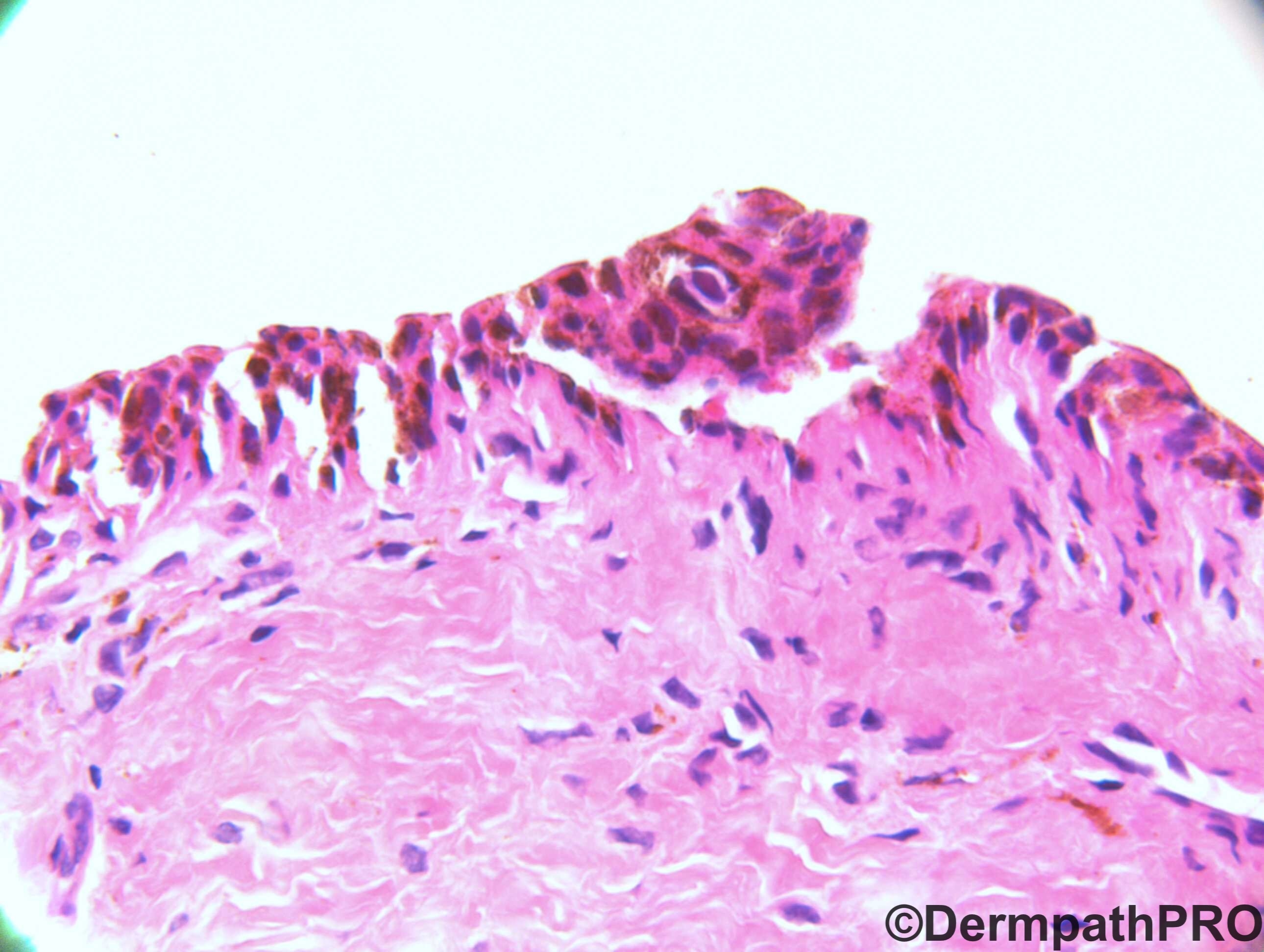
F5. Melanonychia. ?Melanoma. Nail plate and nail bed biopsy


  Report Record

User Feedback


John Zhang

Posted

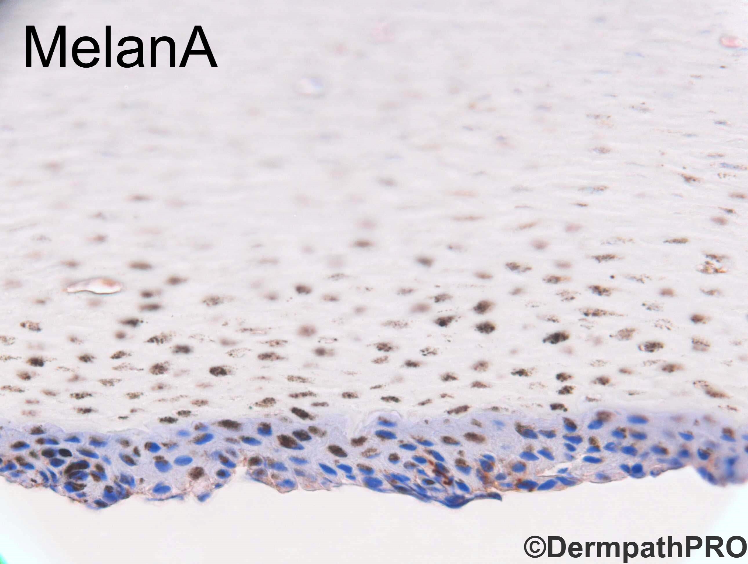
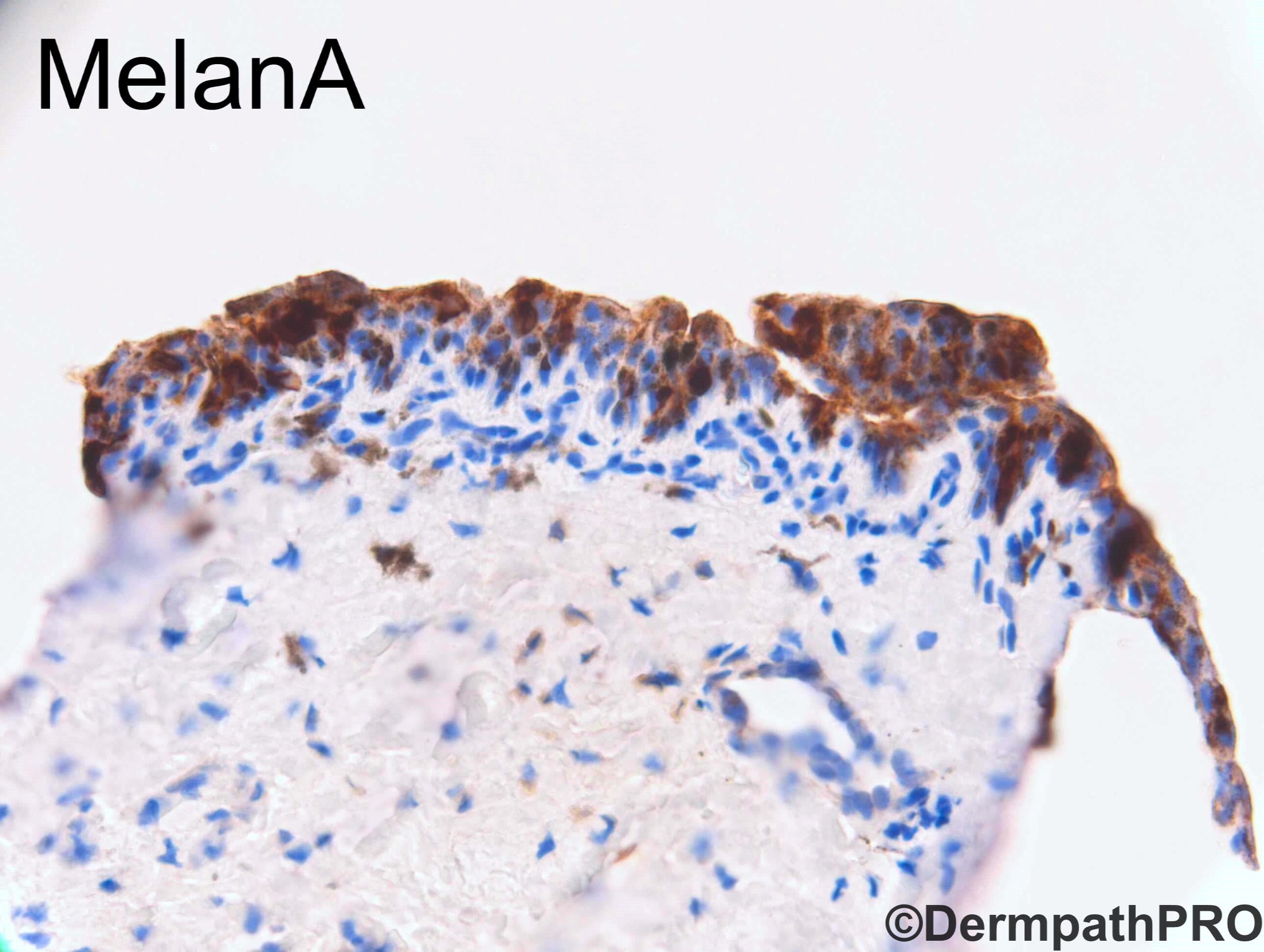
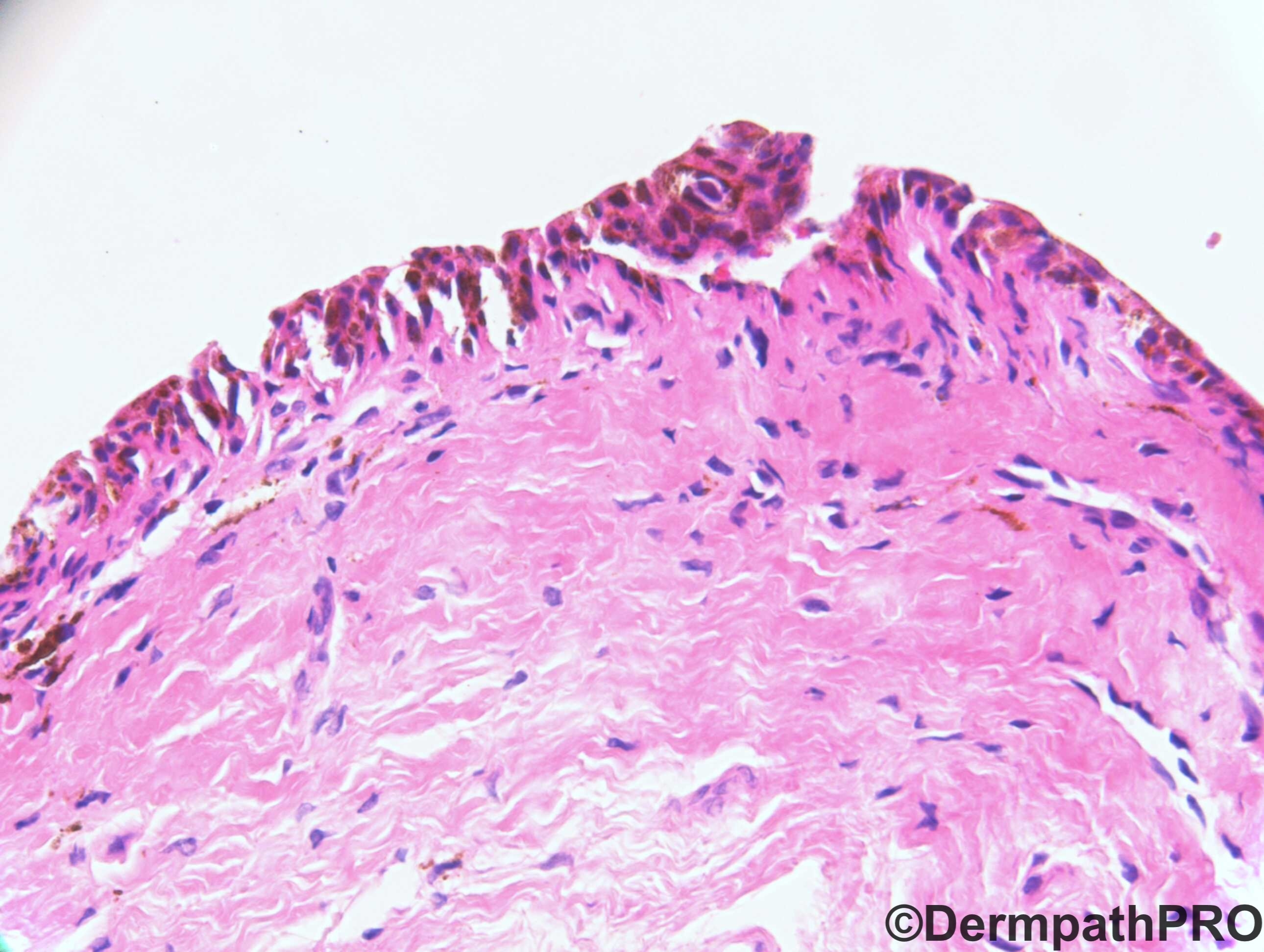
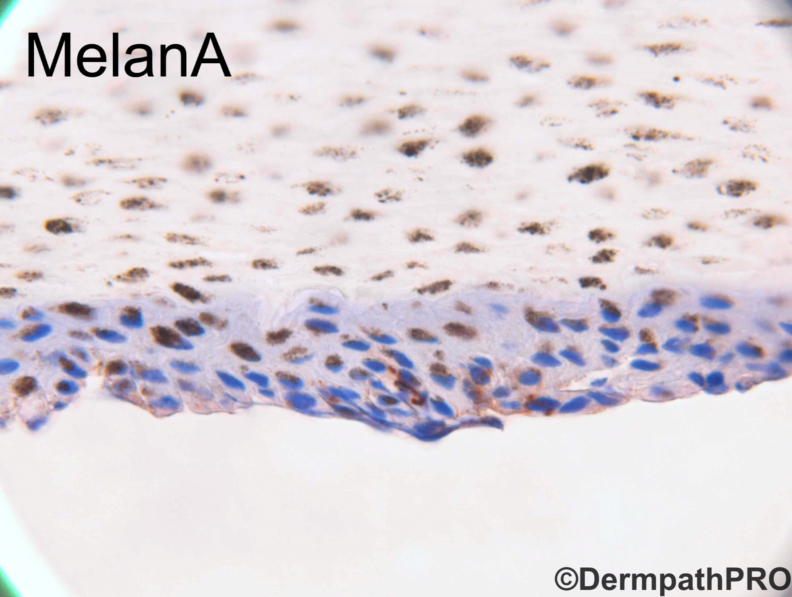
Although in figure 6 the melanocytes reach the surface, from fig. 1&2 I suspect that the real top half of the epidermis is ripped off from the dermal portion of the biopsy in fig. 6, therefore the melanocytes are not really reaching the top. There are pigmented cells in upper portion of the epidermis in figs 5&8, but those pigments are eccentrically located in the cell and they have a greenish hue (unlike the more brownish chromogen in the IHC stains in the lower portion of the epidermis in the same photo). So those are pigmented keratinocytes rather than melanocytes. In addition, Dr. LeBoit said childhood melanomas are in general "vanishingly rare", and myy dermatologists colleagues once told me that there were dermatology big shots who believed that there were no childhood nail melanomas - show him a childhood nail melanoma, he will prove to you that it was an over diagnosis. 

So my diagnosis for this lesion in a 5 year old - junctional nevus, period.

Share this comment


Link to comment
share_externally

John Zhang

Posted

13 minutes ago, John Zhang said:

 and myy dermatologists colleagues once told me that there were dermatology big shots who believed that there were no childhood nail melanomas - show him a childhood nail melanoma, he will prove to you that it was an over diagnosis.

I think the last part in my paragraph about no real childhood nail melanoma was a little too anecdotal. I should only cite published references or my direct personal experience. Sorry.

Share this comment


Link to comment
share_externally

Mohamed khaled

Posted

Not easy case. I wish I could see the dermoscopic picture of this case also for correlation.

Share this comment


Link to comment
share_externally

 Pigmented lesions of the nail always scare me.

Usually I see histologic changes similar to Figure 8, meaning pigmented changes in the nail plate but Melan-A and HMB-45 do not show any melanocytic proliferation. That's the typical picture that I usually encounter. Just don't know what to say in those cases , hopefully Dr. Carr can give us some advice how to deal with such biopsy like in Fig. 1,3,5, and 8 where we see pigment up in the nail plate but no melanocytes in Melan-A.

Figure 6 clearly show some slightly enlarged melanocytes and maybe melanin in the keratinocytes (as John said), highlighted by Melan-A. I really do not appreciate significant increase of enlarged melanocytes in the H&E to warrant the diagnosis of a melanoma in situ. Patient is young (F5) so I would go for simple lentigo.

Share this comment


Link to comment
share_externally

IgorSC

Posted

We have 2 different speciens to analyse in this case. One containing the nail plate plus the superficial portion of the nail matrix, called keratogeneous zone. We can see the ventral portion of the proximal nail fold above as well. If we analyse the nail plate, we can see pigments mostly in the bottom parts of the nail, so the proliferation problably is located in the distal matrix (that produces the inferior part of the nail plate). Melanomas in children are extremely rare (I really do not know about this existence), and you can even see hutchinson sign in children in congenital nevi (in fact, pseudo-hutchinson sign). The other specimen is from the nail matrix. Here it is possible to see melanocytic proliferation and epithelial hyperpigmentation. 75% of LM in children are caused by melanocytic nevi and I believe this is the diagnoisis, better seen with Melan-A. If we consider that the pigmentation is mostly melanin, so this should be also a bening lesion, like a Lentigo. A red chromogen or a immunostain with nuclear staining would be better for this conclusion.

Share this comment


Link to comment
share_externally

Dr. Richard Carr

Posted

Great discussion. I pretty much agree word for word with John, IgorSC and Anh.

This was my report (case was a referral from a children's centre).

Many thanks for sending this case. As is often the case with partial biopsies of the nailbed and nail plate, assessment is exceedingly challenging due to the technical difficulty of cutting sections and the crust artefact that takes place during the procedure. The main abnormality is marked pigmentation within the nail matrix epithelium extending into the surprabasal keratinocytes and up into the nail plate.  I could not identify any definite abnormal melanocytic proliferation.  The differential diagnosis, therefore, includes melanocyte activation and benign lentigo.  I should point out melanoma arising in the nail plate of children is vanishingly rare and I have not got any personal experience of a lesion but I don’t think this is unusual even for experts.

I too heard that many don't believe cases of melanoma in the nail in children at this young age (at the ISDP meeting in Sau Paulo). I also took the opportunity to discuss the general issue with David De Berker (expert clinician in UK for nail diseases) and his admittedly anecdotal comment largely agreed and the general advice was not to take a biopsy! 

Share this comment


Link to comment
share_externally

John Zhang

Posted

16 hours ago, Dr. Richard Carr said:

Great discussion. I pretty much agree word for word with John, IgorSC and Anh.

This was my report (case was a referral from a children's centre).

Many thanks for sending this case. As is often the case with partial biopsies of the nailbed and nail plate, assessment is exceedingly challenging due to the technical difficulty of cutting sections and the crust artefact that takes place during the procedure. The main abnormality is marked pigmentation within the nail matrix epithelium extending into the surprabasal keratinocytes and up into the nail plate.  I could not identify any definite abnormal melanocytic proliferation.  The differential diagnosis, therefore, includes melanocyte activation and benign lentigo.  I should point out melanoma arising in the nail plate of children is vanishingly rare and I have not got any personal experience of a lesion but I don’t think this is unusual even for experts.

I too heard that many don't believe cases of melanoma in the nail in children at this young age (at the ISDP meeting in Sau Paulo). I also took the opportunity to discuss the general issue with David De Berker (expert clinician in UK for nail diseases) and his admittedly anecdotal comment largely agreed and the general advice was not to take a biopsy! 

Dr. Carr I didn’t realize that “vanishingly rare” was actually your phrase! :) thanks for showing the case.

Share this comment


Link to comment
share_externally

Dr. Mona Abdel-Halim

Posted

Great discussion. Reassuring to know that nail unit melanoma in children is "vanishingly rare"! I like this expression. My opinion was melanocyte activation or lentigo too. 

Share this comment


Link to comment
share_externally


Join the conversation

You can post now and register later. If you have an account, sign in now to post with your account.

Guest
Add a comment...

×   Pasted as rich text.   Paste as plain text instead

  Only 75 emoji are allowed.

×   Your link has been automatically embedded.   Display as a link instead

×   Your previous content has been restored.   Clear editor

×   You cannot paste images directly. Upload or insert images from URL.

Loading...

×
×
  • Create New...