Jump to content
In this section we have spot diagnoses posted on a daily basis since June 2010, now over 4000! You can review the archived cases and read the suggested diagnoses by users and the final comment by the contributors.
Case are uploaded each week day by 10 am UK time with the correct diagnosis will generally be posted at 8 pm UK time. Why not view the most recent spot diagnosis and proffer a diagnosis?

Case Number : Case 2329 - 21 May 2019 Posted By: Uma Sundram

Please read the clinical history and view the images by clicking on them before you proffer your diagnosis.
Submitted Date :
   (0 reviews)

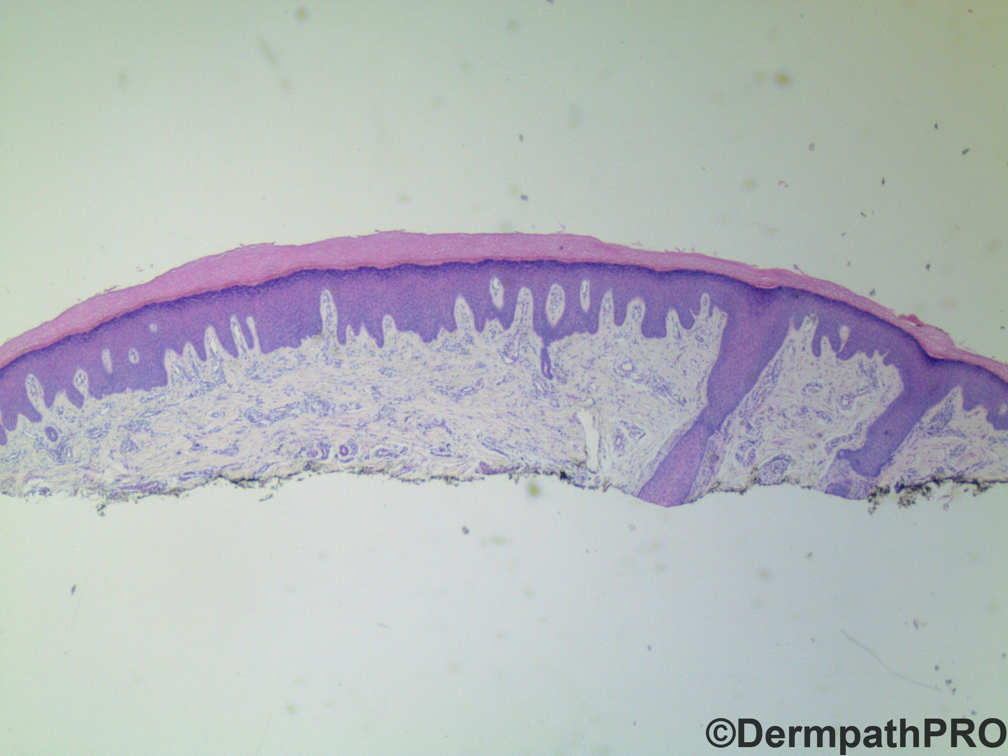
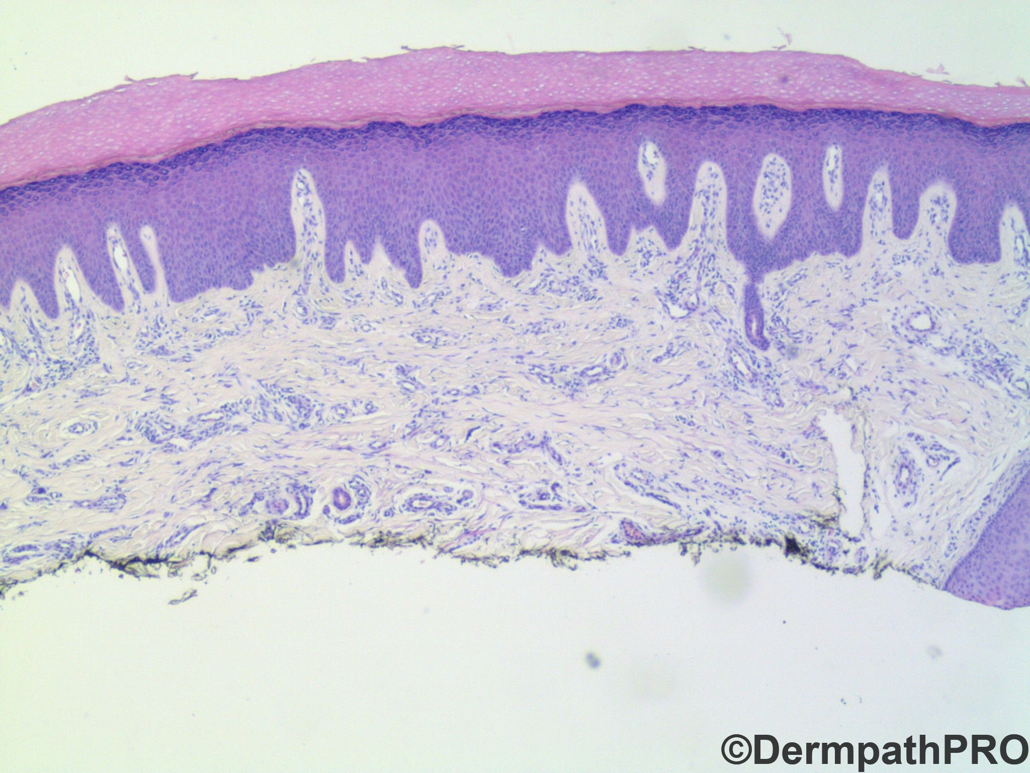
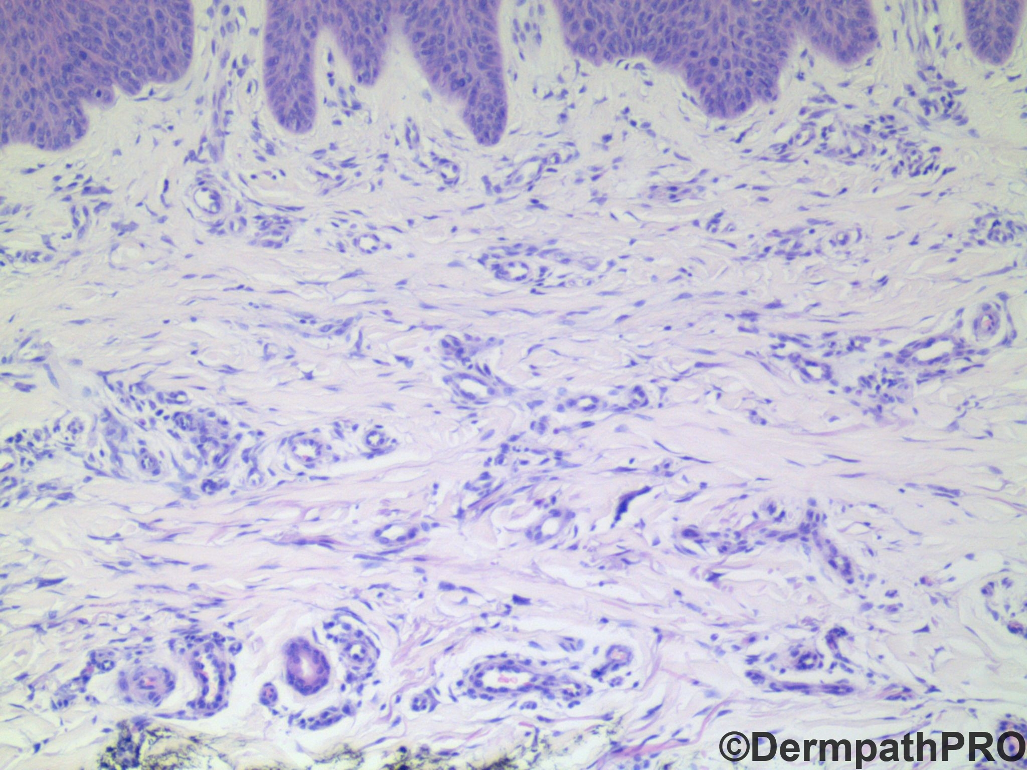
63 year old male with lesion on finger

Edited by Admin_Dermpath


  Report Record

User Feedback


John Zhang

Posted

I favor dermatofibroma, especially given the overlying epidermal hyperplasia. I also thought about interstitial granuloma annulare. Krishnakumar's idea of angiofibroma is interesting. But angiofibromas are usually subcutanous.

Share this comment


Link to comment
share_externally

Alex-Ventura-Leon

Posted

What about Multinucleated Giant Cell Angiohistiocytoma?

Share this comment


Link to comment
share_externally

IgorSC

Posted

Agree with Alex Ventura, this is a multinucleated giant cell angiohistiocytoma. Some argues that dermatofibroma does not exist in the fingers, but I have seen one or two cases very similar to it. I would like to know the opinion of the collegues about this.

Share this comment


Link to comment
share_externally

John Zhang

Posted

Ha ha, I thought about multinucleated cell angiohistiocytoma, too. But I shied away from it because I thought there was only one possible multinucleated cell (in the center of the last photo). Not enough multinucleated cells! 

Share this comment


Link to comment
share_externally

Uma Sundram

Posted

Yes, a case with numerous possibilities. We finally decided on scar like dermatofibroma. Digital DF has been described. This one is unusual because lesional cells are really scarce. There's no wrong answer here; if you can defend it, you can diagnose it! I was also interested in what you all thought about this case.

Share this comment


Link to comment
share_externally

vincenzo

Posted

I was thinking of superficial acral fibromyxoma. 

Share this comment


Link to comment
share_externally

Neil Catterall

Posted

Not sure if you can get acquired elastotic haemangioma on acral skin but the plaque-like spread of vessels can look like this.

Share this comment


Link to comment
share_externally

Uma Sundram

Posted

Great additional points. CD34 is negative, arguing against superficial acral fibromyxoma (but the histology is really good for it). I'm not sure how common acquired elastotic hemangiomas are on acral sites, but I think the vessels tend to be bigger and more prominent (perhaps more clearly a vascular process than this lesion seems to be?). 

Share this comment


Link to comment
share_externally

Uma Sundram

Posted

 I should say, CD34 is negative, somewhat arguing against the diagnosis of superficial acral fibromyxoma, since up to 30% of cases are negative. So, this is a possibility as well.

Share this comment


Link to comment
share_externally


Join the conversation

You can post now and register later. If you have an account, sign in now to post with your account.

Guest
Add a comment...

×   Pasted as rich text.   Paste as plain text instead

  Only 75 emoji are allowed.

×   Your link has been automatically embedded.   Display as a link instead

×   Your previous content has been restored.   Clear editor

×   You cannot paste images directly. Upload or insert images from URL.

Loading...

×
×
  • Create New...