Jump to content
In this section we have spot diagnoses posted on a daily basis since June 2010, now over 4000! You can review the archived cases and read the suggested diagnoses by users and the final comment by the contributors.
Case are uploaded each week day by 10 am UK time with the correct diagnosis will generally be posted at 8 pm UK time. Why not view the most recent spot diagnosis and proffer a diagnosis?

Case Number : Case 2332 - 24 May 2019 Posted By: Dr. Richard Carr

Please read the clinical history and view the images by clicking on them before you proffer your diagnosis.
Submitted Date :
   (0 reviews)

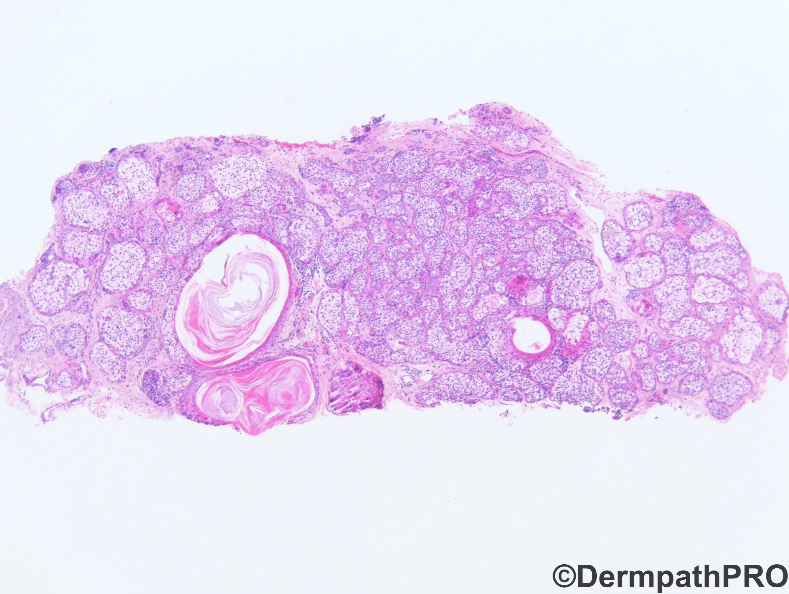
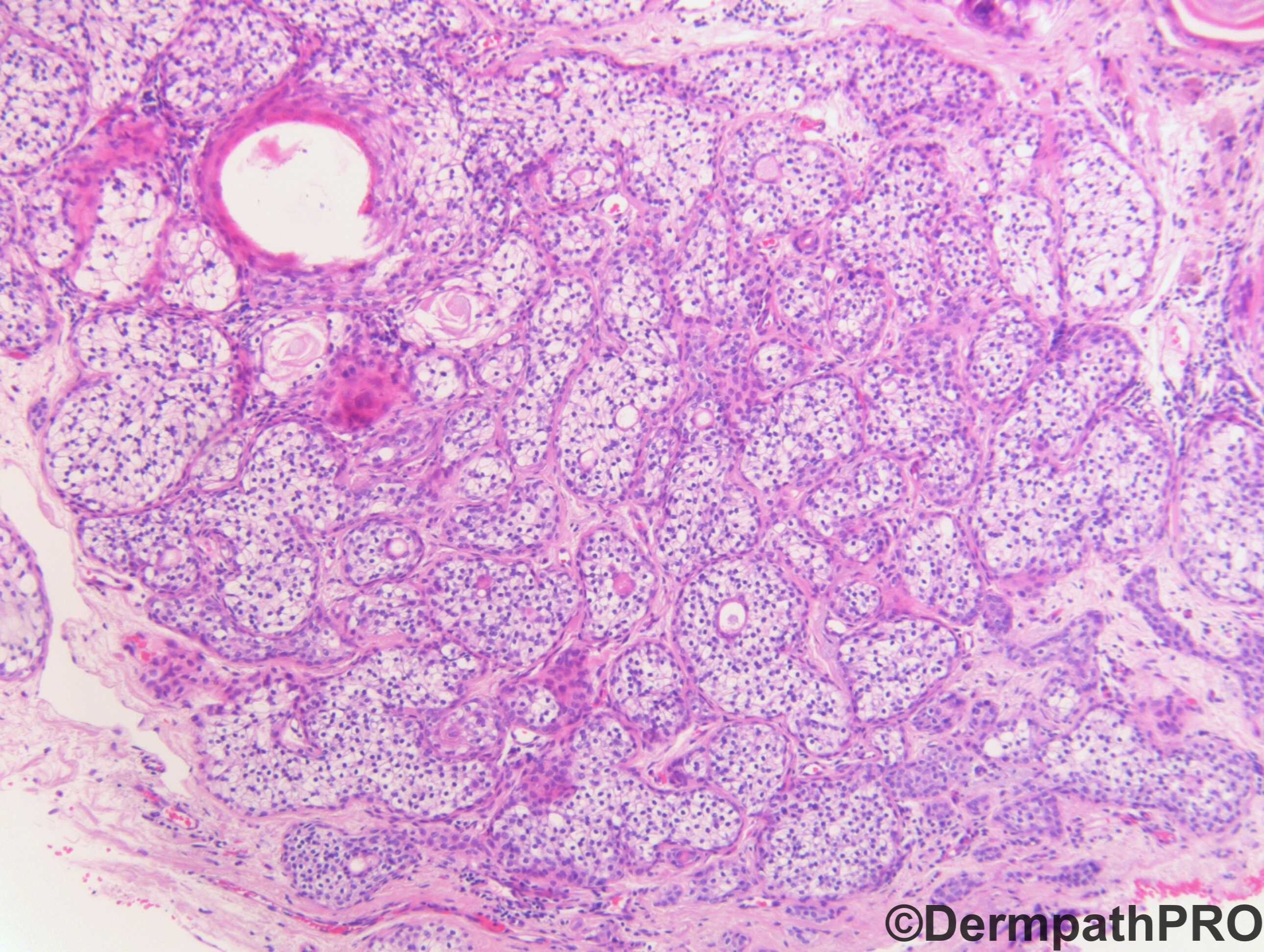
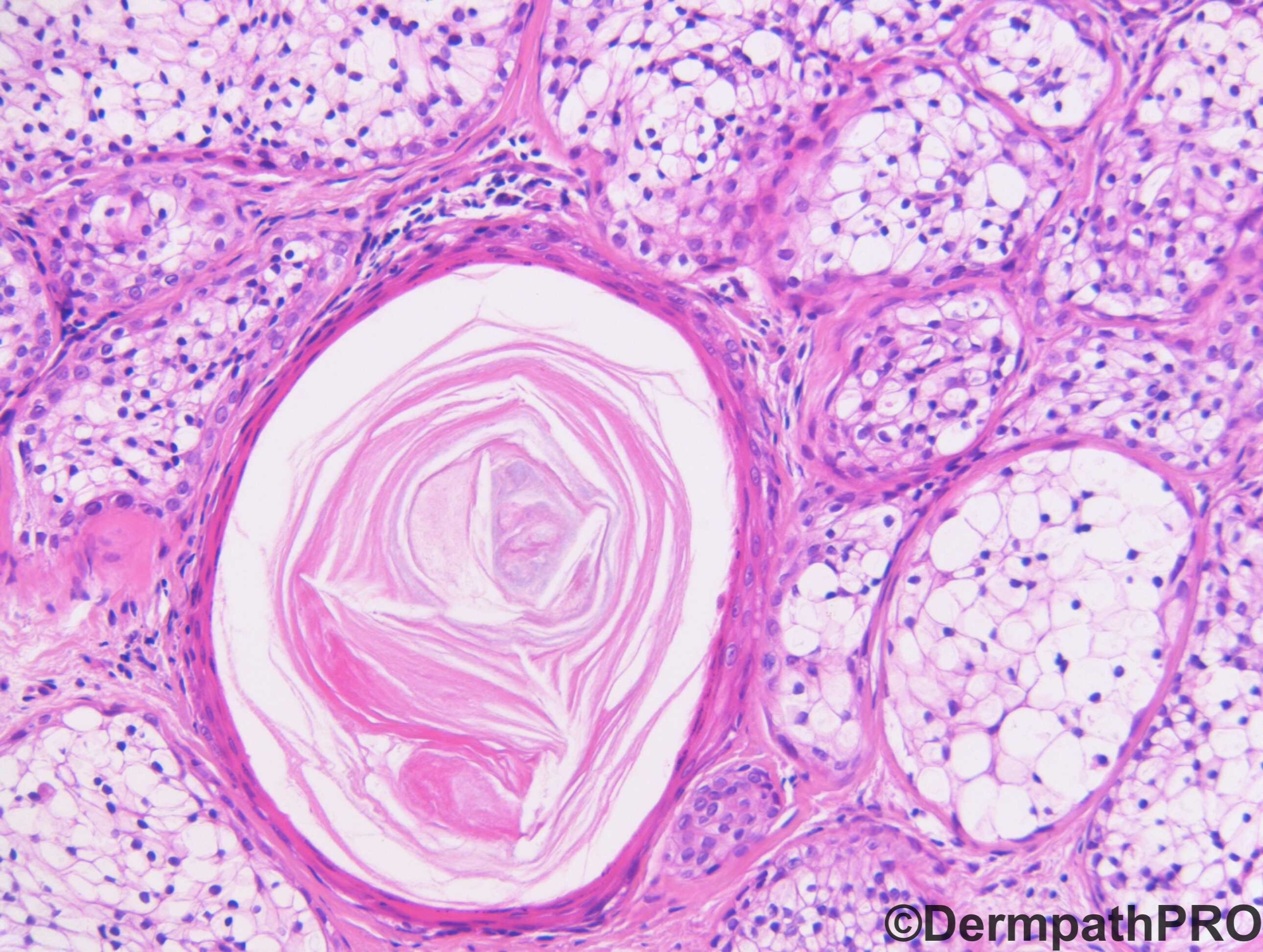
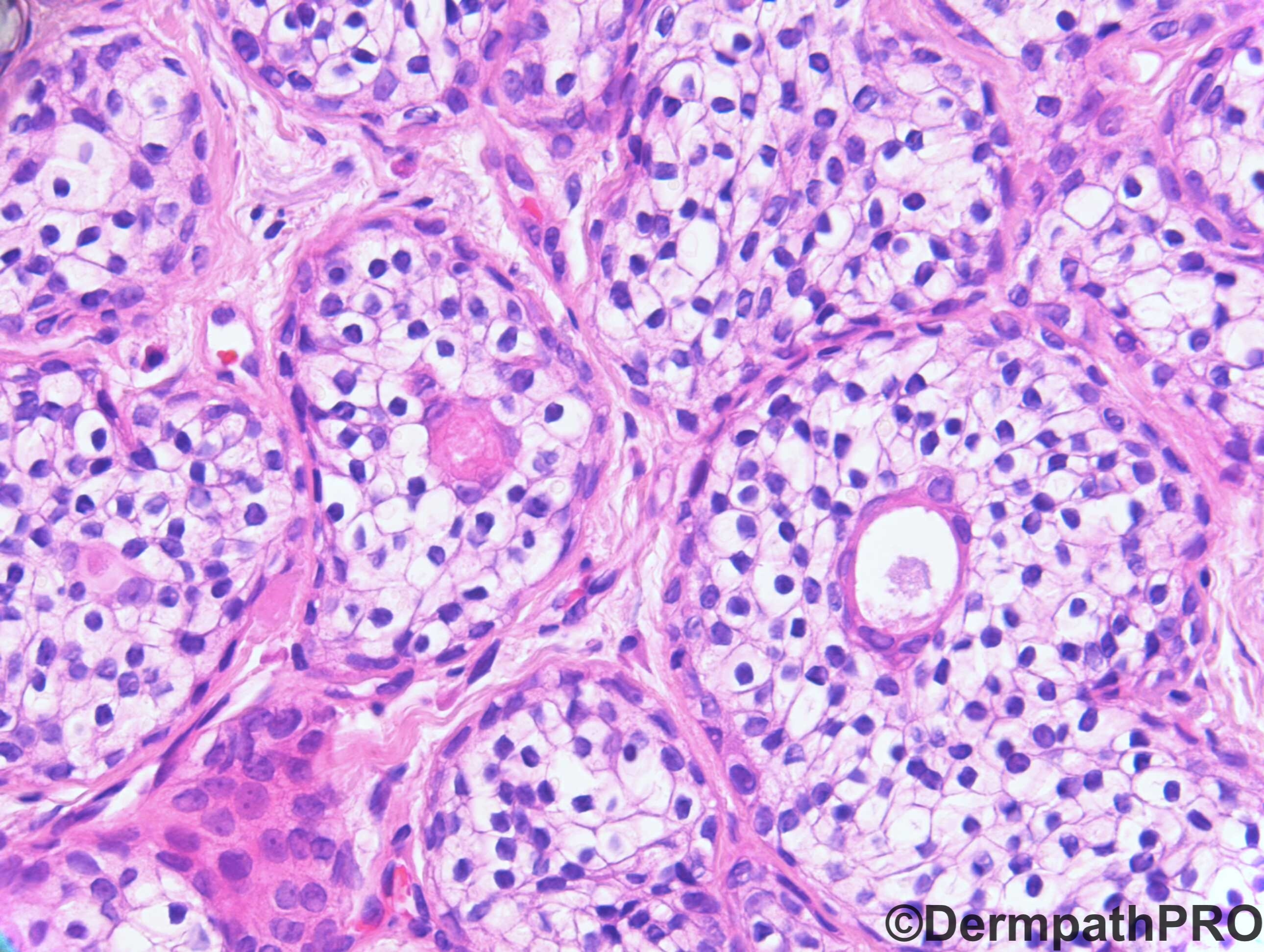
F80. Lower lid. ?Sebaceous cyst, ?Naevus, ?Seborrhoeic keratosis


  Report Record

User Feedback


John Zhang

Posted

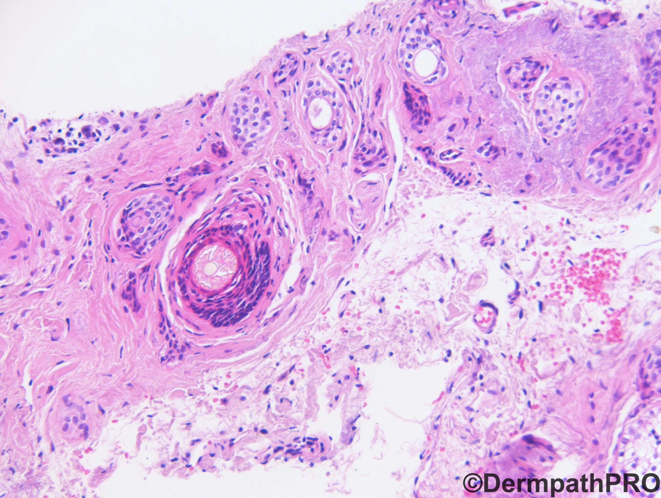
what an interesting lesion! I don't know what it is. But the presence of multiple round lumen, especially in fig 3, makes me think of a sweat gland neoplasm. Also it's the eyelid, I would run neuroendocrine markers chromogranin, synaptophysin to rule out EMPSGC. If these stains are negative, I would run CEA, if the lumen is positive then I will consider hidradenoma or tubular adenoma. But because I am not sure if the periphery is infiltrative, I would recommend excision in case it is a sweat gland carcinoma. If CEA is negative, I will consider metastatic renal cell carcinoma (RCC and PAX8 to confirm).

I feel like that I am firing a shotgun towards the other direction of the target. But hey this forum is so pleasant I dare to have a chit chat...

Share this comment


Link to comment
share_externally

John Zhang

Posted

I don't think it is sebaceous carcinoma. But would run adipophilin if the neuroendocrine markers are negative.

Share this comment


Link to comment
share_externally

IgorSC

Posted

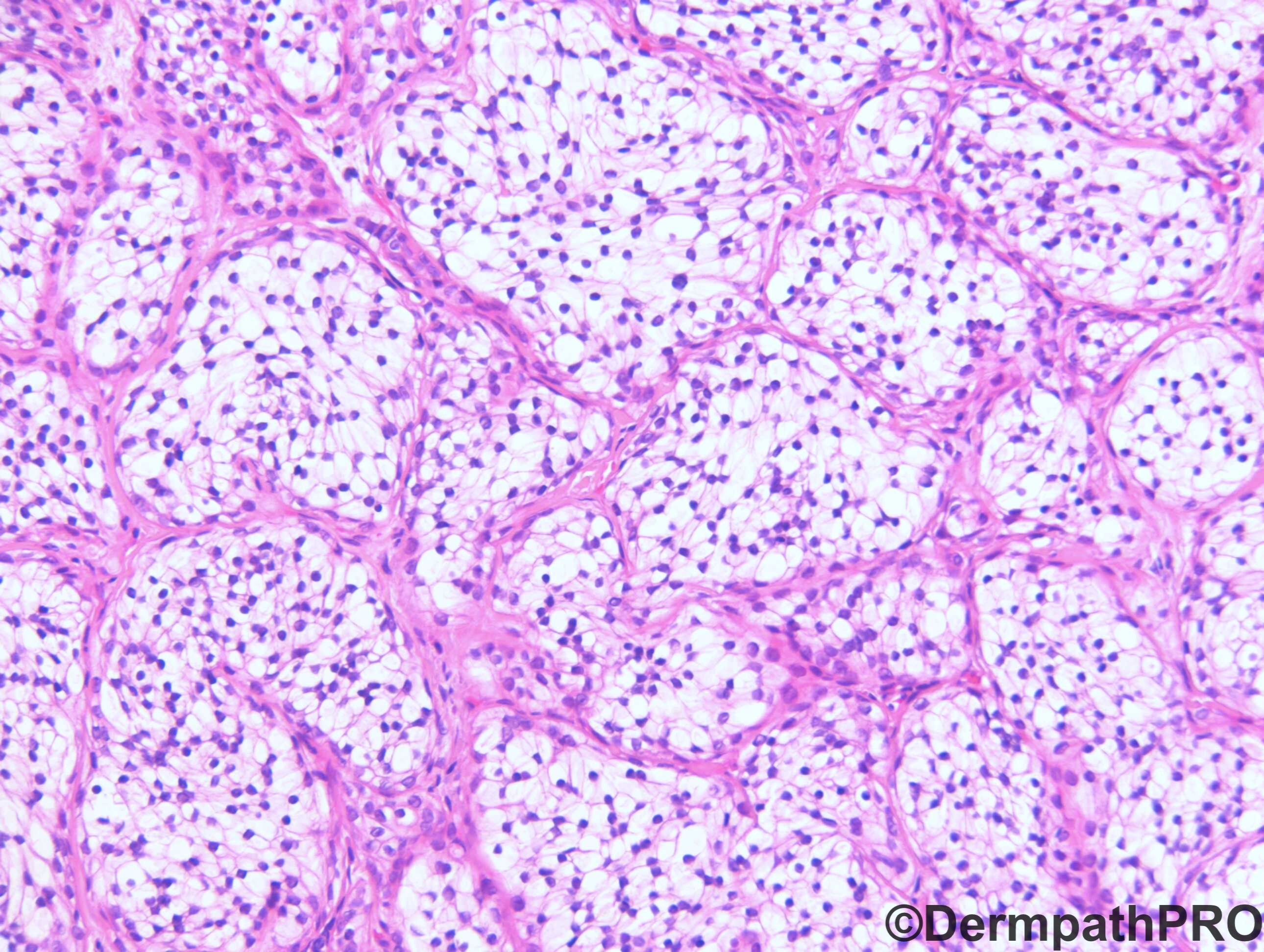
On picture 3 we can see better the true nature of this proliferation. I think this is a syringoma presenting a huge clear cell differentiantion in the cytoplasm (seen better in the other pictures). So, this is a clear cell syringoma.

Share this comment


Link to comment
share_externally

vincenzo

Posted

Tricky and intriguing case! The differentiation seems follicular-trichilemmal. I see something reminiscent of trichoadenoma and something reminiscent of desmoplastic trichilemmoma. ?Mixed clear cell trichoadenoma-trichilemmoma?

Share this comment


Link to comment
share_externally

Krishnakumar subramanian

Posted

looks like a benign tumor more for trichelemmoma like 

do not know

Share this comment


Link to comment
share_externally

Completely agree with Igor's observation. Clear cell syringoma.

Share this comment


Link to comment
share_externally

Neil Catterall

Posted

Would also agree with Igor based on site and obvious syringoma in 3rd pic.

Share this comment


Link to comment
share_externally

Krishnakumar subramanian

Posted

agree, looks like clear cell syringoma

Share this comment


Link to comment
share_externally

Dr. Richard Carr

Posted

I think there are two main possibilites i.e. Syringoma, clear cells with unusually expansile solid lobules or dermal duct tumour, clear cell variant. I guess it is academic. Certainly the lack of typical poroid elements and location favour the former.

https://www.ncbi.nlm.nih.gov/pubmed/18042069

Share this comment


Link to comment
share_externally

Krishnakumar subramanian

Posted

Thanks a lot sir, for interesting case to learn pattern diagnosis

Share this comment


Link to comment
share_externally

Dr. Mona Abdel-Halim

Posted

What a beautiful case!


Share this comment


Link to comment
share_externally


Join the conversation

You can post now and register later. If you have an account, sign in now to post with your account.

Guest
Add a comment...

×   Pasted as rich text.   Paste as plain text instead

  Only 75 emoji are allowed.

×   Your link has been automatically embedded.   Display as a link instead

×   Your previous content has been restored.   Clear editor

×   You cannot paste images directly. Upload or insert images from URL.

Loading...

×
×
  • Create New...