Jump to content
In this section we have spot diagnoses posted on a daily basis since June 2010, now over 4000! You can review the archived cases and read the suggested diagnoses by users and the final comment by the contributors.
Case are uploaded each week day by 10 am UK time with the correct diagnosis will generally be posted at 8 pm UK time. Why not view the most recent spot diagnosis and proffer a diagnosis?

Case Number : Case 2334 - 28 May 2019 Posted By: Uma Sundram

Please read the clinical history and view the images by clicking on them before you proffer your diagnosis.
Submitted Date :
   (0 reviews)

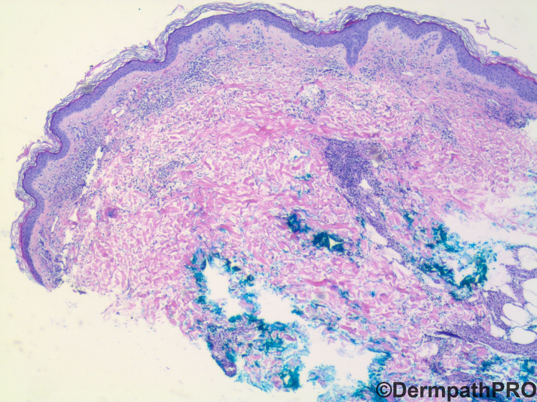
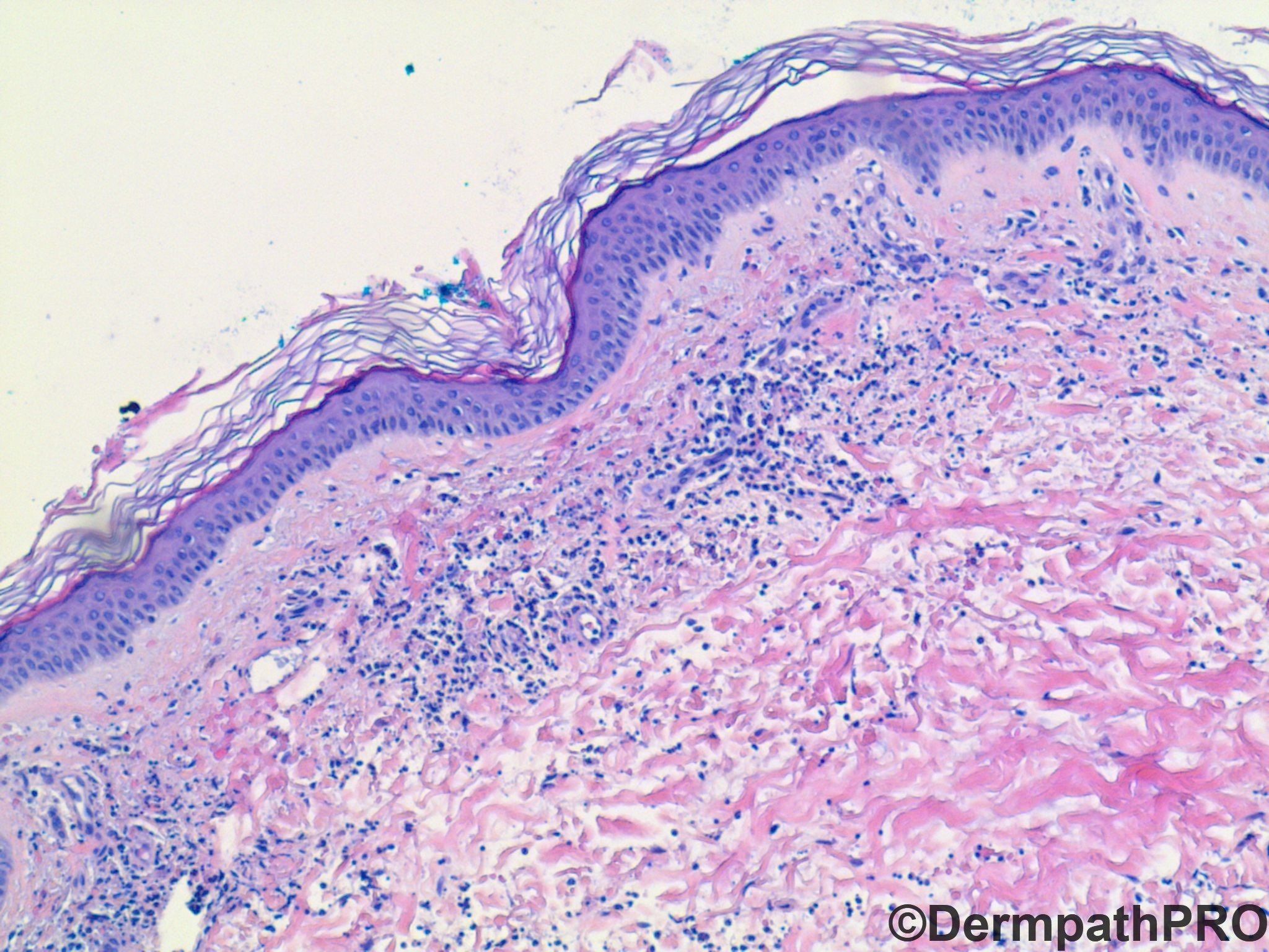
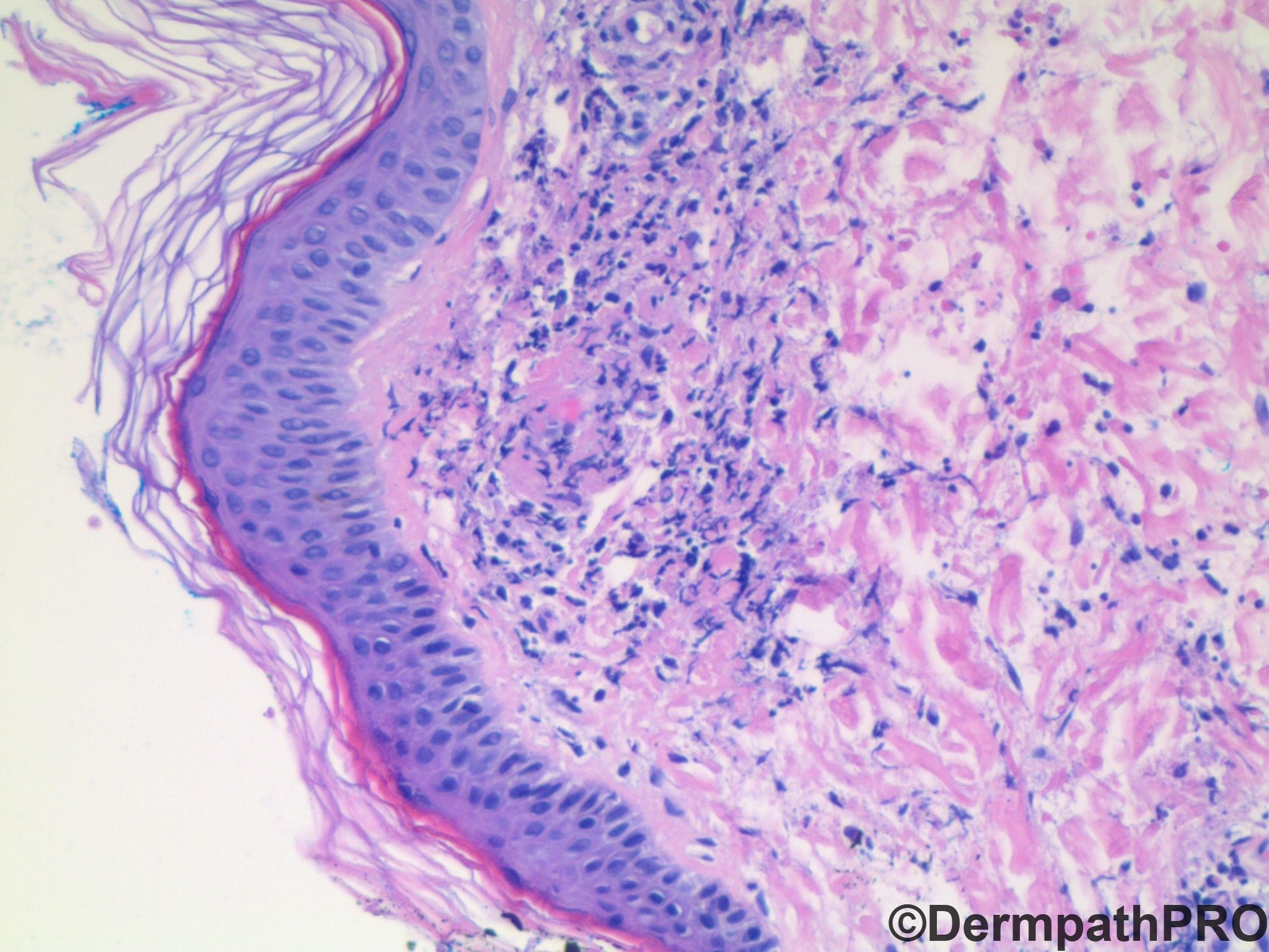
66 year old female with new onset purpuric rash on upper thighs, fading over a day. Now with new purpuric rash on bilateral upper arms; also involving palms. Perivascular IgA is positive. What is your differential diagnosis?


  Report Record

User Feedback


vincenzo

Posted

Leukocytoclastic vasculitis + perivascular IgA = Henoch-Schonlein Purpura. Actually that's all I can think of right now, but I'm sure there are many other options.

Share this comment


Link to comment
share_externally

Krishnakumar subramanian

Posted

epidermis and dermis

dermal vessel wall damage with neutrophils and neutrophil debri

DIF showing perivascular IgA I am surprised

correct me, I thought that IgA would deposit in the vessel walls

so also the age. the other thing is any history of bowel disturbances, and palmar rashes

I would like to keep atypical Dermatitis herpetiformis


Share this comment


Link to comment
share_externally

John Zhang

Posted

22 hours ago, vincenzo said:

Leukocytoclastic vasculitis + perivascular IgA = Henoch-Schonlein Purpura. Actually that's all I can think of right now, but I'm sure there are many other options.

Agree with Vincenzo.

Share this comment


Link to comment
share_externally

IgorSC

Posted

I can´t think of anything else but IgA vasculitis of adults. Maybe I am missing something... waiting for the conclusion.

Share this comment


Link to comment
share_externally

Alex-Ventura-Leon

Posted

Agree with the comments above. Waiting to learn more differentials.

Share this comment


Link to comment
share_externally

Uma Sundram

Posted

Yes, I gave you all an easy one this time, but also wanted to point out that we are learning a lot now about IgA vasculitis as a community of practitioners. The patient came in with acute kidney injury and the rash developed on the legs first. The rash on the legs faded and a similar rash appeared on the arms with palmar involvement. What was called Henoch Schonlein purpura is now called IgA vasculitis. It is vastly more common in children than in adults. Interestingly, this patient had kidney and skin findings, but not GI or other organ involvement. She also had brittle diabetes which complicated the kidney findings. An excellent paper from cleveland clinic shows that the presence of perivascular and vessel wall deposition of IgA correlated very well with the clinical diagnosis of IgA vasculitis (HSP) in the context of LCV (Feasel P, 2018). I had not realized that this triad (clinical findings + IgA deposition + LCV) was so powerful, so wanted to share it with all of you. Also important to know that the bx has to be done fairly early (within 48 hrs of rash development) as IgA fades quickly.

Share this comment


Link to comment
share_externally

On 30/05/2019 at 09:45, Uma Sundram said:

Yes, I gave you all an easy one this time, but also wanted to point out that we are learning a lot now about IgA vasculitis as a community of practitioners. The patient came in with acute kidney injury and the rash developed on the legs first. The rash on the legs faded and a similar rash appeared on the arms with palmar involvement. What was called Henoch Schonlein purpura is now called IgA vasculitis. It is vastly more common in children than in adults. Interestingly, this patient had kidney and skin findings, but not GI or other organ involvement. She also had brittle diabetes which complicated the kidney findings. An excellent paper from cleveland clinic shows that the presence of perivascular and vessel wall deposition of IgA correlated very well with the clinical diagnosis of IgA vasculitis (HSP) in the context of LCV (Feasel P, 2018). I had not realized that this triad (clinical findings + IgA deposition + LCV) was so powerful, so wanted to share it with all of you. Also important to know that the bx has to be done fairly early (within 48 hrs of rash development) as IgA fades quickly.

Beautiful case. Thanks!

Share this comment


Link to comment
share_externally


Join the conversation

You can post now and register later. If you have an account, sign in now to post with your account.

Guest
Add a comment...

×   Pasted as rich text.   Paste as plain text instead

  Only 75 emoji are allowed.

×   Your link has been automatically embedded.   Display as a link instead

×   Your previous content has been restored.   Clear editor

×   You cannot paste images directly. Upload or insert images from URL.

Loading...

×
×
  • Create New...