Jump to content
In this section we have spot diagnoses posted on a daily basis since June 2010, now over 4000! You can review the archived cases and read the suggested diagnoses by users and the final comment by the contributors.
Case are uploaded each week day by 10 am UK time with the correct diagnosis will generally be posted at 8 pm UK time. Why not view the most recent spot diagnosis and proffer a diagnosis?

Case Number : Case 2438 - 6 November 2019 Posted By: Saleem Taibjee

Please read the clinical history and view the images by clicking on them before you proffer your diagnosis.
Submitted Date :
   (0 reviews)

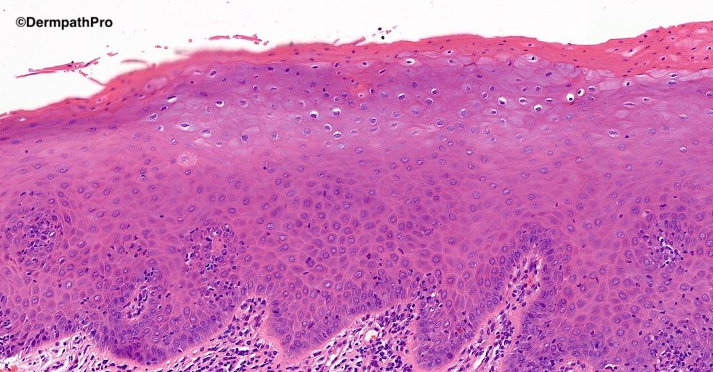
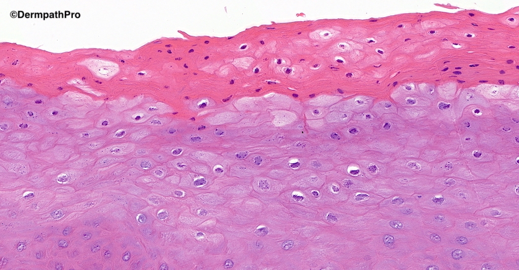
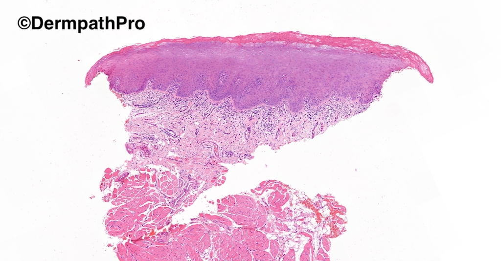
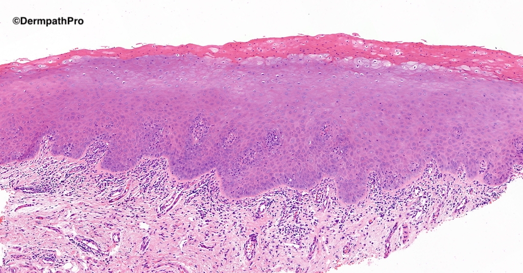
60M Asymptomatic white patches on lateral borders of tongue. Smoker. ?lichen planus ?frictional keratosis

  • Like 2
  Report Record

User Feedback


Victor Delgado

Posted (edited)

Leukoedema.

Edited by Victor Delgado

Share this comment


Link to comment
share_externally

Acanthosis and koilocyte-like ballooning of cells are the features that suggest oral hairy leukoplakia. Lateral border of the tongue is a characteristic site. 

Share this comment


Link to comment
share_externally

17 hours ago, Anil Patki said:

Acanthosis and koilocyte-like ballooning of cells are the features that suggest oral hairy leukoplakia. Lateral border of the tongue is a characteristic site. 

Agree. Nuclear marination and beading of chromatine are enough typical. 

Share this comment


Link to comment
share_externally

White sponge nevus , Leukodema, and Verruciform xanthoma  are additional considerations but agree on oral hairy leukoplakia.

HIV testing is recommended.

Share this comment


Link to comment
share_externally

Saleem Taibjee

Posted

I am a dermatopathologist and dermatologist, but this was an oral biopsy which came my way (and has put me off straying into oral cases!). Although I didn't know what this was, fortunately I had the sense to ask for a second opinion.

This was the opinion from Prof Eddy Odell:

This is mildly lichenoid.  In the correct clinical context and in a middle aged patient, this could be mild lichenoid process, but there are cleared keratinocytes layered in with the parakeratin, so this may be an Oral Hairy Leukoplakia, as these are now increasing found in patients and not associated with any immunodeficiency of any kind.  It needs to have an EBV in situ for EBER done to exclude EBV, only then could it be ascribed to a lichenoid process (which in a patient this age as a new diagnosis would most likely be a drug reaction).

As a result I requested EBER, shown below, confirming Oral Hairy Leukoplakia. The comment from Prof Odell that this seems to be increasingly found in immunocompetent patients is particularly interesting.32571_20.0x EBV.jpg

32571_40.0x EBV.jpg

Share this comment


Link to comment
share_externally


Join the conversation

You can post now and register later. If you have an account, sign in now to post with your account.

Guest
Add a comment...

×   Pasted as rich text.   Paste as plain text instead

  Only 75 emoji are allowed.

×   Your link has been automatically embedded.   Display as a link instead

×   Your previous content has been restored.   Clear editor

×   You cannot paste images directly. Upload or insert images from URL.

Loading...

×
×
  • Create New...