Jump to content
In this section we have spot diagnoses posted on a daily basis since June 2010, now over 4000! You can review the archived cases and read the suggested diagnoses by users and the final comment by the contributors.
Case are uploaded each week day by 10 am UK time with the correct diagnosis will generally be posted at 8 pm UK time. Why not view the most recent spot diagnosis and proffer a diagnosis?

Case Number : Case 2440 - 8 November 2019 Posted By: Dr. Richard Carr

Please read the clinical history and view the images by clicking on them before you proffer your diagnosis.
Submitted Date :
   (0 reviews)

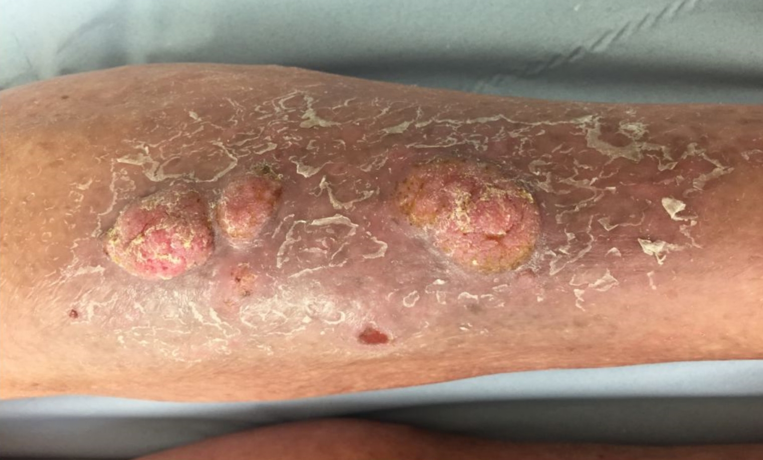
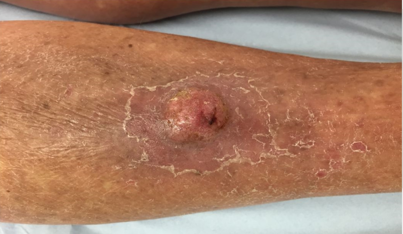
M65. 6/52 hx of multiple warty nodules both shins ?reactive squamous hyperplasia ?other


  • Like 2
  Report Record

User Feedback


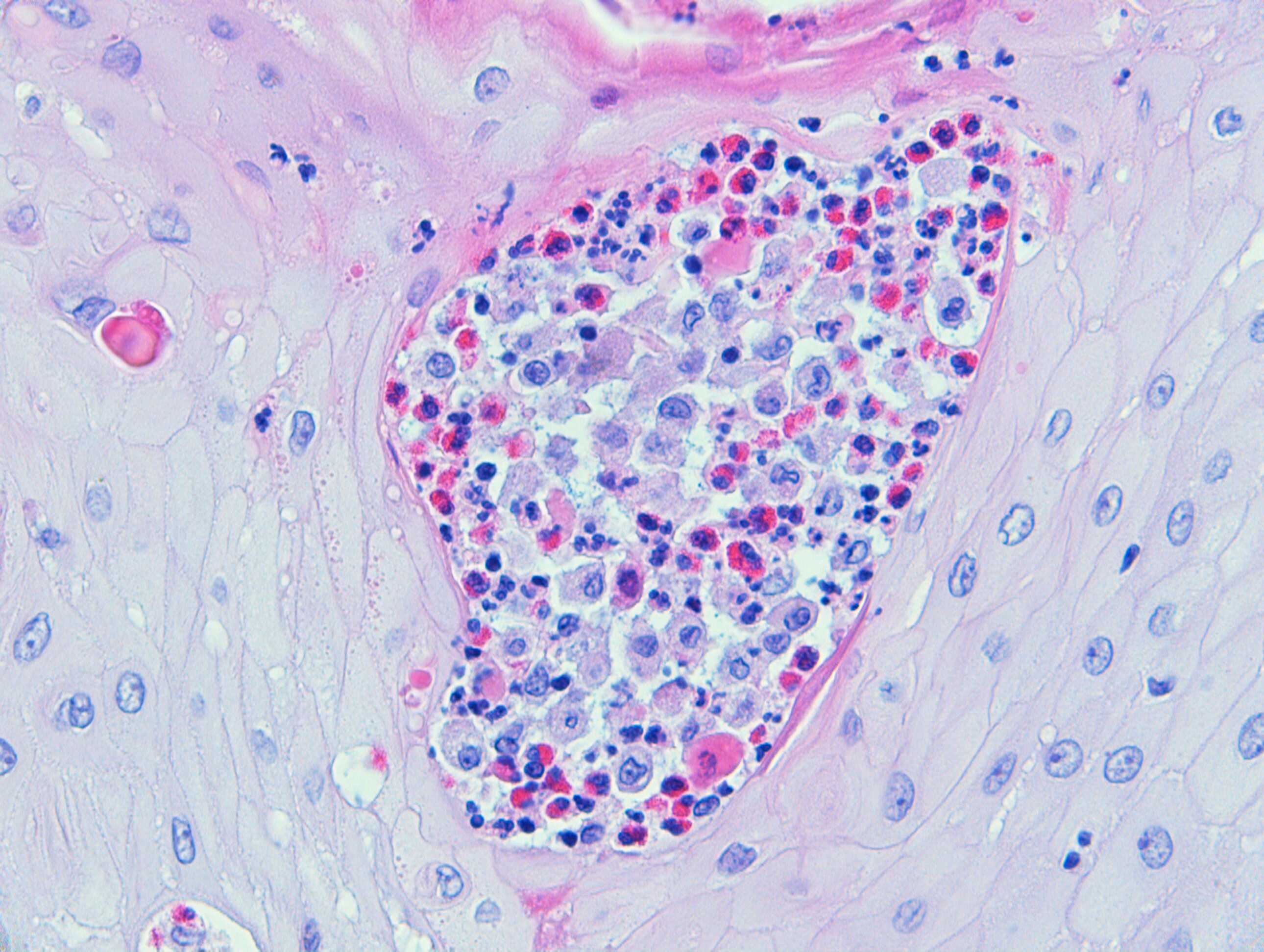
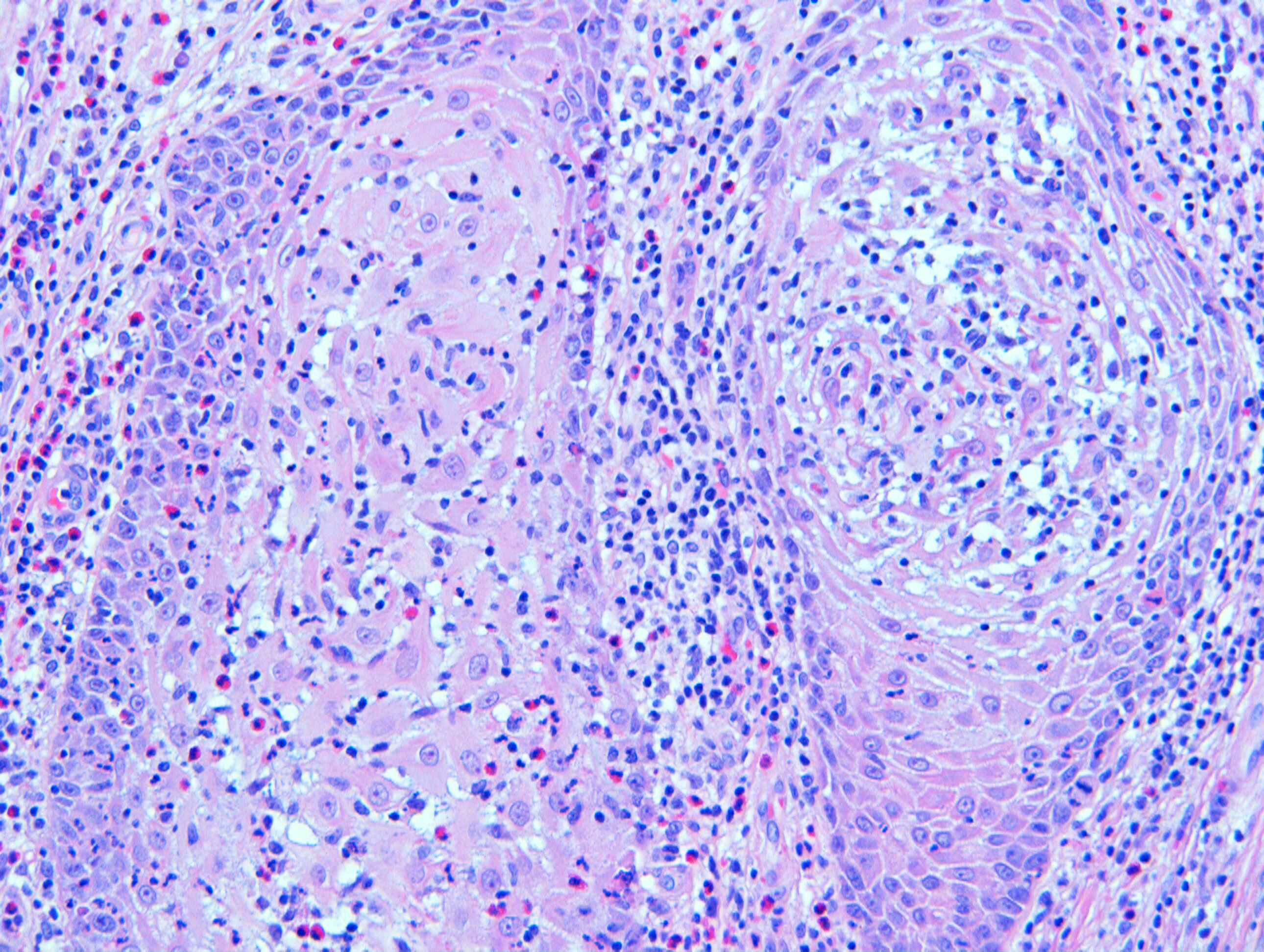
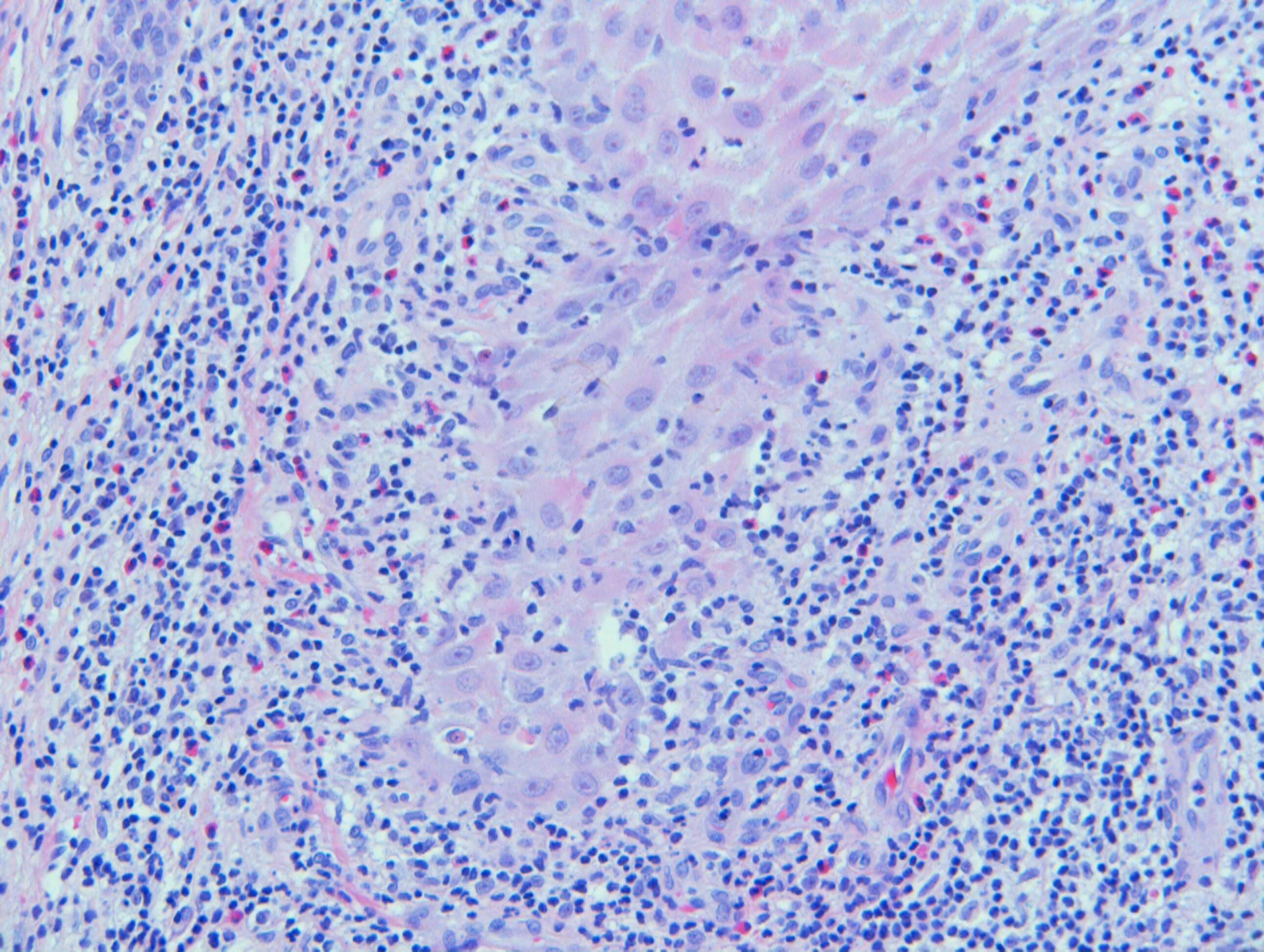
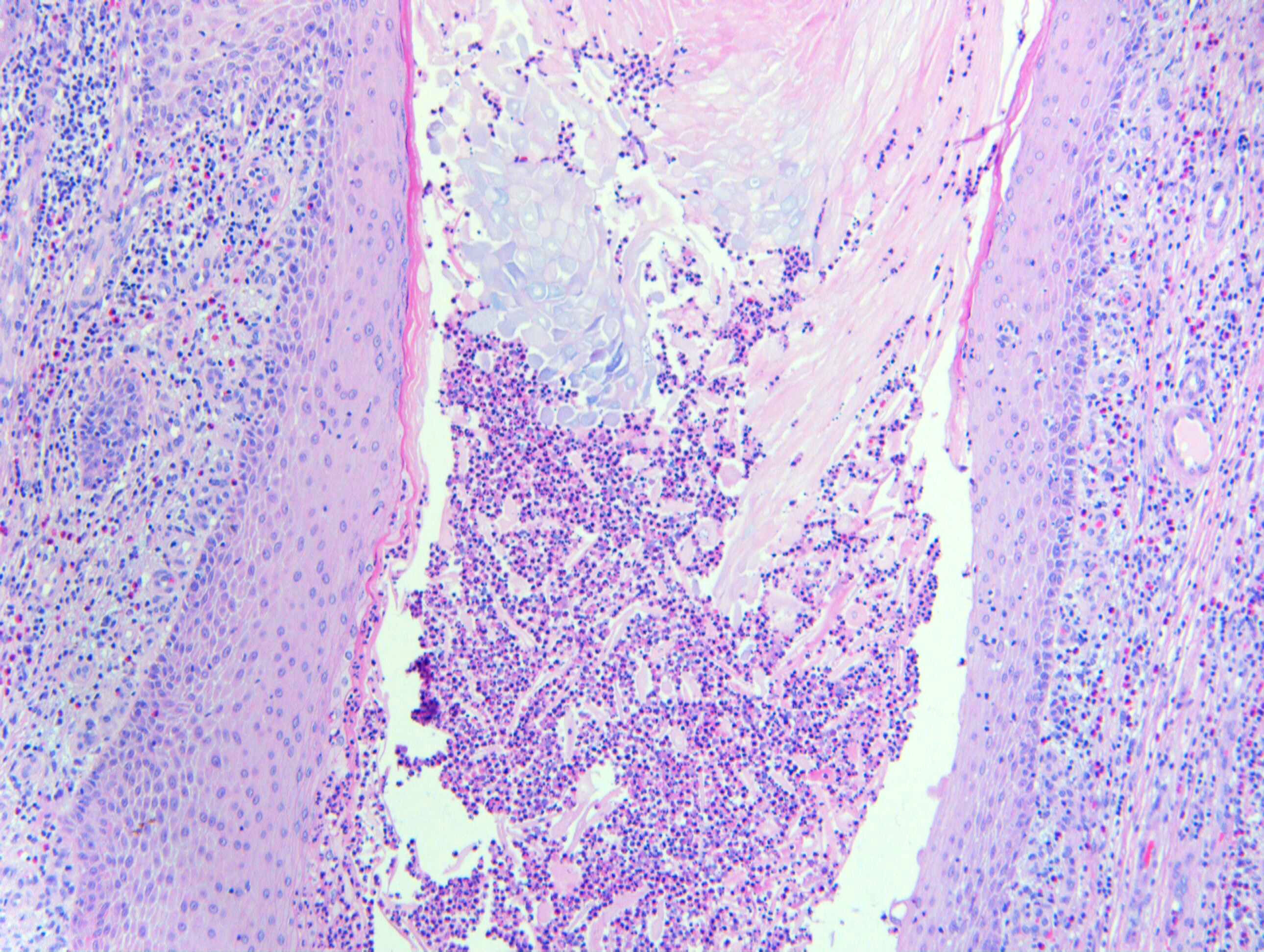
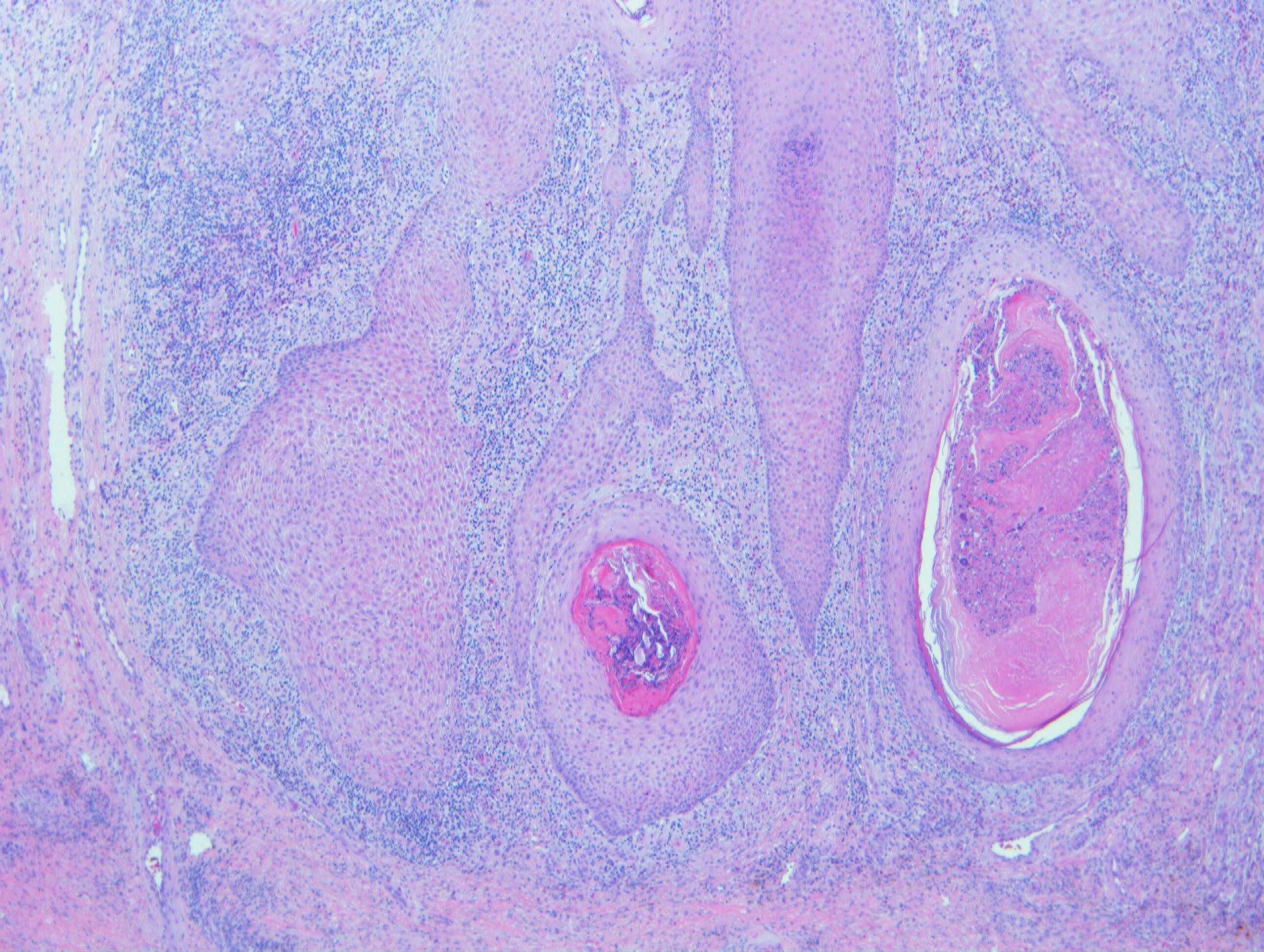
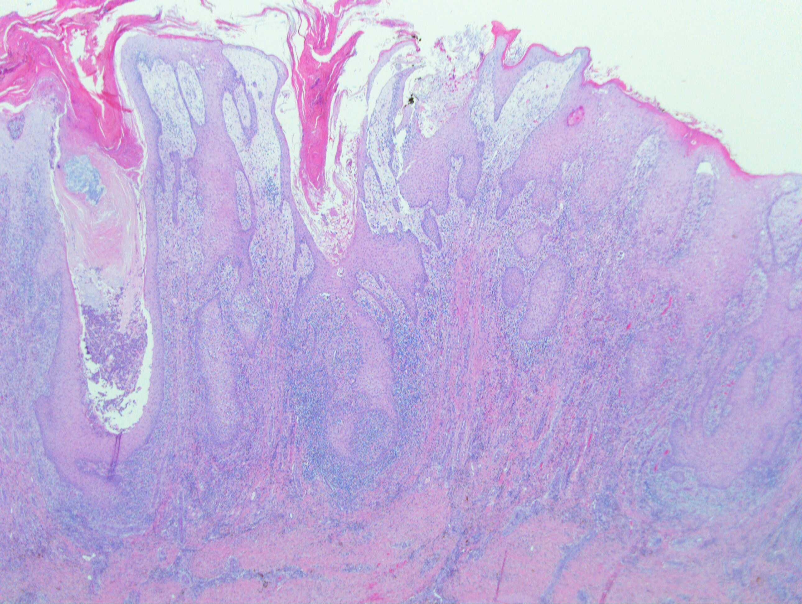
 Hyperkeratosis, irregular acanthosis amounting to psuedoepitheliomatous hyperplasia and dermal infiltrate of lymphocytes and eosinophils are the salient features. Clinical picture and histology are suggestive of prurigo nodularis. The scaling around the nodules seen in clinical pictures and prominence of eosinophils in the infiltrate suggest that in addition, there is probably contact sensitization to some topically applied preparation. 

Share this comment


Link to comment
share_externally

?Something cowpox-like or ORF?infection in a countryside worker...

Share this comment


Link to comment
share_externally

Krishnakumar subramanian

Posted

sir I wonder whether the patient was exposed to bromides etc

I would like to rule our halogenodermas

are the lesions elsewhere/acute onset


the other DD is Pemphigoid nodular

Share this comment


Link to comment
share_externally

Many challenges lately. I see a Pseudoepitheliomatous hyperplasia along many mixed abscesses, many of them showing neutrophils and eosinophils. Do not see atypia. Special stains are necessary fot microorganisms, like fungus or mycobacteria. Leishmaniosis can do the same pattern of inflamation. More clinical information is necessary as we can see this pattern of inflamation in cases more uncommon like Halogenoderma (iododerma, bromoderma and fluoroderma). Halogenoderma can appear in patients after use of iodine contrast, use of anesthesics, etc.

Share this comment


Link to comment
share_externally

Dr. Richard Carr

Posted

This patient has chronic obstructive pulmonary disease and is on the following medications:

Alendronic acid, doxazocin, lisinopril, Evacal D3 D, amlodipine and the inhalers tiotropium bromide and Sirdupla (salmeterol 25mcg/fluticasone 250mcg) and salbutamol 200mcg, all which he had been on for many years. Does this help?

Share this comment


Link to comment
share_externally

Ever seen before a Halogenoderma case. Great case and congratulation to all my Colleagues. 

Share this comment


Link to comment
share_externally

Great. The tiotropium bromide should be the causative agent. Bromoderma.

Share this comment


Link to comment
share_externally

Amazing case! Nodular papillomatous bromoderma. Occurs due to replacement of chlorine by bromine in the tissues. Doesn't disappear quickly after stopping bromide consumption but responds to intravenous administration of normal saline. Thanks for sharing! 

Share this comment


Link to comment
share_externally

Dr. Richard Carr

Posted

This was one of those cases in which we used a Sherlockian (as in Sherlock Holmes) approach. Once we have excluded all the common and less common possibilities (infections, lichenoid, neoplastic, granulomatous etc.) for pseudoepitheliomatous hyperplasia (PEH) there is only one diagnosis that fits, all be it very rare, and that is halogenoderma (neutrophils and eosinophils microabscesses in the absence of other known causes of PEH). In fact we used the rather wonderful review by Zayour and Lazova on pseudoepitheliomatous hyperplasia and there was only a single diagnosis with this combination of negative and positive findings. Once we had a possible diagnosis we took the simple step of going through the chemical structures of all the medications and the only ones that contained halogens were the inhalers!!!  We published this case as halogenoderma due to inhalers containing both bromine and flourine (fluticasone has fluouride ions). We believe this is the first such case in the literature although there was a mention in an old medical textbook and a comment that fluoroderma was more common on the legs. We postulated the location in our patient be due in part to poor venous return in a patient with COPD who is likely to spend much of the day sat chair bound. Steroids helped flatten the lesions. Would you believe one year later I colleague brought me a similar histology and after a few seconds I asked them was the patient COPD on inhalers. Answer yes. At that moment all the hairs on the back of my neck stood up!  The latter patient had had the lesion for 10 years and had previously be considered to have a carcinoma. Sometimes doing this specialty provides wonderful opportunities to make incredible diagnoses. Well done all for taking this challenge!!! I strongly suspect this condition is under-recognised but I'm still waiting for a 3rd case.

I note that if you type halogenoderma and inhalers into pubmed our paper does not come up (probably because of the title). One case does come up on a google search and may be our case  but I'm not 100% sure as I don't have access to the PDF but they appear to cite out paper.


Reactions Weekly

December 2018, Volume 1730, Issue 1, pp 185–185| Cite as

Ipratropium bromide/salmeterol/fluticasone propionate/tiotropium bromide

Halogenoderma: case report
Case report
First Online: 01 December 2018


Clin Exp Dermatol. 2018 Dec;43(8):959-961. doi: 10.1111/ced.13664. Epub 2018 Jun 4.

An unusual case of multiple nodules on the lower legs.


Am J Dermatopathol. 2011 Apr;33(2):112-22; quiz 123-6. doi: 10.1097/DAD.0b013e3181fcfb47.

Pseudoepitheliomatous hyperplasia: a review.

Author information

1
Dermatology and Pathology, Yale Dermatopathology Laboratory, Department of Dermatology, Yale University School of Medicine, New Haven, CT 06520, USA.

Abstract

Pseudoepitheliomatous hyperplasia (PEH) is a benign condition, characterized by hyperplasia of the epidermis and adnexal epithelium, closely simulating squamous cell carcinoma. PEH may be present in a number of conditions characterized by prolonged inflammation and/or chronic infection, as well as in association with many cutaneous neoplasms. Herein, we review different inflammatory, infectious, and neoplastic skin diseases, in which florid epidermal hyperplasia is a prominent histopathologic feature, and introduce a systematic approach in the interpretation of PEH.

Share this comment


Link to comment
share_externally


Join the conversation

You can post now and register later. If you have an account, sign in now to post with your account.

Guest
Add a comment...

×   Pasted as rich text.   Paste as plain text instead

  Only 75 emoji are allowed.

×   Your link has been automatically embedded.   Display as a link instead

×   Your previous content has been restored.   Clear editor

×   You cannot paste images directly. Upload or insert images from URL.

Loading...

×
×
  • Create New...