Jump to content
In this section we have spot diagnoses posted on a daily basis since June 2010, now over 4000! You can review the archived cases and read the suggested diagnoses by users and the final comment by the contributors.
Case are uploaded each week day by 10 am UK time with the correct diagnosis will generally be posted at 8 pm UK time. Why not view the most recent spot diagnosis and proffer a diagnosis?

Case Number : Case 2444 - 14 November 2019 Posted By: Saleem Taibjee

Please read the clinical history and view the images by clicking on them before you proffer your diagnosis.
Submitted Date :
   (0 reviews)

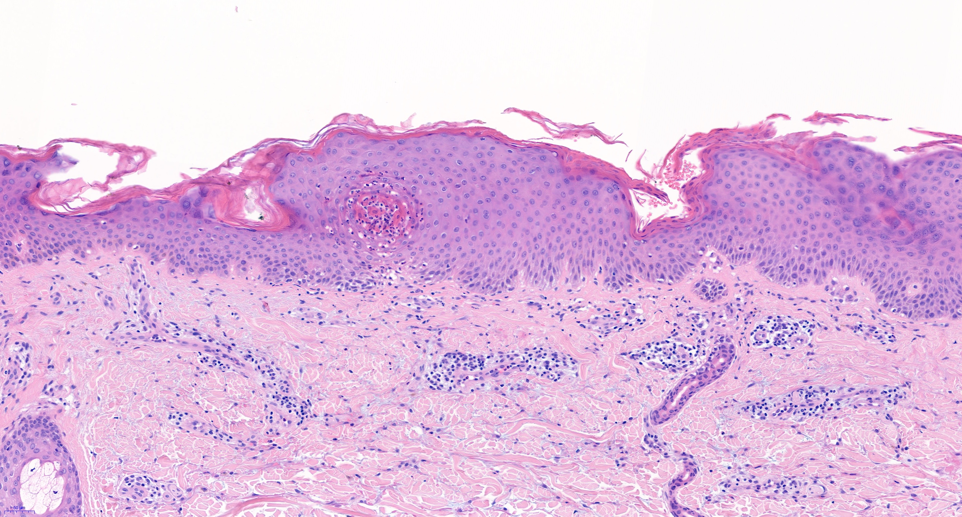
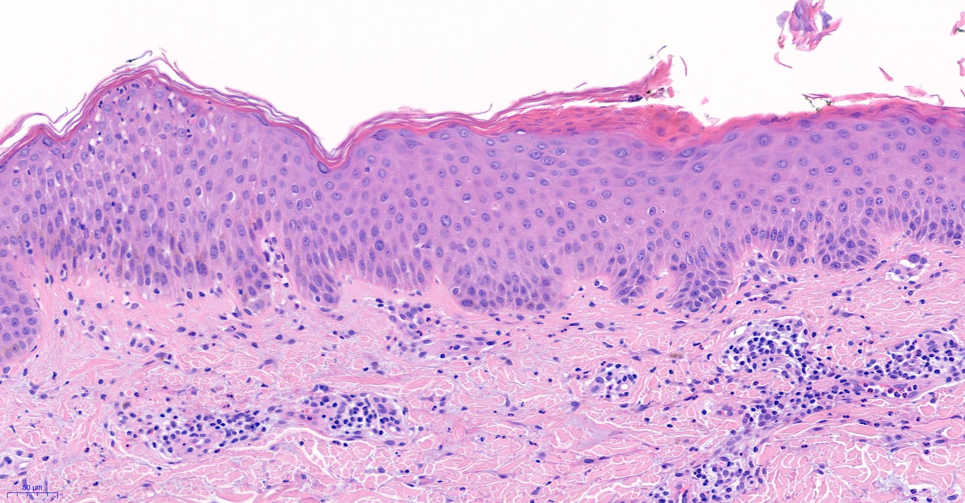
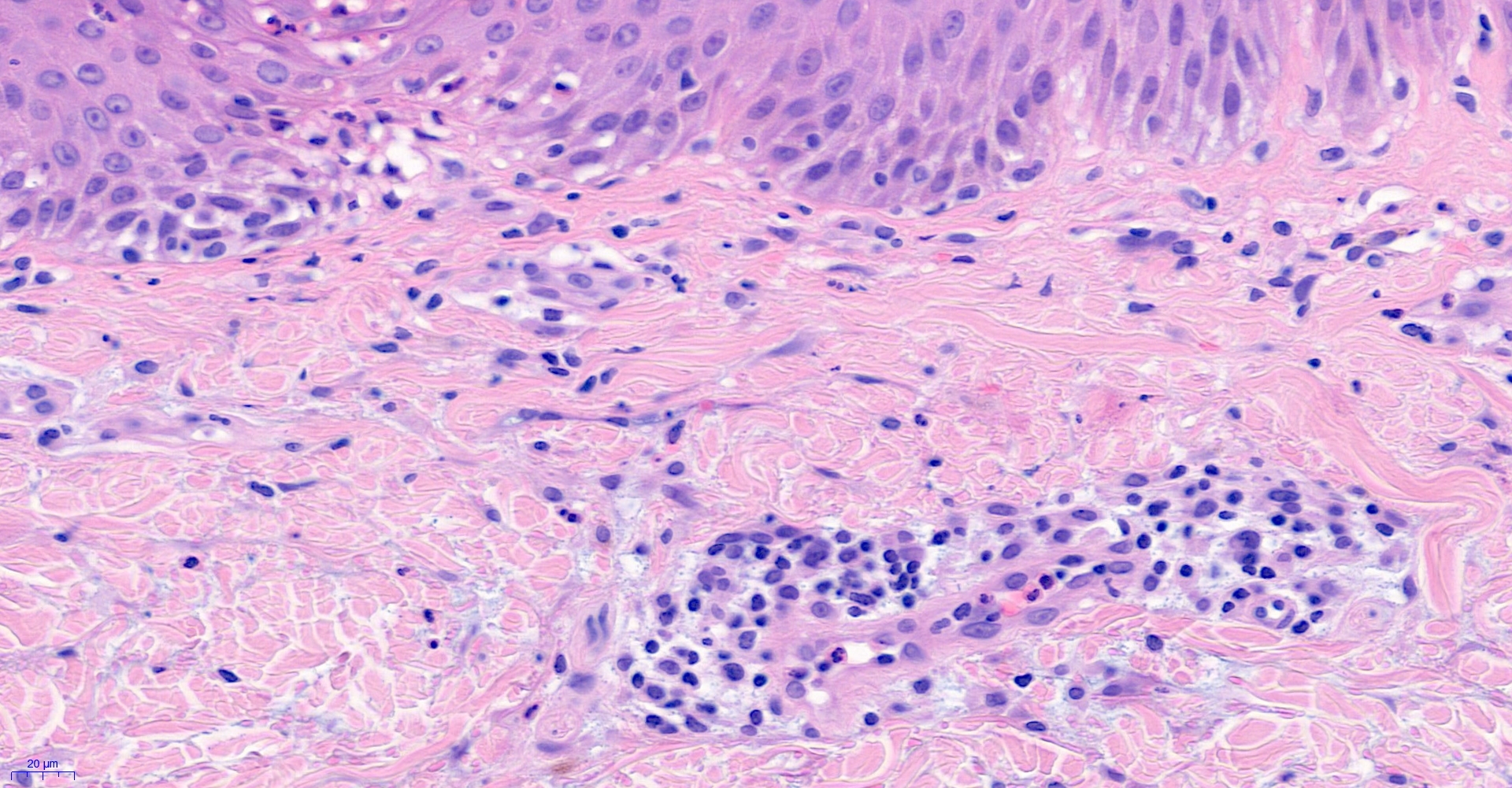
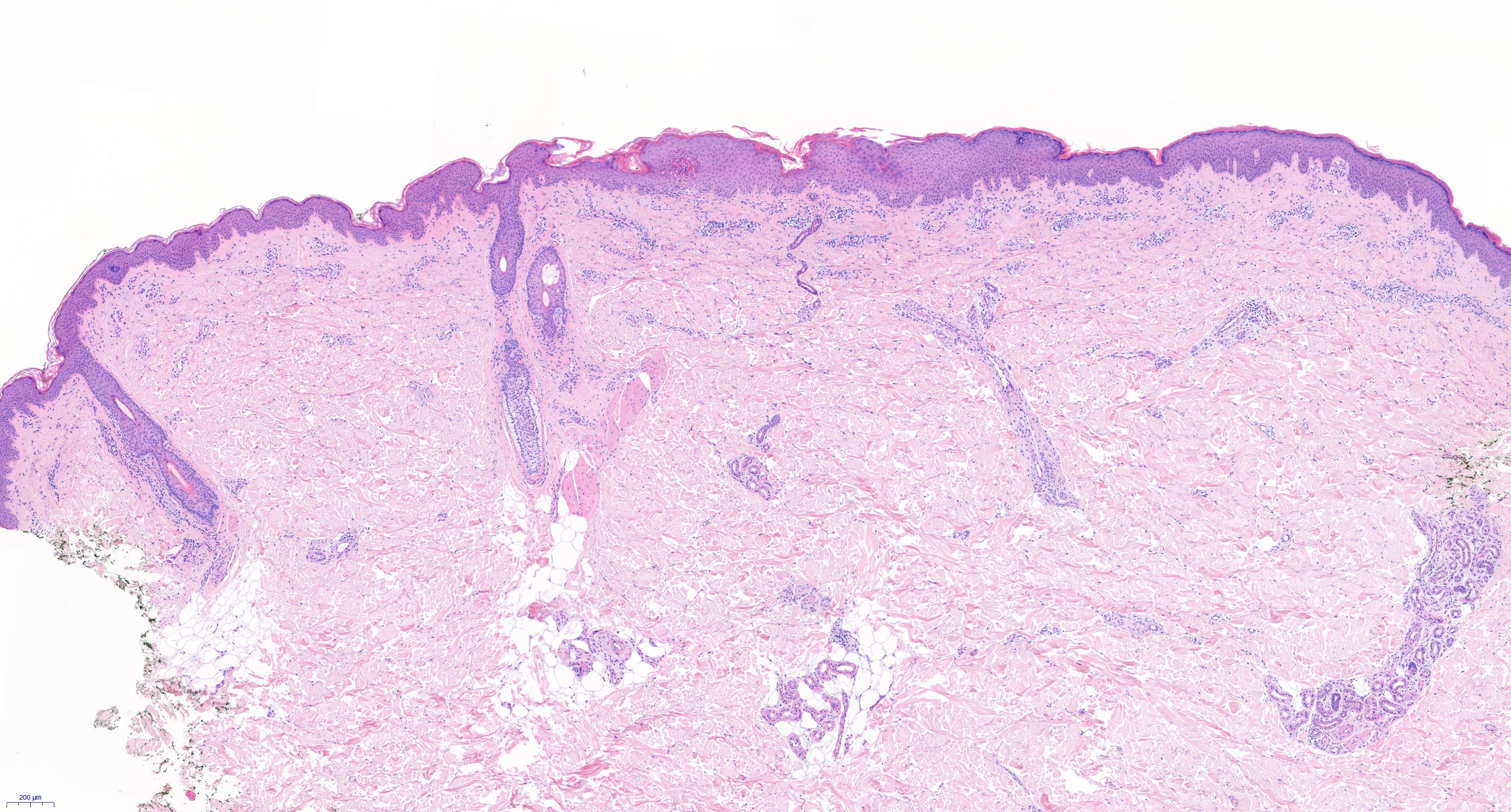
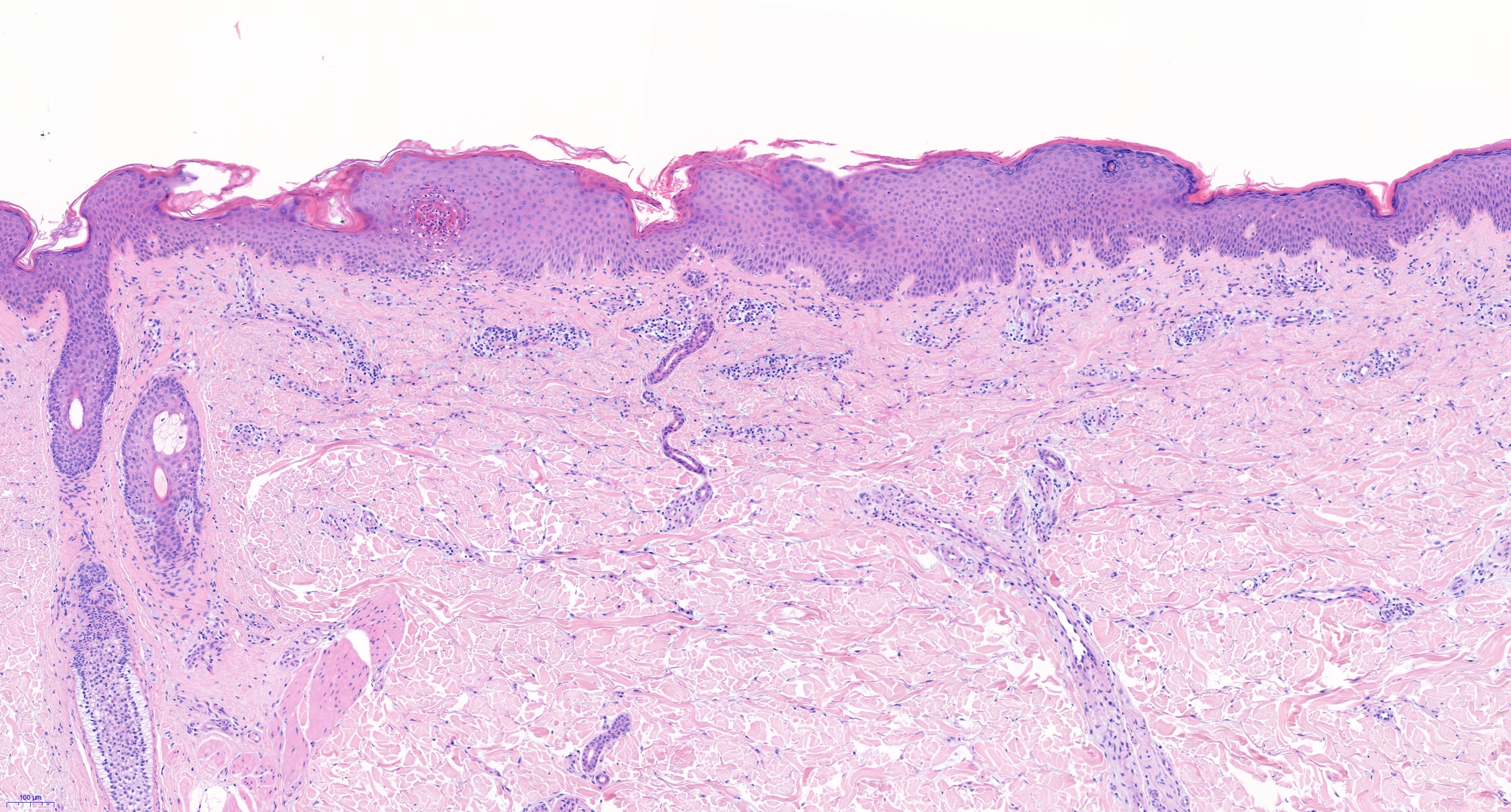
67 year old man with persistent plaques right flank and buttocks. Lack of response to psoriasis creams.

  • Like 1
  Report Record

User Feedback


First thing I would do is a PAS stain and look for hypha (tinea incognito).

Share this comment


Link to comment
share_externally

Krishnakumar subramanian

Posted

neutrophilic spongiosis

PAS stain to rule out dermatophytosis

Share this comment


Link to comment
share_externally

Many neutrophils and capillaries on tip of dermal papilla make me think of a psoriasis ( maybe drug resistant). 

Share this comment


Link to comment
share_externally

If dermatophytosis is ruled out by PAS stain, pityriasis rubra pilaris should be considered in the differential diagnosis as there's irregular acanthosis and parakeratosis alternating with orthokeratosis

Share this comment


Link to comment
share_externally

Victor Delgado

Posted

Psoriasis and Dermatophytosis are great differentials, I´ll suggest nutritional deficiency aswell, there is a little bit of vacuolar degeneration and a few necrotic keratinocytes so we should think on drugs Rx aswell. Clinical correlation.

Share this comment


Link to comment
share_externally

Last figure:

looks like a little vacuolar interface as Victor mentioned. There appears an increase of interstitial mucin. What are the  perivascular cells? Some are lymphocytes but many are not. Mast cells?

Share this comment


Link to comment
share_externally

Krishnakumar subramanian

Posted

sir once we something like mucin in dermis I would like to rule out dermatomyositis

however there would be other clinical parameters and lab support

Share this comment


Link to comment
share_externally

Saleem Taibjee

Posted

You guys are good! Yes this was tinea incognito. The PAS stain and striking clinical appearance is shown below, before and after treatment with oral antifungals.11252_40.0x b PAS.jpg11252_37.0x PAS.jpgTinea incognito - case Mr CC.jpegTinea incognito(1).JPG


Share this comment


Link to comment
share_externally

Krishnakumar subramanian

Posted

sir wonderful

common things are common

Share this comment


Link to comment
share_externally


Join the conversation

You can post now and register later. If you have an account, sign in now to post with your account.

Guest
Add a comment...

×   Pasted as rich text.   Paste as plain text instead

  Only 75 emoji are allowed.

×   Your link has been automatically embedded.   Display as a link instead

×   Your previous content has been restored.   Clear editor

×   You cannot paste images directly. Upload or insert images from URL.

Loading...

×
×
  • Create New...