Jump to content
In this section we have spot diagnoses posted on a daily basis since June 2010, now over 4000! You can review the archived cases and read the suggested diagnoses by users and the final comment by the contributors.
Case are uploaded each week day by 10 am UK time with the correct diagnosis will generally be posted at 8 pm UK time. Why not view the most recent spot diagnosis and proffer a diagnosis?

Case Number : Case 2455 - 29 November 2019 Posted By: Dr. Richard Carr

Please read the clinical history and view the images by clicking on them before you proffer your diagnosis.
Submitted Date :
   (0 reviews)

M35. Spot Diagnosis. c/o Dr Neil Catterall


  Report Record

User Feedback


Krishnakumar subramanian

Posted

necrolytic lesions with dermal edema

acrodermatitis enteropathica/necrolytic acral erythema

Share this comment


Link to comment
share_externally

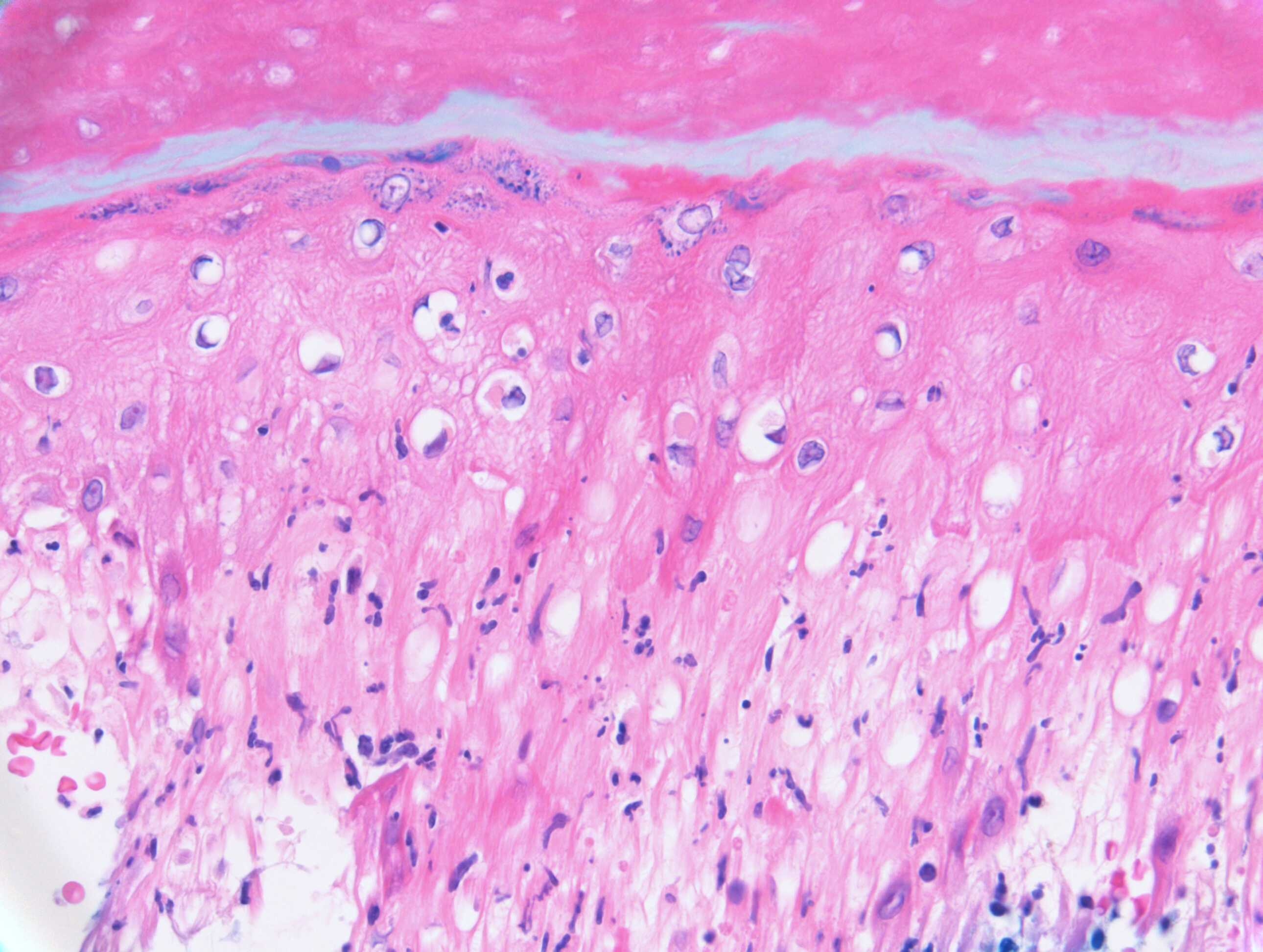
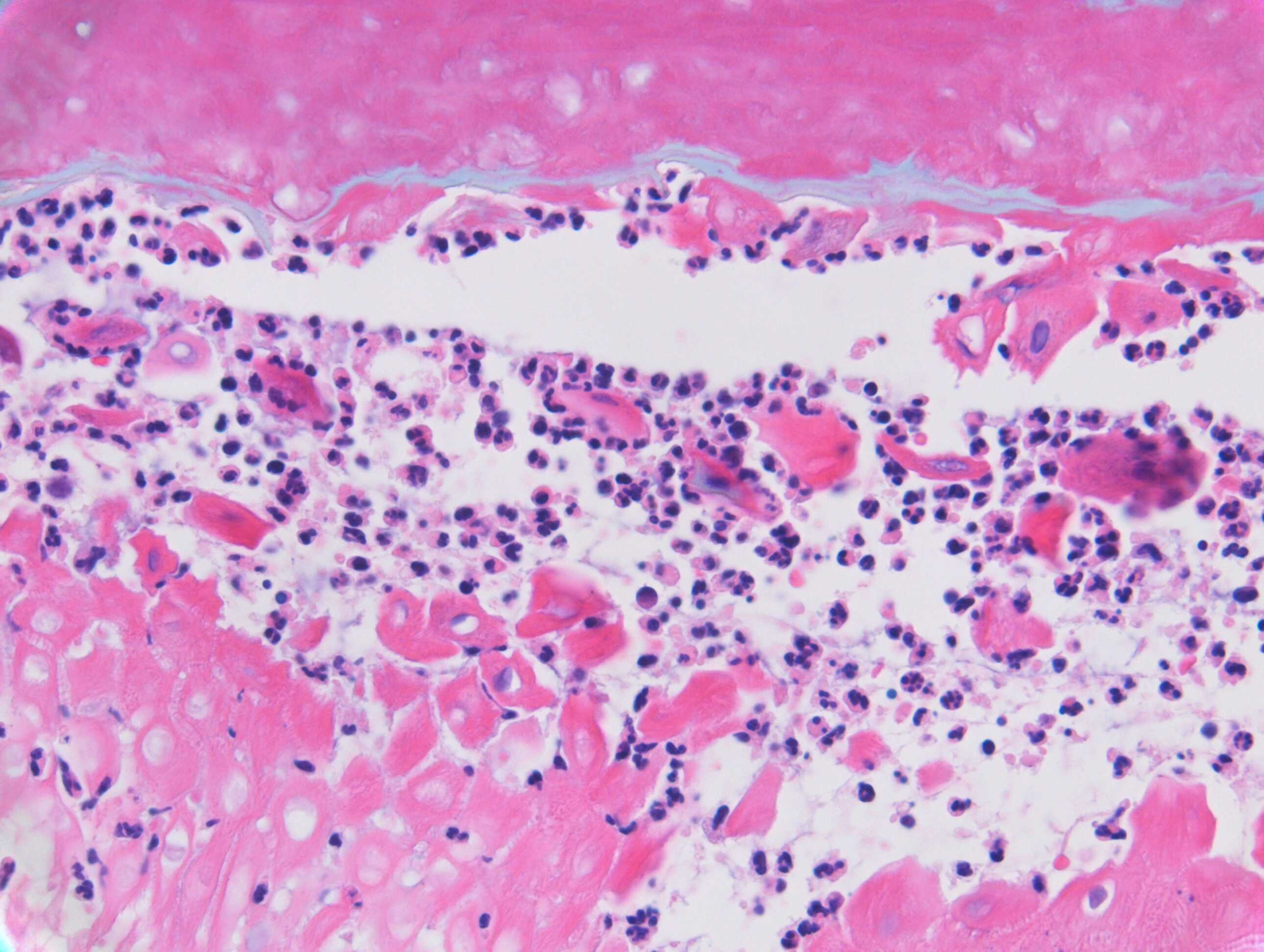
Acral skin with prominent dermal edema and polymorphs. Early stage of pernio. 

Share this comment


Link to comment
share_externally

Herpes/papular acrodermatitis(gianotti Crosti)...

Share this comment


Link to comment
share_externally

Saman Fatah

Posted

Difficult in the absence of further clinical informations, once infective cause excluded other differentials are acral neutrophilic dermatoses or acral pustular psoriasis. 

Will wait, it would be interesting to know what the final feedback is.     

Share this comment


Link to comment
share_externally

Saman Fatah

Posted

Vincenzo.... When I mentioned exclusion of an infective cause that meant to include viral causes particularly Coxsackie A6 "atypical" hand, foot and mouth disease, agree why not? we do PCR for entrovirus on the blister fluid and that can help the diagnosis easily if one suspect it clinically rather than a biopsy..

Anil ... Neutrophilic dermatoses can affect palms and soles especially in those with haematological malignancies but it is rare and there are few anecdotal cases already published.  Neutrophilic Dermatoses of Hands/ Dorsal hands is another acral variant which is more familar to many. It is very unlikely in this cases.

Share this comment


Link to comment
share_externally

Dr. Richard Carr

Posted

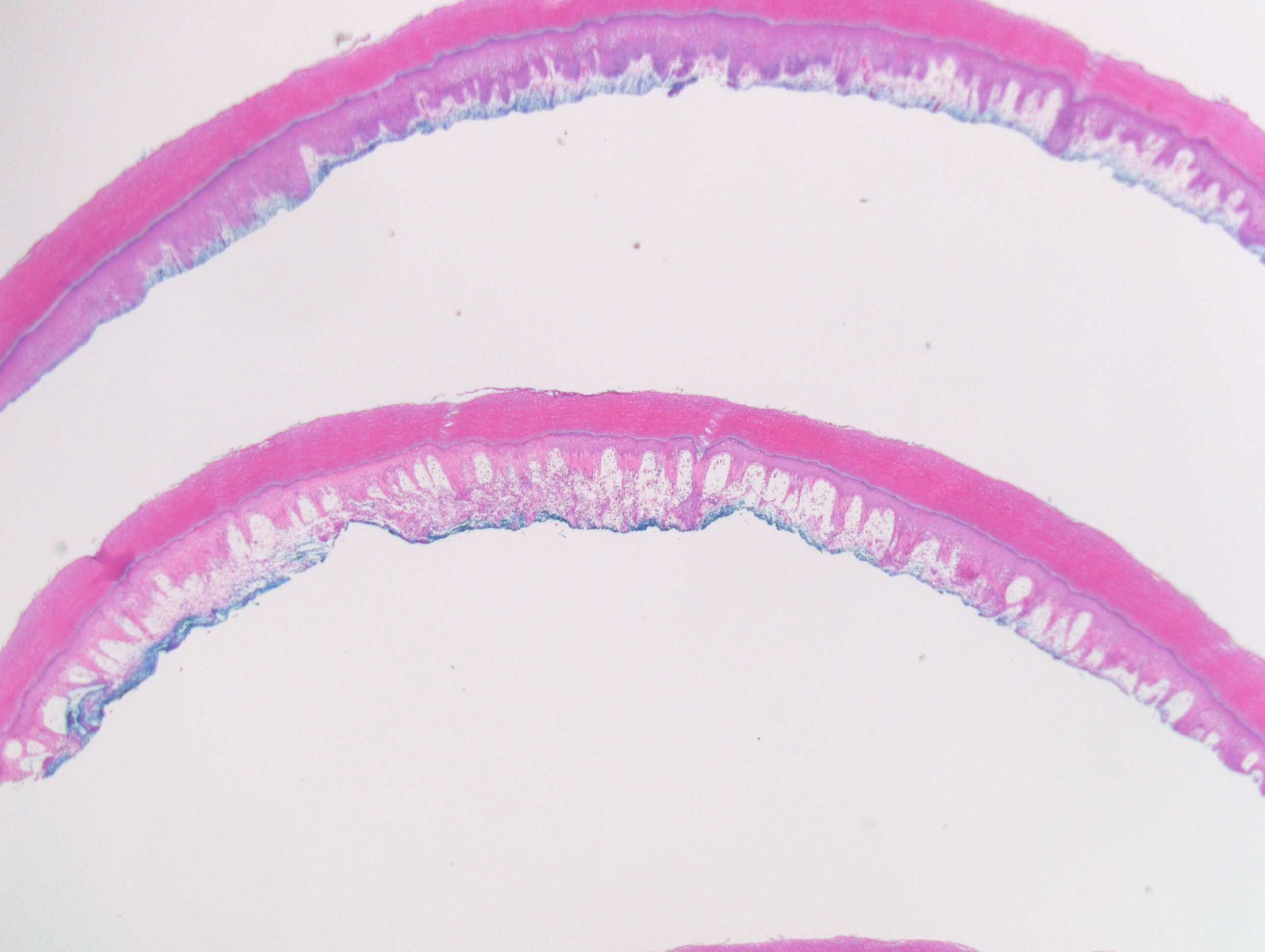
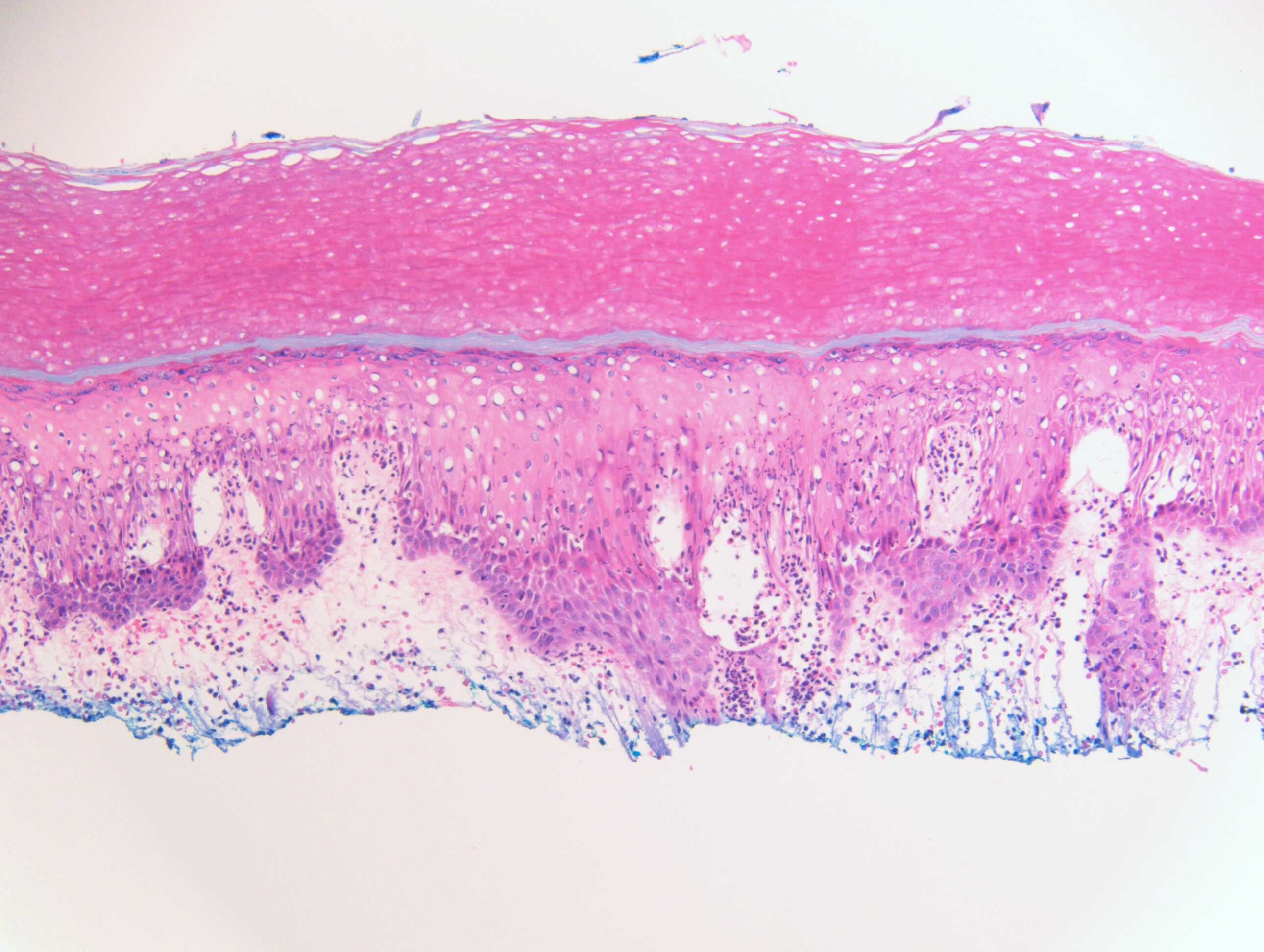
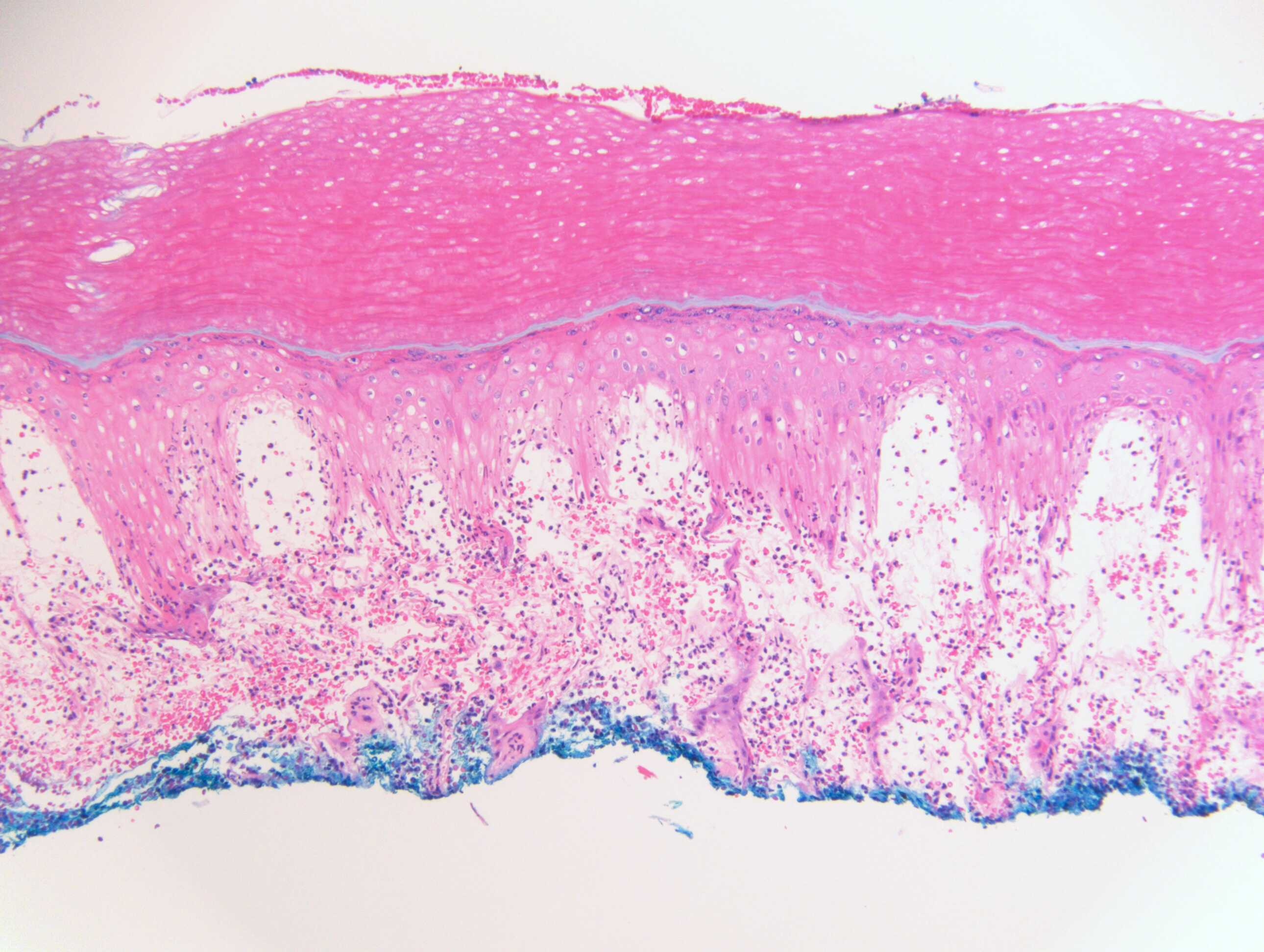
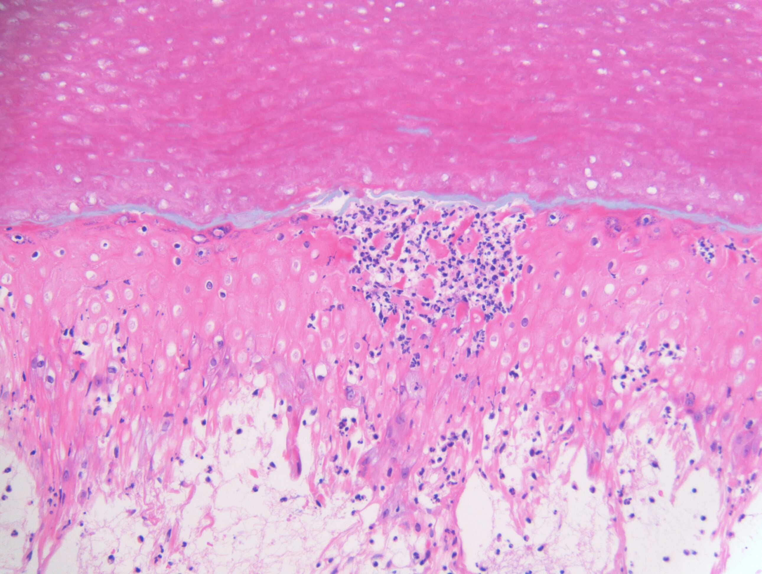
This is a case of Orf. Note the intracytoplasmic small eosinophilic cytoplasmic inclusions in the lower left image.

Share this comment


Link to comment
share_externally

Saman Fatah

Posted

Thanks very much for the feedback and educating us (particularly  Clinicians who are interested to learn more about dermatopathology). 

Share this comment


Link to comment
share_externally


Join the conversation

You can post now and register later. If you have an account, sign in now to post with your account.

Guest
Add a comment...

×   Pasted as rich text.   Paste as plain text instead

  Only 75 emoji are allowed.

×   Your link has been automatically embedded.   Display as a link instead

×   Your previous content has been restored.   Clear editor

×   You cannot paste images directly. Upload or insert images from URL.

Loading...

×
×
  • Create New...