Jump to content
In this section we have spot diagnoses posted on a daily basis since June 2010, now over 4000! You can review the archived cases and read the suggested diagnoses by users and the final comment by the contributors.
Case are uploaded each week day by 10 am UK time with the correct diagnosis will generally be posted at 8 pm UK time. Why not view the most recent spot diagnosis and proffer a diagnosis?

Case Number : Case 2430- 24 October 2019 Posted By: Saleem Taibjee

Please read the clinical history and view the images by clicking on them before you proffer your diagnosis.
Submitted Date :
   (0 reviews)

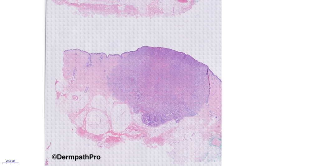
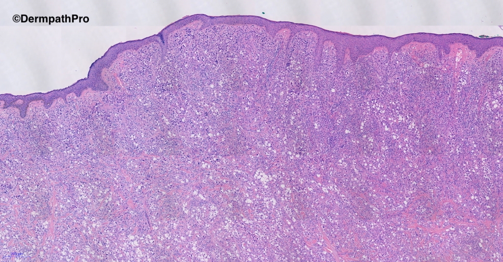
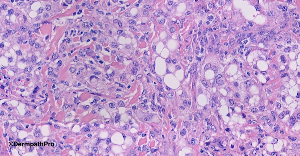
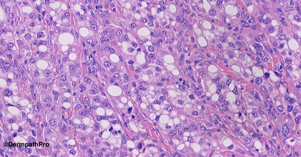
76M, nodule on posterior neck.


  • Like 1
  Report Record

User Feedback


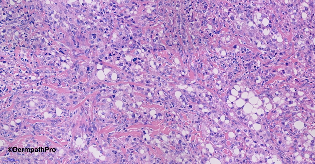
Don't really know the differentiation of this tumor. Epithelioid cells with lipoblast or signet ring cell cytologic appearance. Quite atypical. Definitely very difficult case requiring some immunohistochemical studies to determine the line of differentiation.

Share this comment


Link to comment
share_externally

Pleomorphic liposarcoma or clear cell variant of atypical fibroxanthoma

Share this comment


Link to comment
share_externally

Saleem Taibjee

Posted

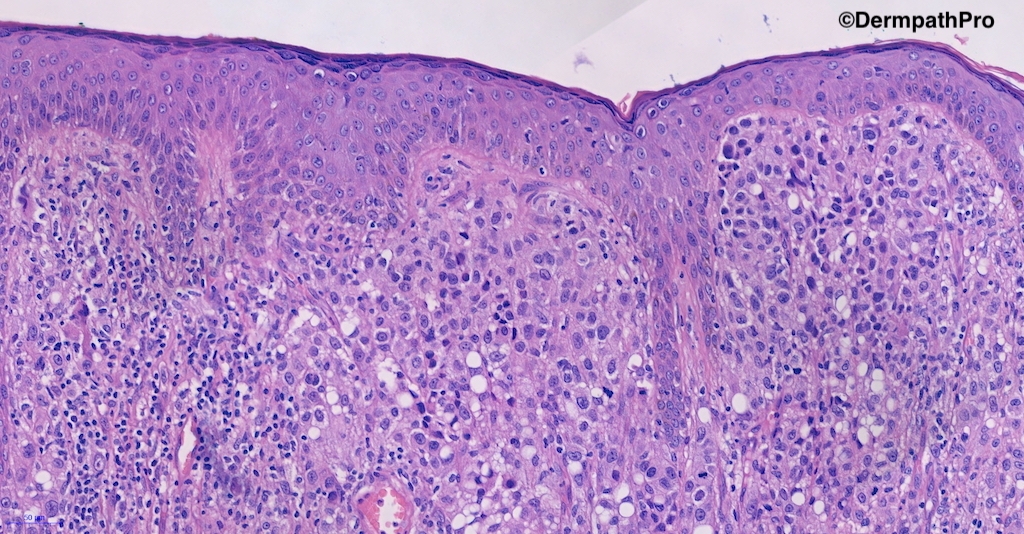
Great to see the range of responses, illustrating the difficulty of the case. It was originally signed out by the pathologists at another centre as favouring liposarcoma, which although extremely uncommon as a primary dermal tumour, is somewhat understandable. It was S100 positive (but Melan-A and HMB45 negative), and the pathologists at the Royal Marsden Sarcoma centre to which the case was subsequently referred, then recognised that this is signet ring cell / clear cell change within melanoma.

The understandable pitfall here is that there is no junctional component.

I think the original pathologists had not appreciated that the patient had a previous melanoma 2 years prior at an anatomical site not too far away.

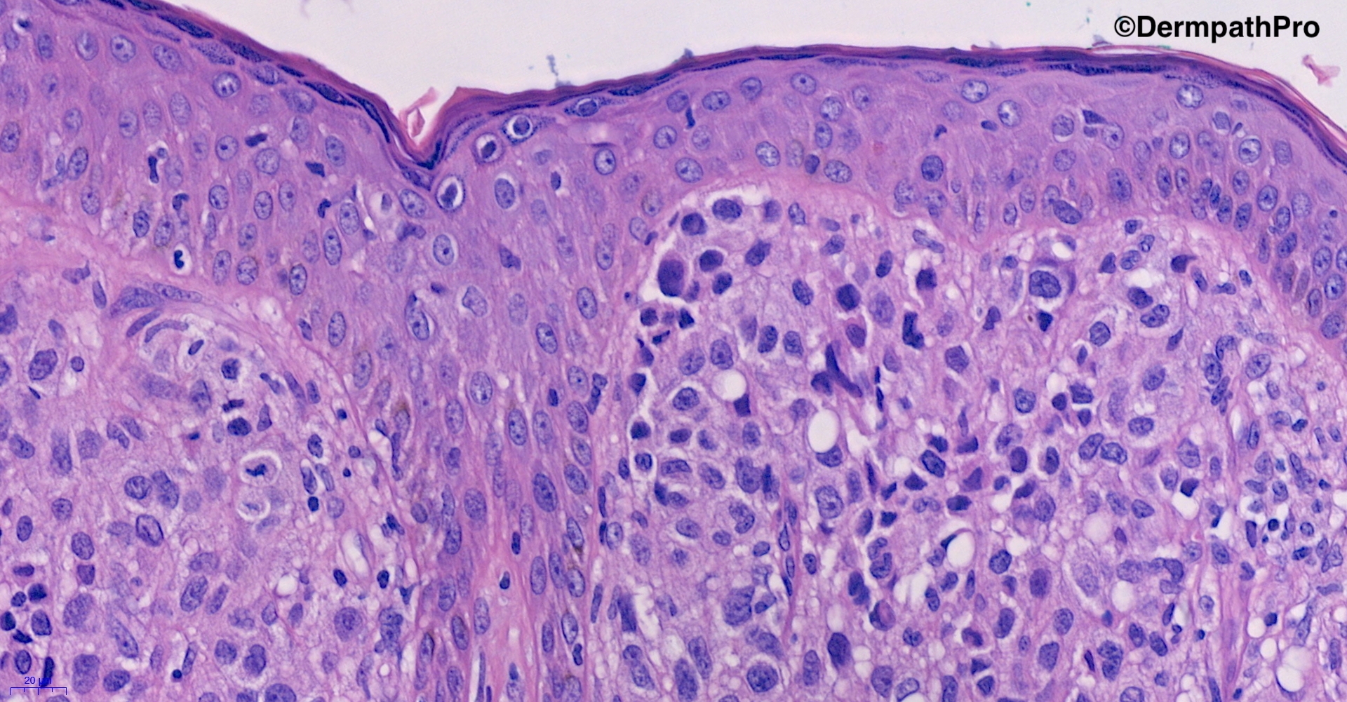
I was recently asked to review this case to arrange BAP1 immunohistochemistry (the patient also has mesothelioma), and with the benefit of hindsight, there are areas of signet ring cell change within the original melanoma (see images below), further confirming that the current specimen is indeed a local metastasis. Both specimens are BRAF V600E mutant on IHC also. This is a valuable case in reminding us on 3 learning points 1. to check on previous specimens for a patient when we sign out unusual or difficult cases 2. the mantra that uncommon morphologies of common(er) tumours are generally more likely than very rare entities 3. think of melanoma metastasis when the junctional component is minimal or absent. By the way, BAP1 was preserved throughout.45449_5.0x.jpg45449_40.0x.jpg

Share this comment


Link to comment
share_externally

Great case Dr Saleem and very valuable  and excellent comments.


Share this comment


Link to comment
share_externally

Krishnakumar subramanian

Posted

Thanks Dr Saleem for sharing this teaching case, one clarification how far we need to stick on to melanoma is S 100 is positive, but HMB45 and Melan A is negative

if this case did not have a prior history of melanoma how shall we need to proceed, can I know, what panel of markers we need to do

Share this comment


Link to comment
share_externally

Saleem Taibjee

Posted

Hello Krishnakumar

This is a good question. I have seen a few examples of melanoma now which were S100 positive, and HMB45 and Melan-A negative. I remember one distinct case in which I had already signed out the case as melanoma, and had a shock when the expected confirmation by immunohistochemistry (Melan-A) was negative. The patient had already been informed of the diagnosis and was due for further surgery that same week. I had a panic, and wondered if I had made a wrong diagnosis (perhaps SCC or something else?!) and contacted the clinician, but panic was over when the subsequent S100 was positive.

Of course, in the end it is all about context. Atypical/pleomorphic S100 positive tumours arising in sun-damaged skin will be in probability most likely melanoma. And we are all aware that desmoplastic melanoma, in particular, may be S100 and SOX10 positive, but Melan-A and HMB45 negative. I have also observed that the invasive component in acral lentiginous melanoma is often rather spindle cell and S100 positive, but negative for the other traditional melanocytic markers.

The counterpart is ensuring not to misinterpret the background S100 positive dendritic/Langerhans cells. I have seen examples of AFX with very large numbers of such cells.

BW

Saleem

Share this comment


Link to comment
share_externally


Join the conversation

You can post now and register later. If you have an account, sign in now to post with your account.

Guest
Add a comment...

×   Pasted as rich text.   Paste as plain text instead

  Only 75 emoji are allowed.

×   Your link has been automatically embedded.   Display as a link instead

×   Your previous content has been restored.   Clear editor

×   You cannot paste images directly. Upload or insert images from URL.

Loading...

×
×
  • Create New...