Jump to content
In this section we have spot diagnoses posted on a daily basis since June 2010, now over 4000! You can review the archived cases and read the suggested diagnoses by users and the final comment by the contributors.
Case are uploaded each week day by 10 am UK time with the correct diagnosis will generally be posted at 8 pm UK time. Why not view the most recent spot diagnosis and proffer a diagnosis?

Case Number : Case 2411 - 26 September 2019 Posted By: Iskander H. Chaudhry

Please read the clinical history and view the images by clicking on them before you proffer your diagnosis.
Submitted Date :
   (0 reviews)

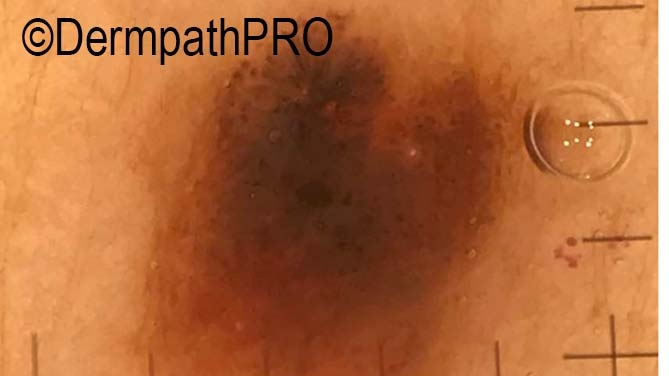
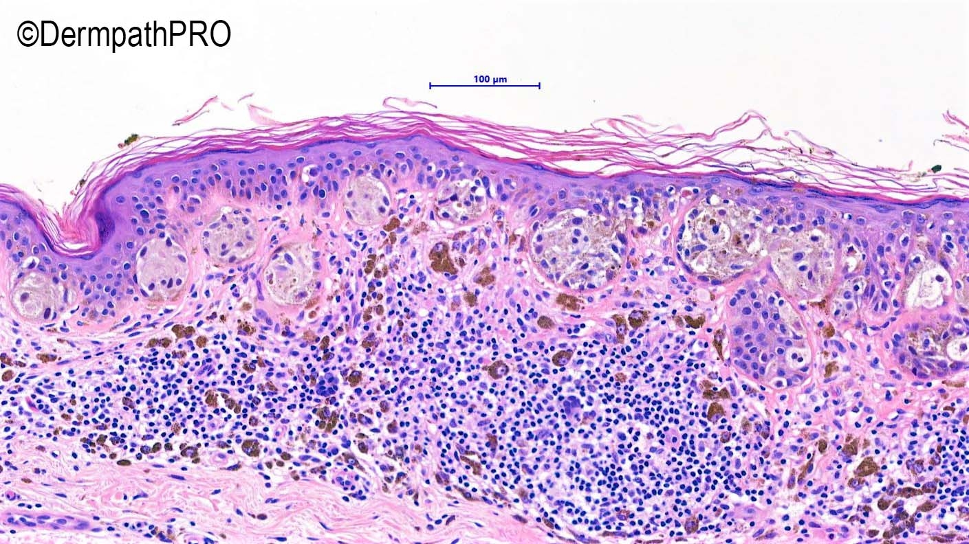
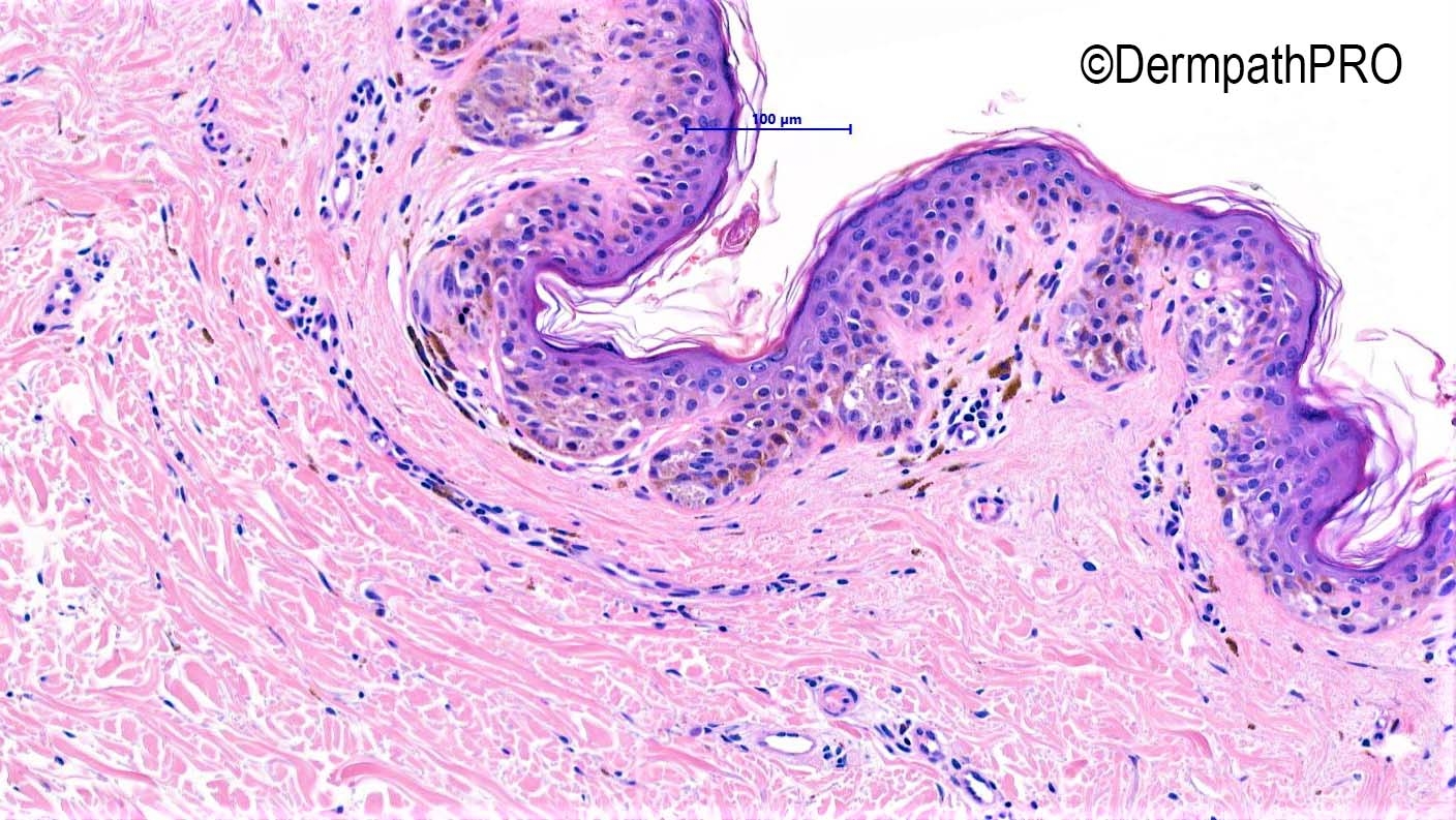
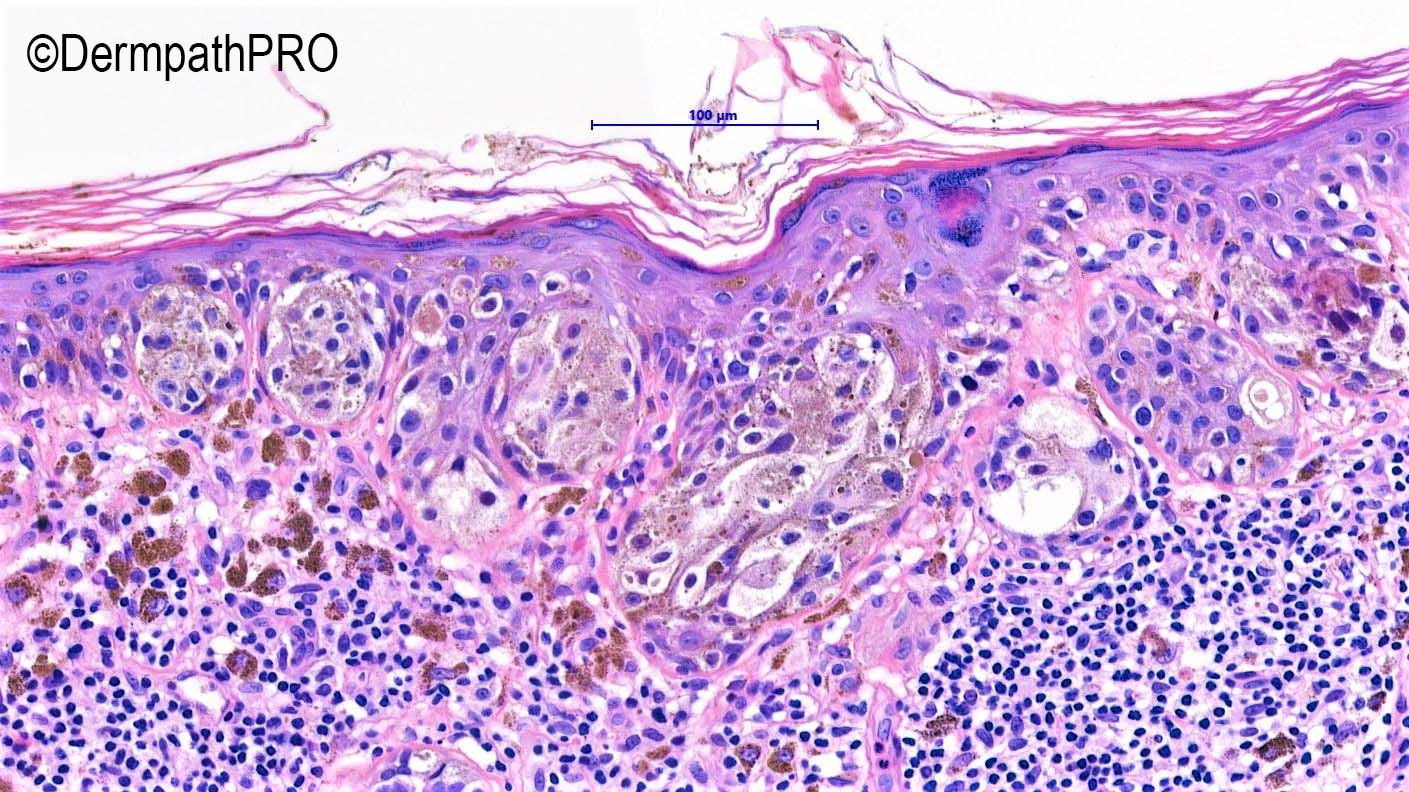
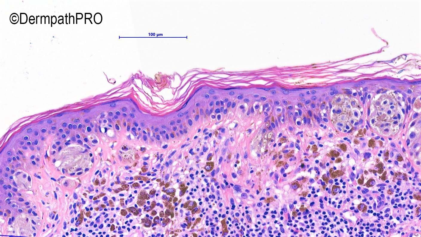
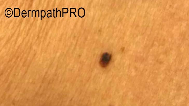
68F, Mole longstanding on right breast - no changes - no bleeds. ( found on examination ). Dermatoscope: large part in black homogeneous irregular?

Edited by Admin_Dermpath


  Report Record

User Feedback


Difficult. There is fibrosis in dermis( unusual in HN), and clinically doesnt fit. There are also definitive shoulder spreading of melanocytes. Favor Melanoma. 

Share this comment


Link to comment
share_externally

I favor a dysplastic nevus - no junctional confluent growth or substantial pagetoid spread. 

Share this comment


Link to comment
share_externally

Atypical nevus with halo reaction (Halo nevus).Follow up is recommended after complete excision.

Share this comment


Link to comment
share_externally

Very atypical epithelioid melanocytic proliferation, I would favor a malignant melanoma.

Share this comment


Link to comment
share_externally

msofopoulos

Posted

I would call it either a dysplastic nevus or a nevus of special site in case its on the milkline

Share this comment


Link to comment
share_externally

Dr. Richard Carr

Posted

Hard to classify thin lesion, atypical, some uncertainty, but favouring benign. May have a deep penetrating type clone (Beta-catenin would be of interest). Provided it's fully excised that is the end of it for me. 

Share this comment


Link to comment
share_externally

Admin_Dermpath

Posted

Dear All 

Yes this was a difficult case which I took a consensus opinion on and this is the final report! 


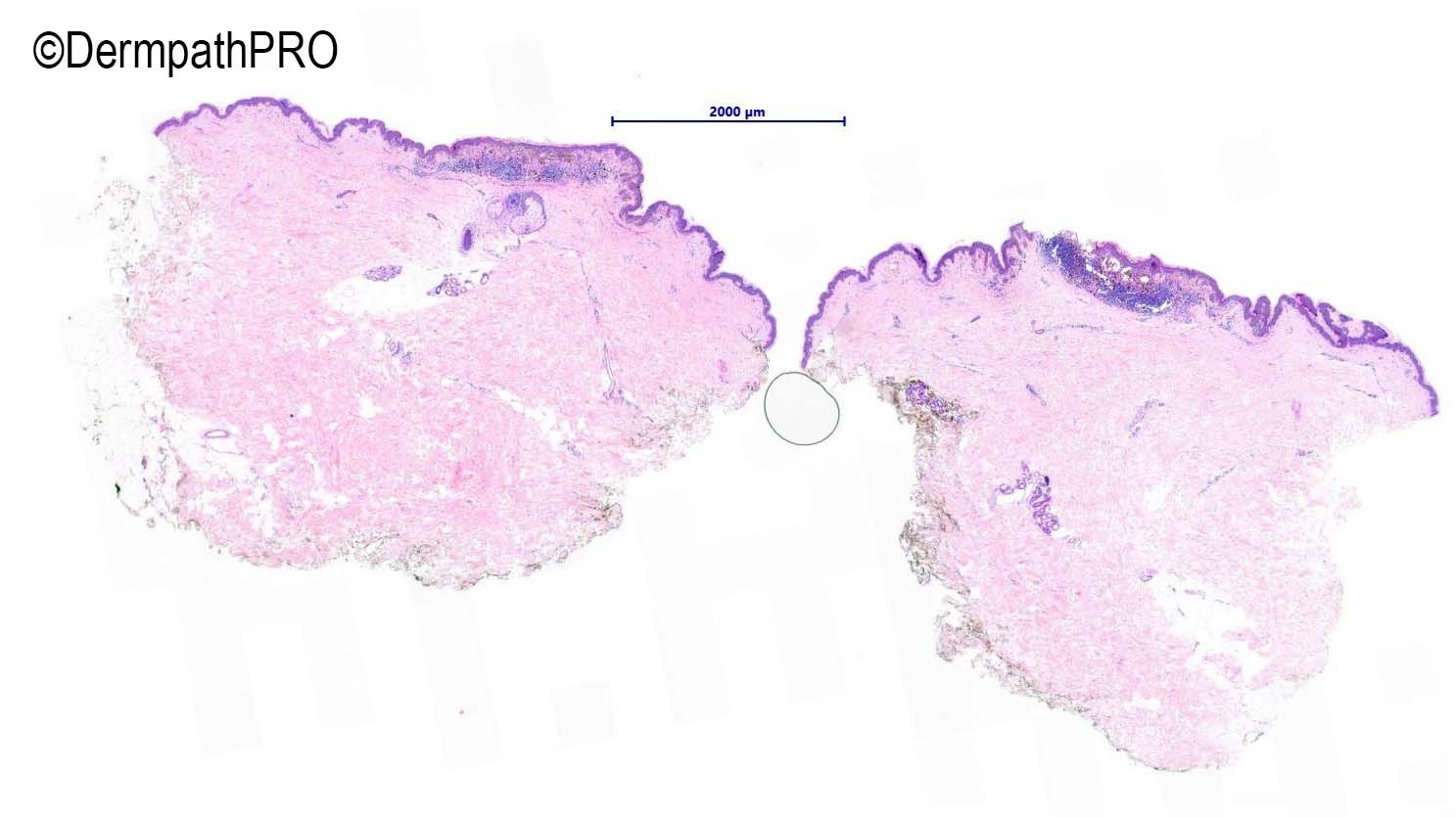
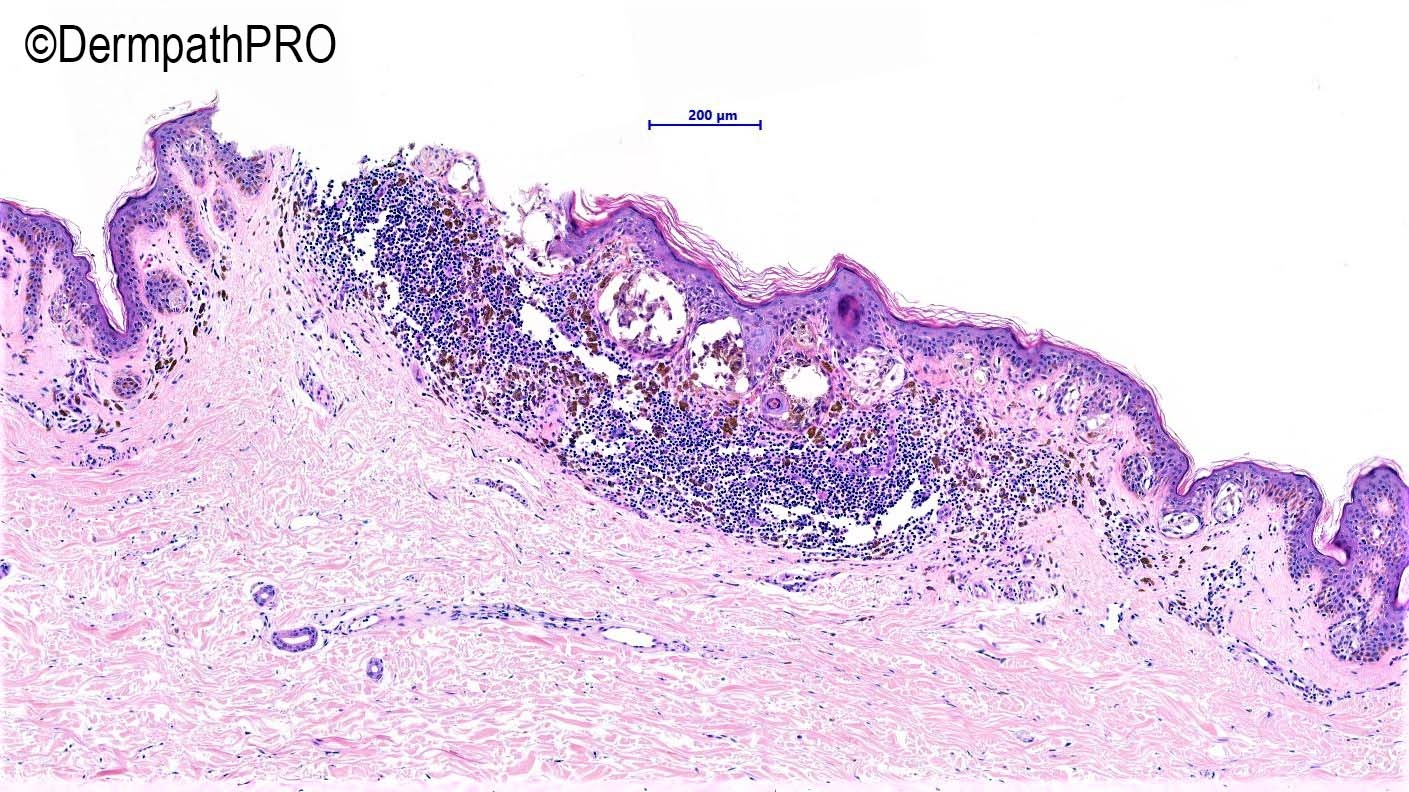
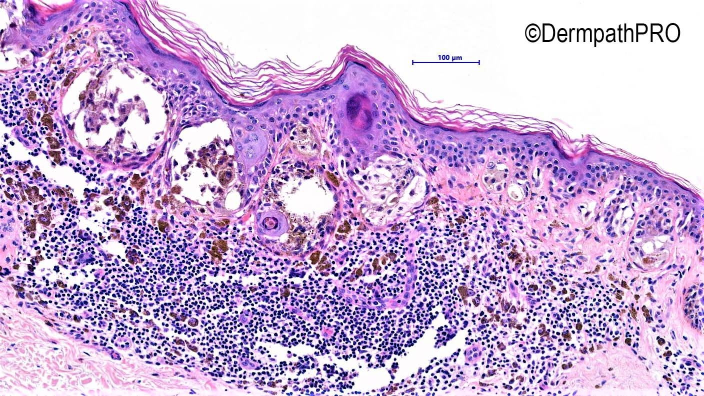
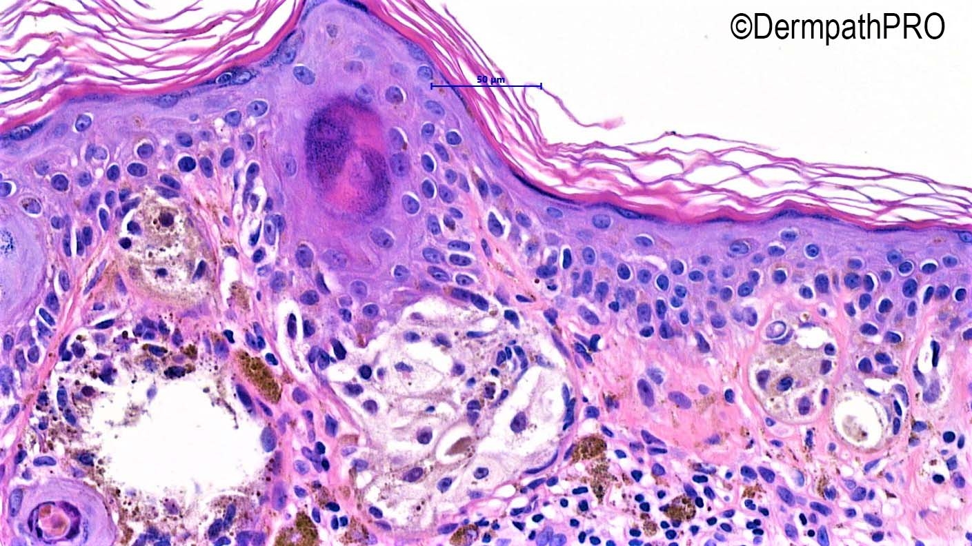
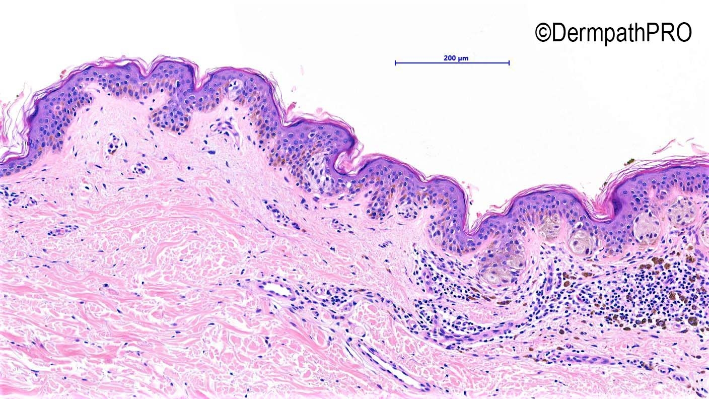
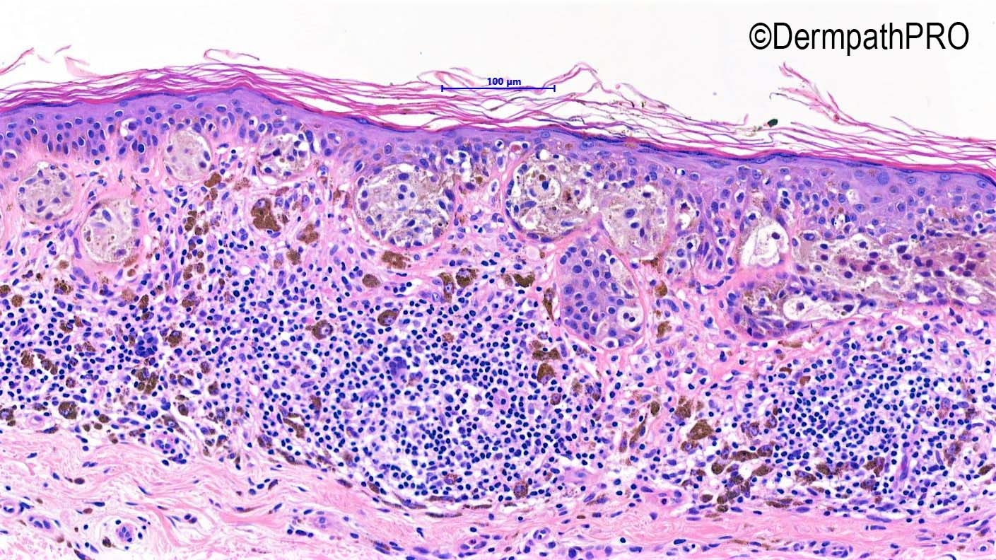
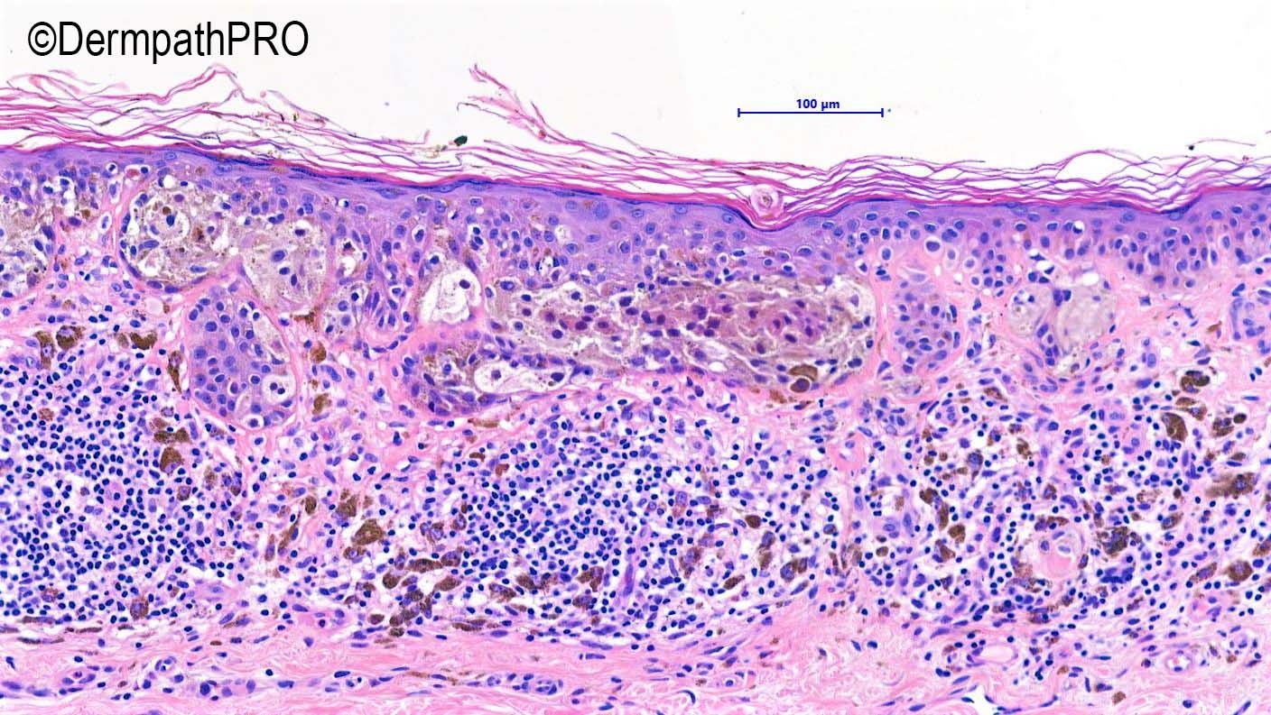
Skin with an asymmetrical compound melanocytic lesion. This melanocytic lesion is predominantly junctional with a few single melanocytic cells in the upper dermis amongst the inflammatory infiltrate. It is small (4mm in maximum diameter) and slightly raised. The junctional component is predominantly nested and the nests are mostly placed at the bottom of the rete ridges with sparing of the suprapapillary epidermis. The component cells are epithelioid, cytological atypia is random and mitotic figures are absent. There is no significant single cell ascent, there is a little lateral spread, there is minor and focal epidermal consumption and no ulceration.


The features are insufficient for a diagnosis of melanoma in situ and the appearances are those of a low to high grade (moderate to severely) dysplastic naevus, with regression; despite the lack of lamellar fibroplasia and elongation of rete is minimal. The lesion is completely excised and lies 1.7 mm from the lateral resection margin. 


Iskander

Share this comment


Link to comment
share_externally


Join the conversation

You can post now and register later. If you have an account, sign in now to post with your account.

Guest
Add a comment...

×   Pasted as rich text.   Paste as plain text instead

  Only 75 emoji are allowed.

×   Your link has been automatically embedded.   Display as a link instead

×   Your previous content has been restored.   Clear editor

×   You cannot paste images directly. Upload or insert images from URL.

Loading...

×
×
  • Create New...