Jump to content
In this section we have spot diagnoses posted on a daily basis since June 2010, now over 4000! You can review the archived cases and read the suggested diagnoses by users and the final comment by the contributors.
Case are uploaded each week day by 10 am UK time with the correct diagnosis will generally be posted at 8 pm UK time. Why not view the most recent spot diagnosis and proffer a diagnosis?

Case Number : Case 2551 - 16 April 2020 Posted By: Saleem Taibjee

Please read the clinical history and view the images by clicking on them before you proffer your diagnosis.
Submitted Date :
   (0 reviews)

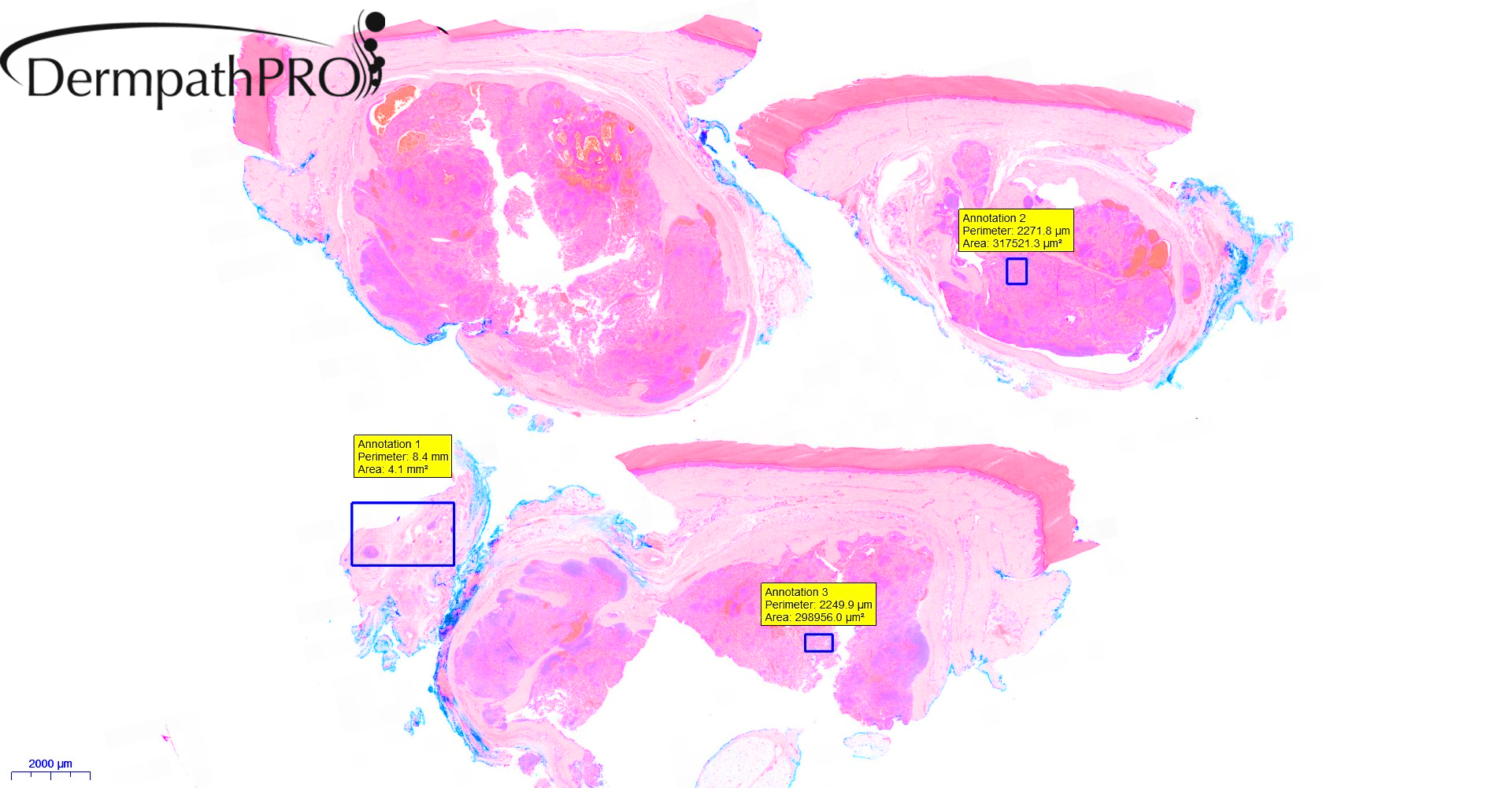
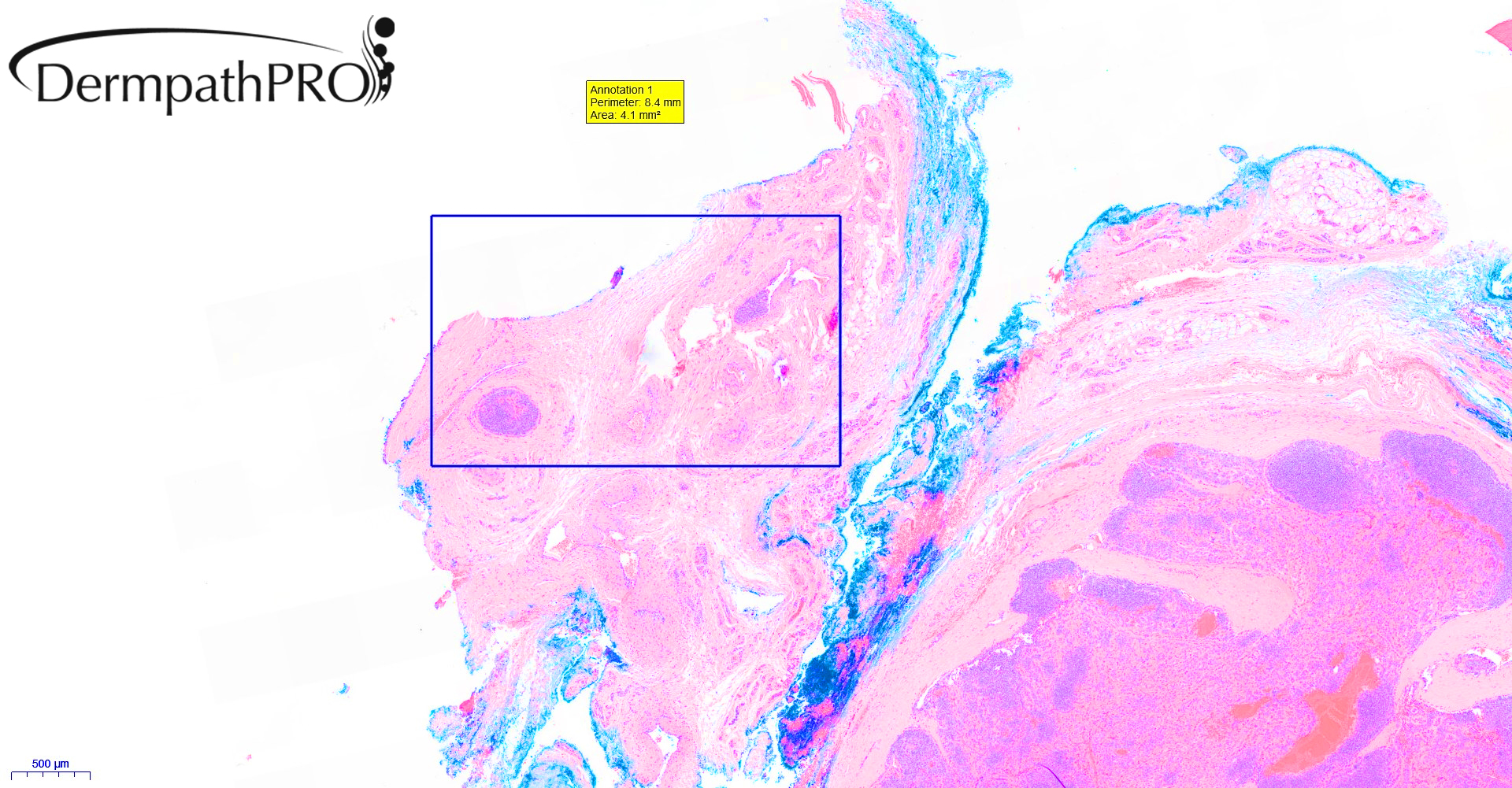
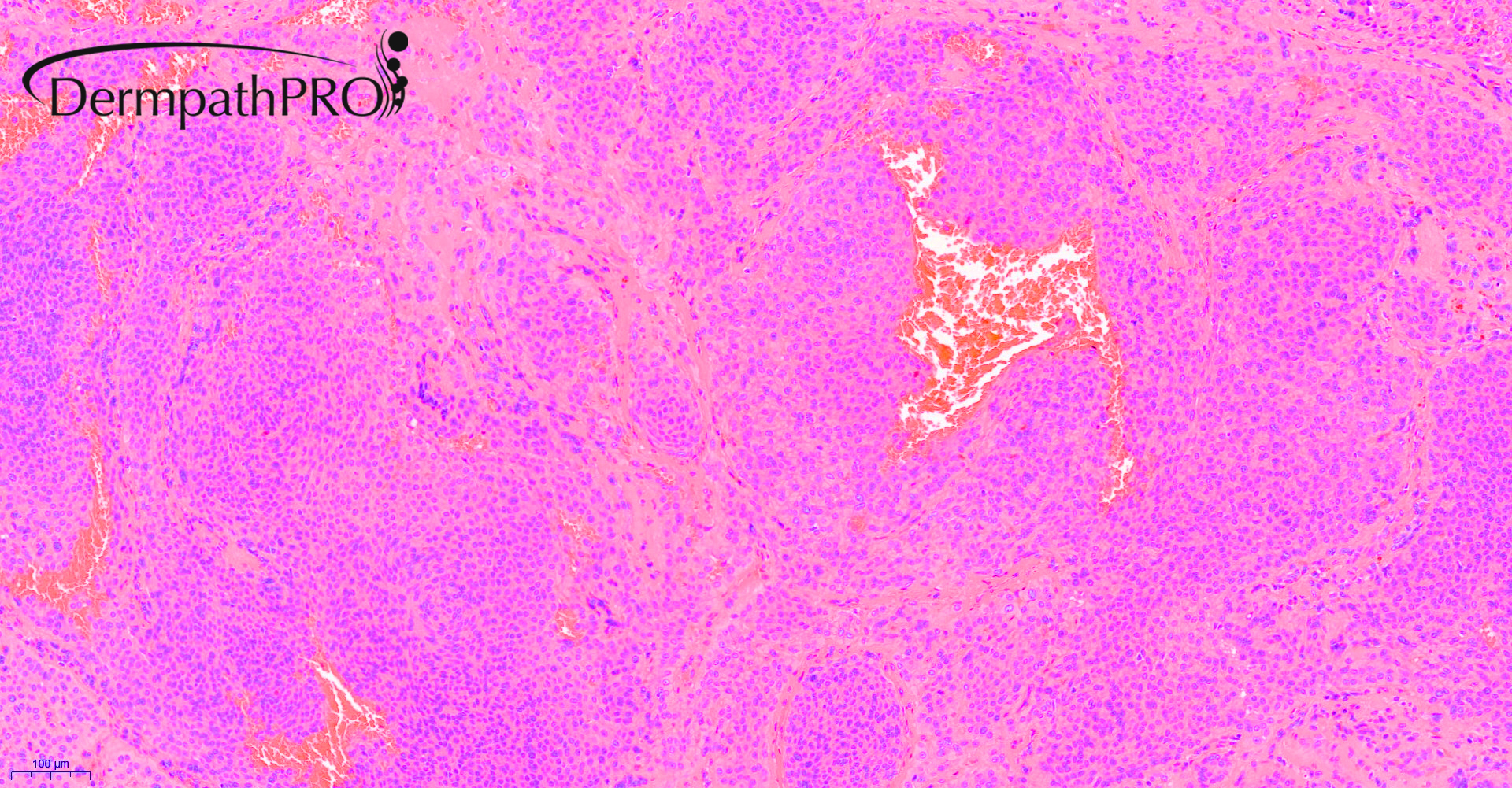
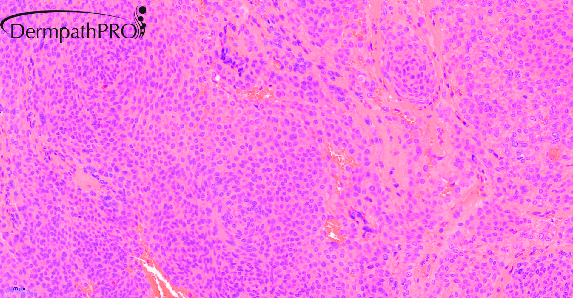
73F, lump on sole of right foot enlarging past 12 months.


  Report Record

User Feedback


Meenakshi Batrani

Posted

Mixed tumor- epithelial and myoepithelial (hyaline/plasamcytoid cells) predominant with scant stroma, and intravascular nests (which can be seen in mixed tumor)

Share this comment


Link to comment
share_externally

13 hours ago, Meenakshi Batrani said:

Mixed tumor- epithelial and myoepithelial (hyaline/plasamcytoid cells) predominant with scant stroma, and intravascular nests (which can be seen in mixed tumor)

Thought the same. Maybe I’m wrong and mixed tumor is the right diagnosis. 

Share this comment


Link to comment
share_externally

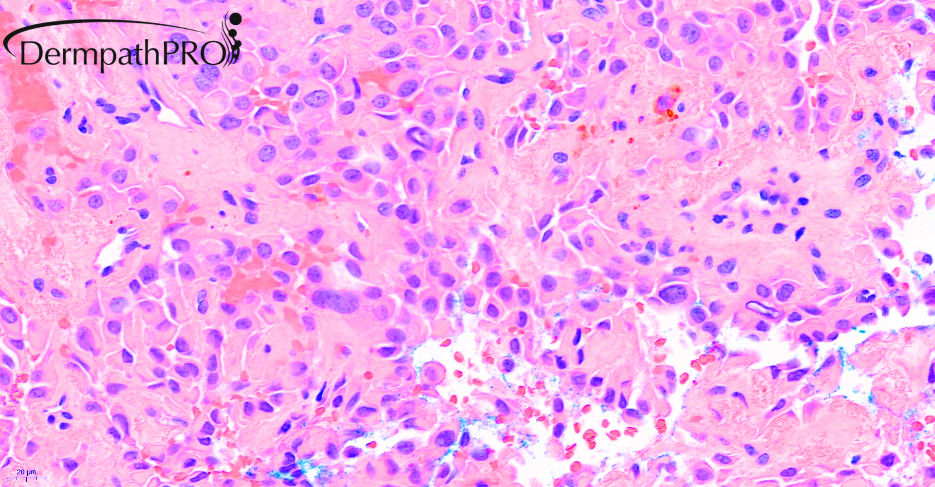
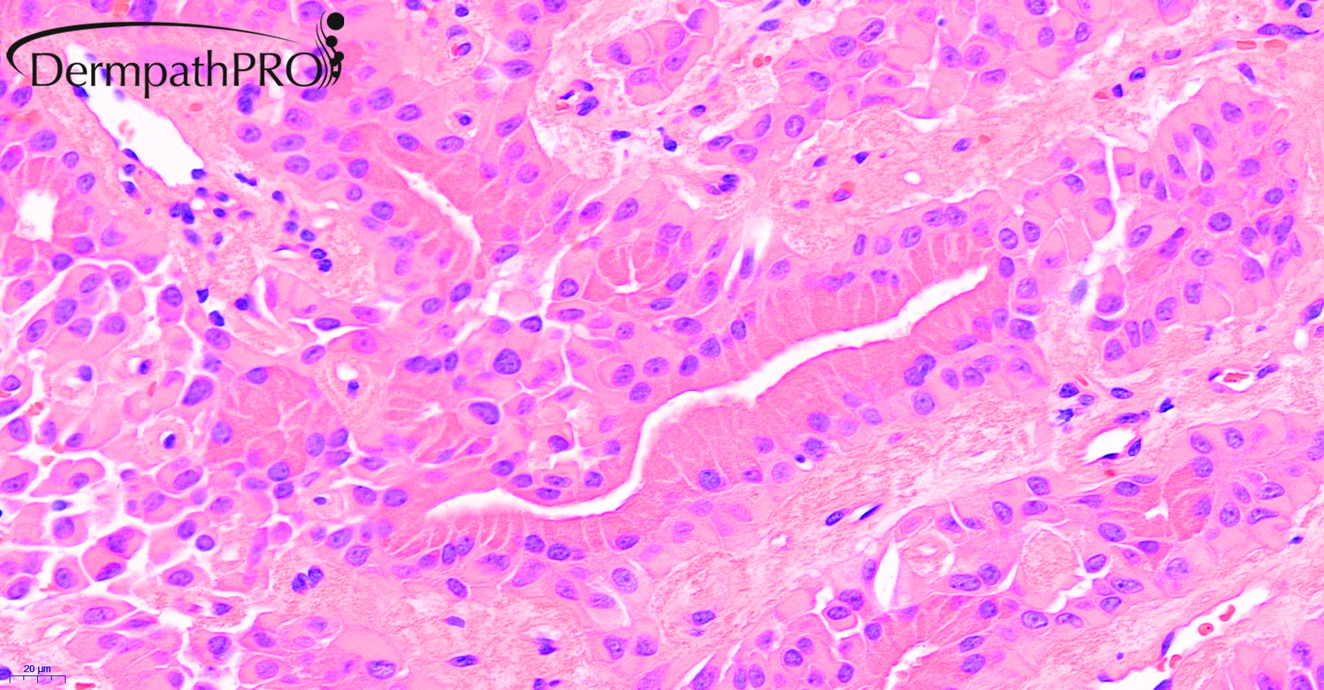
Looks like there  are 2 distinct areas in the tumor:

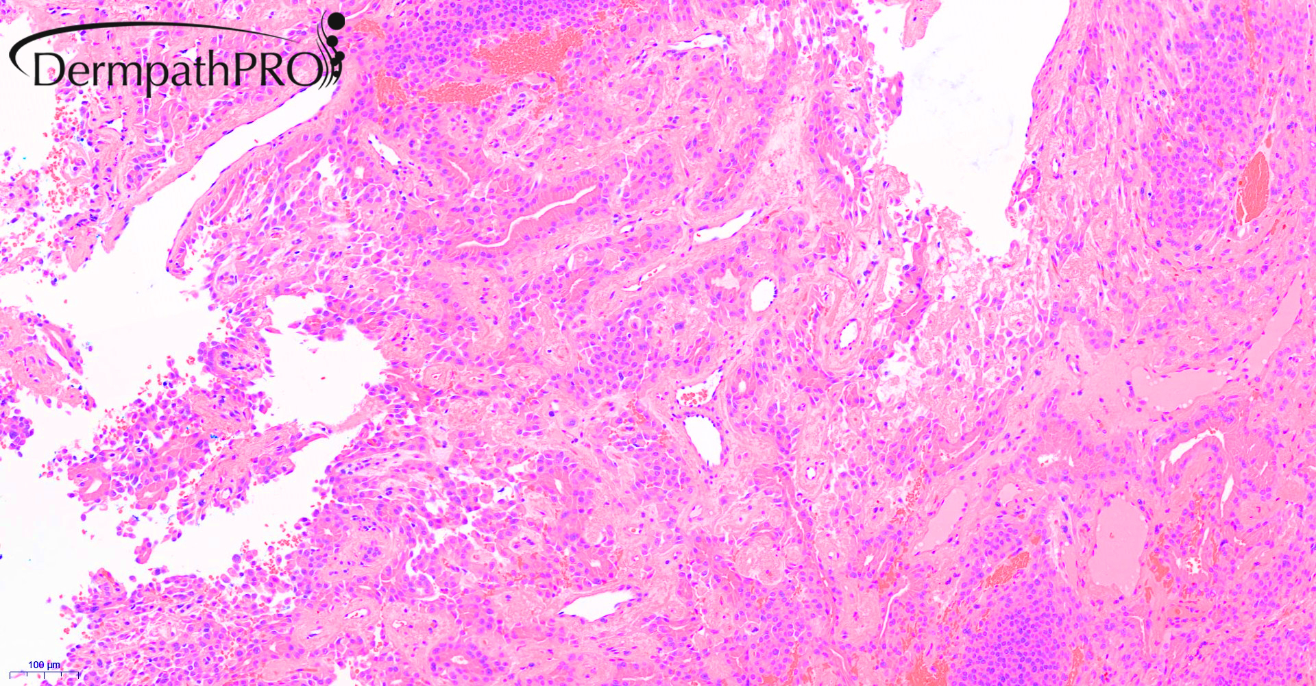
The last 2 figures show a biphasic tumor composed of peripheral myoepithelial cells, some with a plasmacytoid cytology. The inner luminal layer is composed of columnar cells with probably apocrine differentiation. No cytologic atypia or mitotic activity seen. There is no chondromyxoid stroma to support the diagnosis of a mixed tumor. In the breast, this type of tumor is called adenomyoepithelioma (if benign). Since there is no benign counter part in the salivary glands or lung, it's called epithelial-myoepithelial carcinoma.

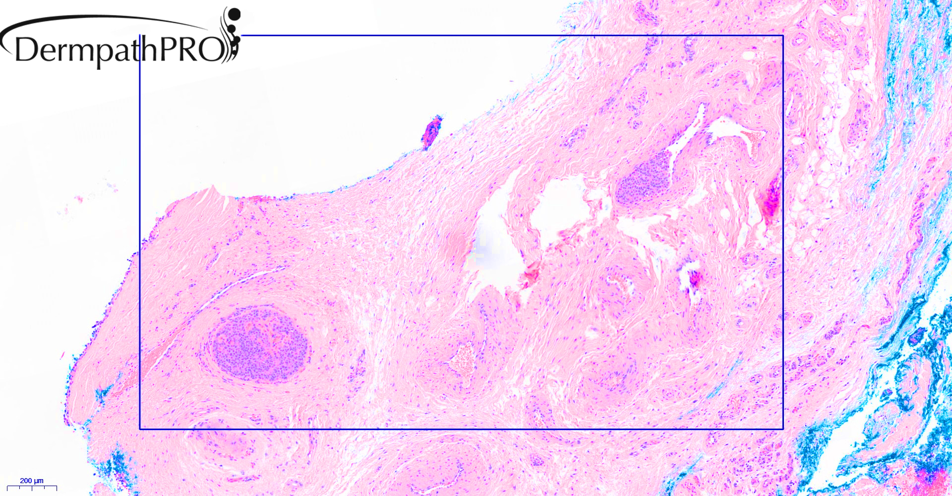
The majority of the tumor looks like a poroma and is composed of "poroid" cells (Figure 4 and 5). However, there are also some plasmacytoid cells in the poroma-like component, particularly in the right upper corner of figure 5. One nest is seen in a vessel (which always worrisome, vascular invasion?).

Now the question how to classify this tumor depends on whether the poroid cells are epithelial cells such as in a true poroma or myoepithelial cells. We will need immunohistochemical studies to answer that question (cytokeratinAE1/AE3, S-100, SOX-10, smooth muscle actin, p63 or other myoepithelial markers)

Share this comment


Link to comment
share_externally

Ductal formation could be encountered in soft tissue myoepithelioma. There is vascular invasion, but I'm not sure how to explain this.

Share this comment


Link to comment
share_externally

Saleem Taibjee

Posted

Thanks for this extremely interesting range of comments. I found this case very tricky, and also shared it with Richard Carr. We had a similar conclusion.

The immunostains showed diffuse positive staining with CK5/6 and CK7. Ducts were nicely highlighted by CEA and EMA. p63 was virtually completely negative.

I felt that the poroid component was the more significant, and therefore that the lesion is more akin to hidradenoma territory, but I can see how the images suggest a plasmacytoid myoepithelial component. The lack of p63 staining goes against that. But I would have expected some degree of staining with p63 even if hidradenoma, and this has confused me. The clearly identifiable focus of lymphovascular invasion at the periphery is also concerning. We know that even benign hidradenomas may occasionally show this, and even 'benign metastasis' is described in this context -  see https://www.ncbi.nlm.nih.gov/pubmed/23095508. But even so, this led us to move this into the atypical hidradenoma category, mainly to emphasise the need for clinical follow-up.

Share this comment


Link to comment
share_externally

Dr. Mona Abdel-Halim

Posted

Very interesting lesion! I was between poroid hidradenoma and mixed tumor

Share this comment


Link to comment
share_externally


Join the conversation

You can post now and register later. If you have an account, sign in now to post with your account.

Guest
Add a comment...

×   Pasted as rich text.   Paste as plain text instead

  Only 75 emoji are allowed.

×   Your link has been automatically embedded.   Display as a link instead

×   Your previous content has been restored.   Clear editor

×   You cannot paste images directly. Upload or insert images from URL.

Loading...

×
×
  • Create New...