Jump to content
In this section we have spot diagnoses posted on a daily basis since June 2010, now over 4000! You can review the archived cases and read the suggested diagnoses by users and the final comment by the contributors.
Case are uploaded each week day by 10 am UK time with the correct diagnosis will generally be posted at 8 pm UK time. Why not view the most recent spot diagnosis and proffer a diagnosis?

Case Number : Case 2552 - 17 April 2020 Posted By: Dr. Richard Carr

Please read the clinical history and view the images by clicking on them before you proffer your diagnosis.
Submitted Date :
   (0 reviews)

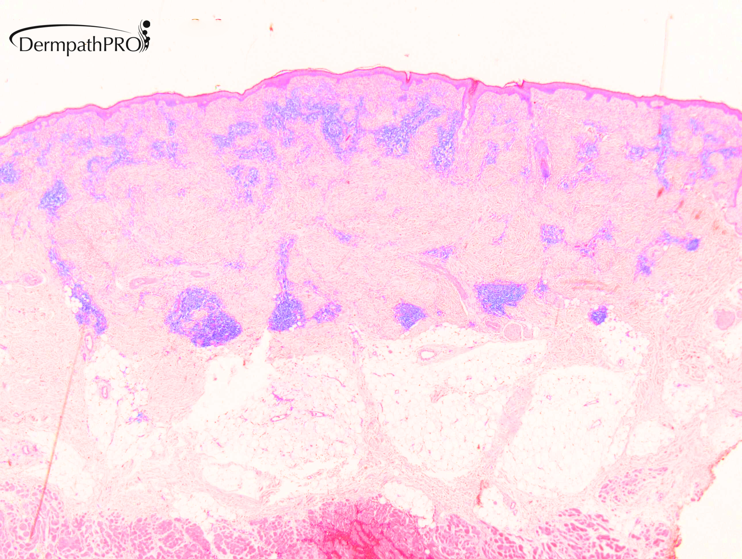
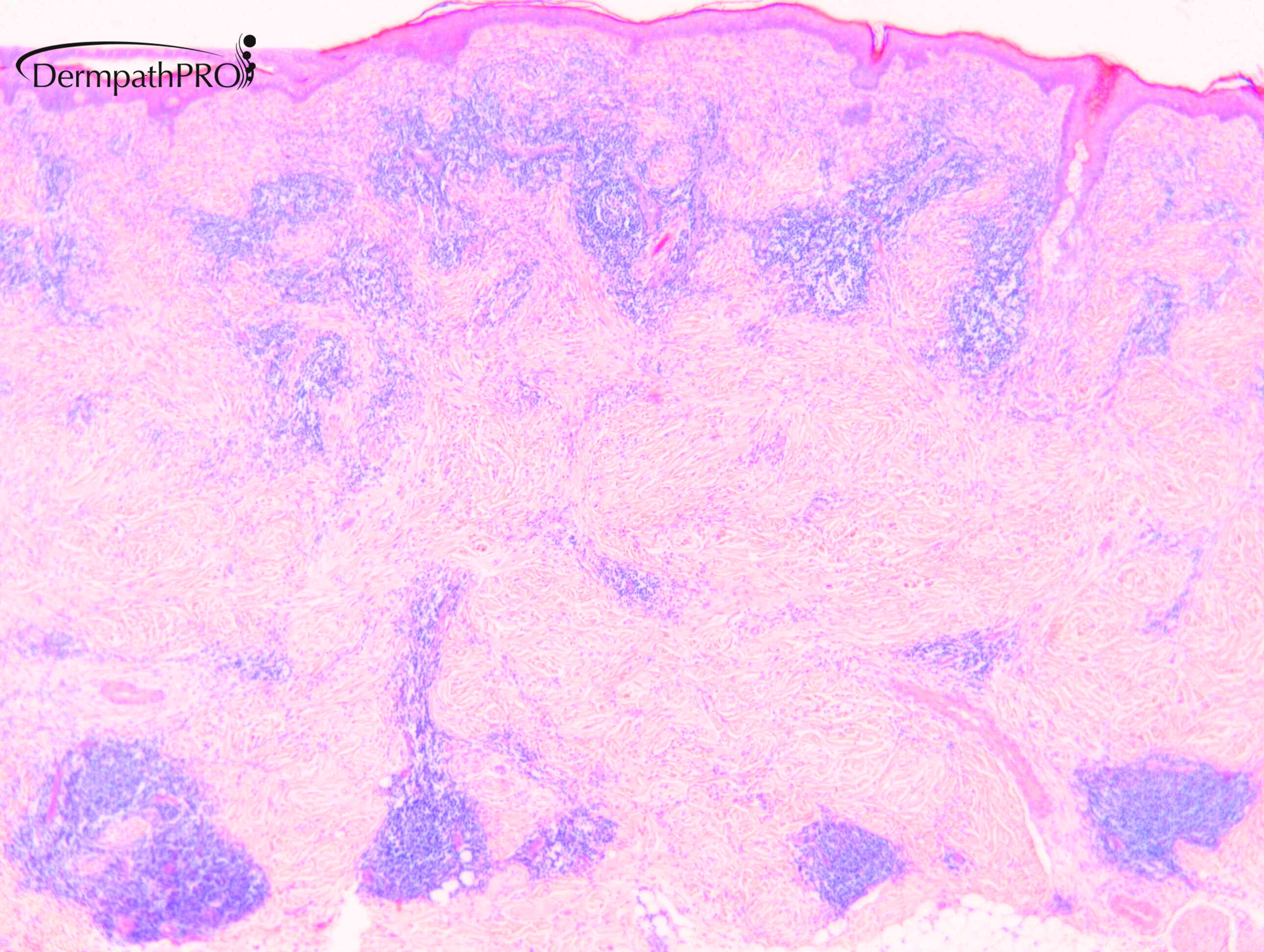
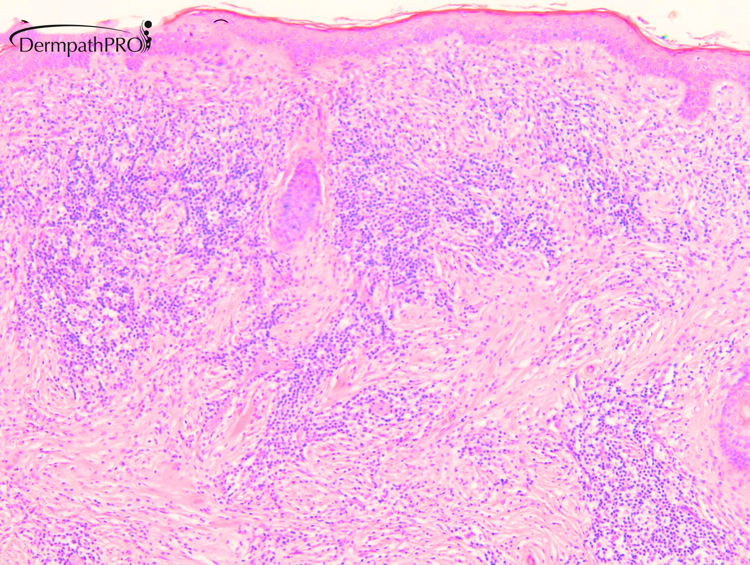
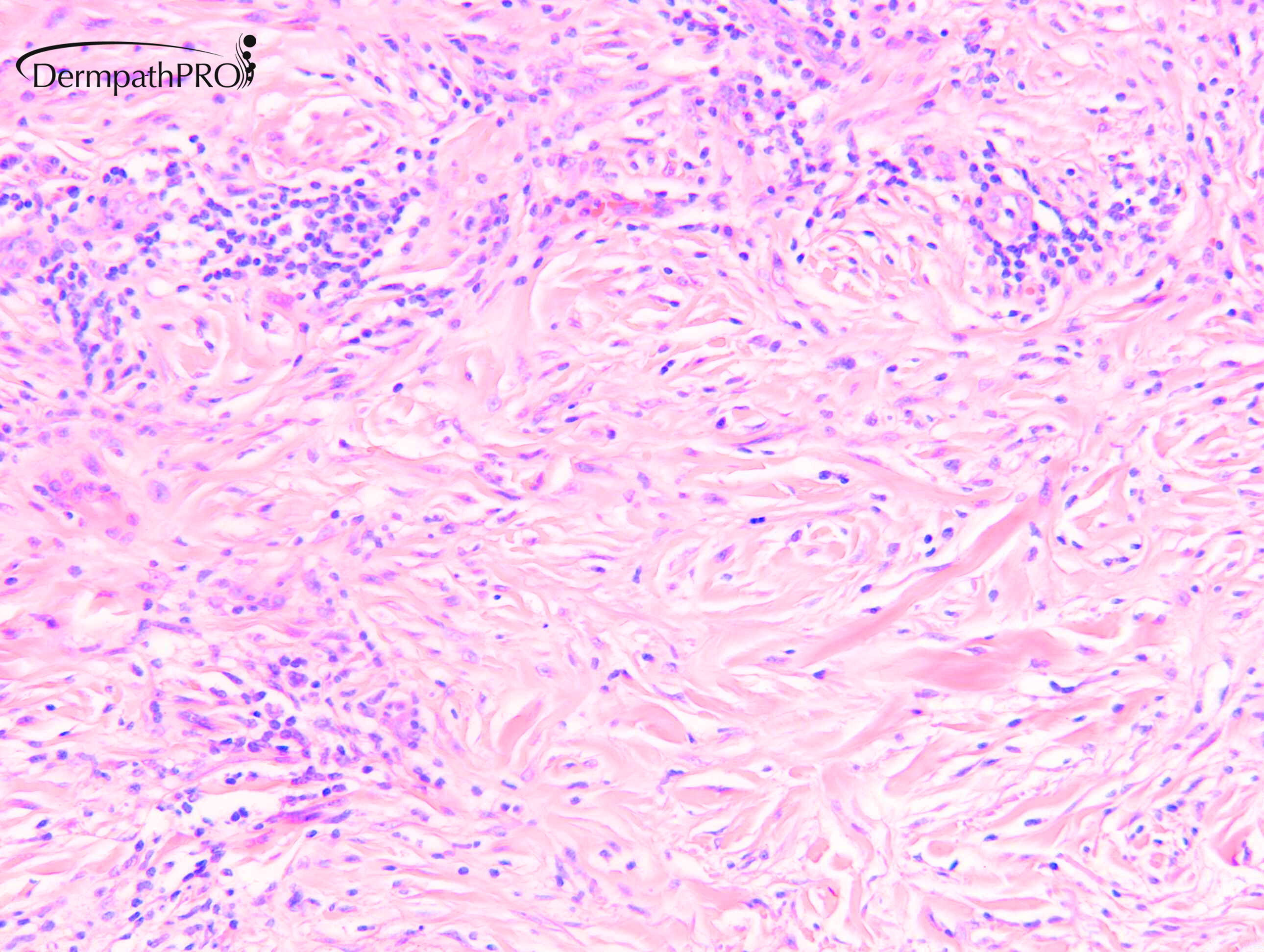
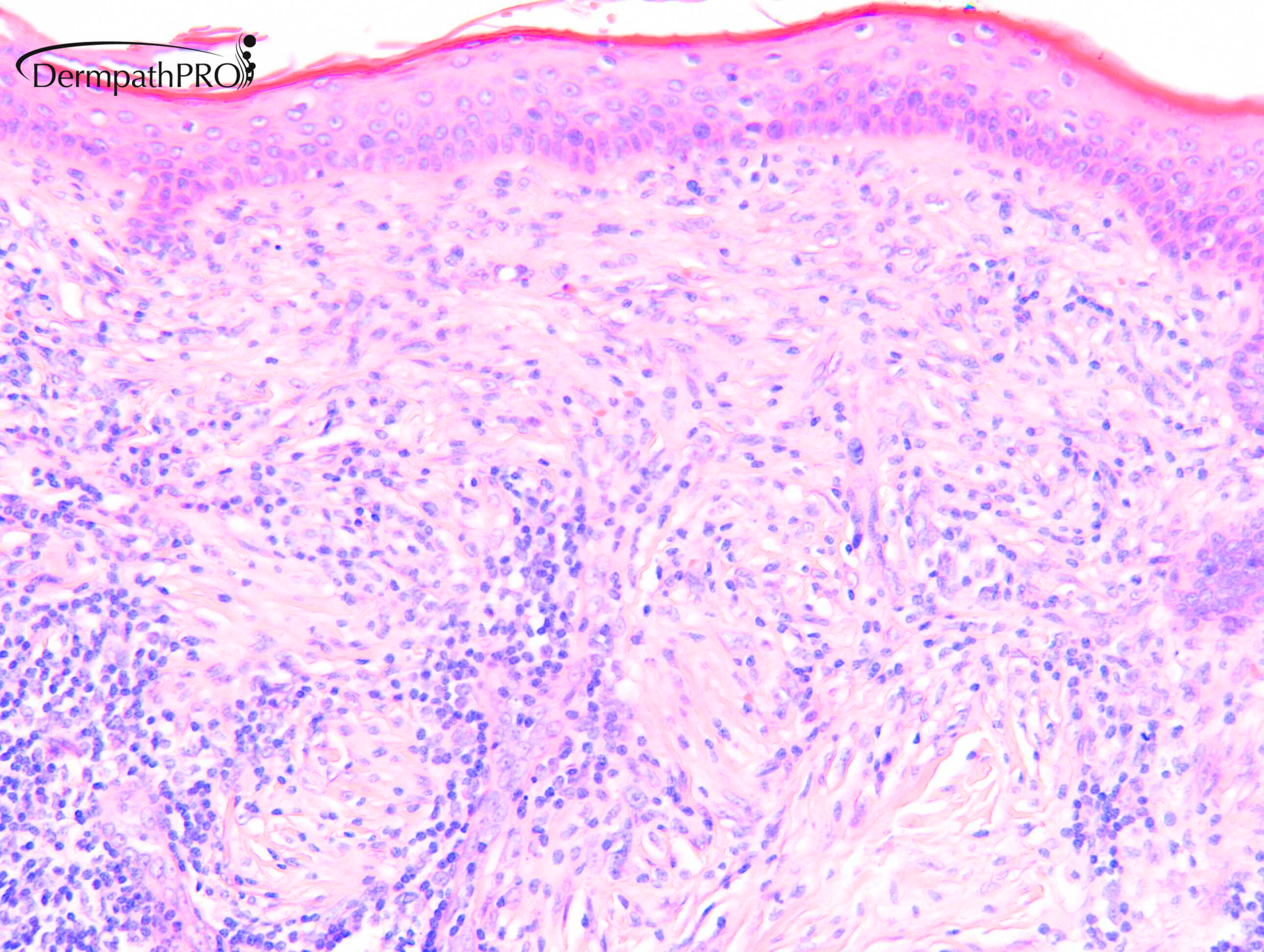
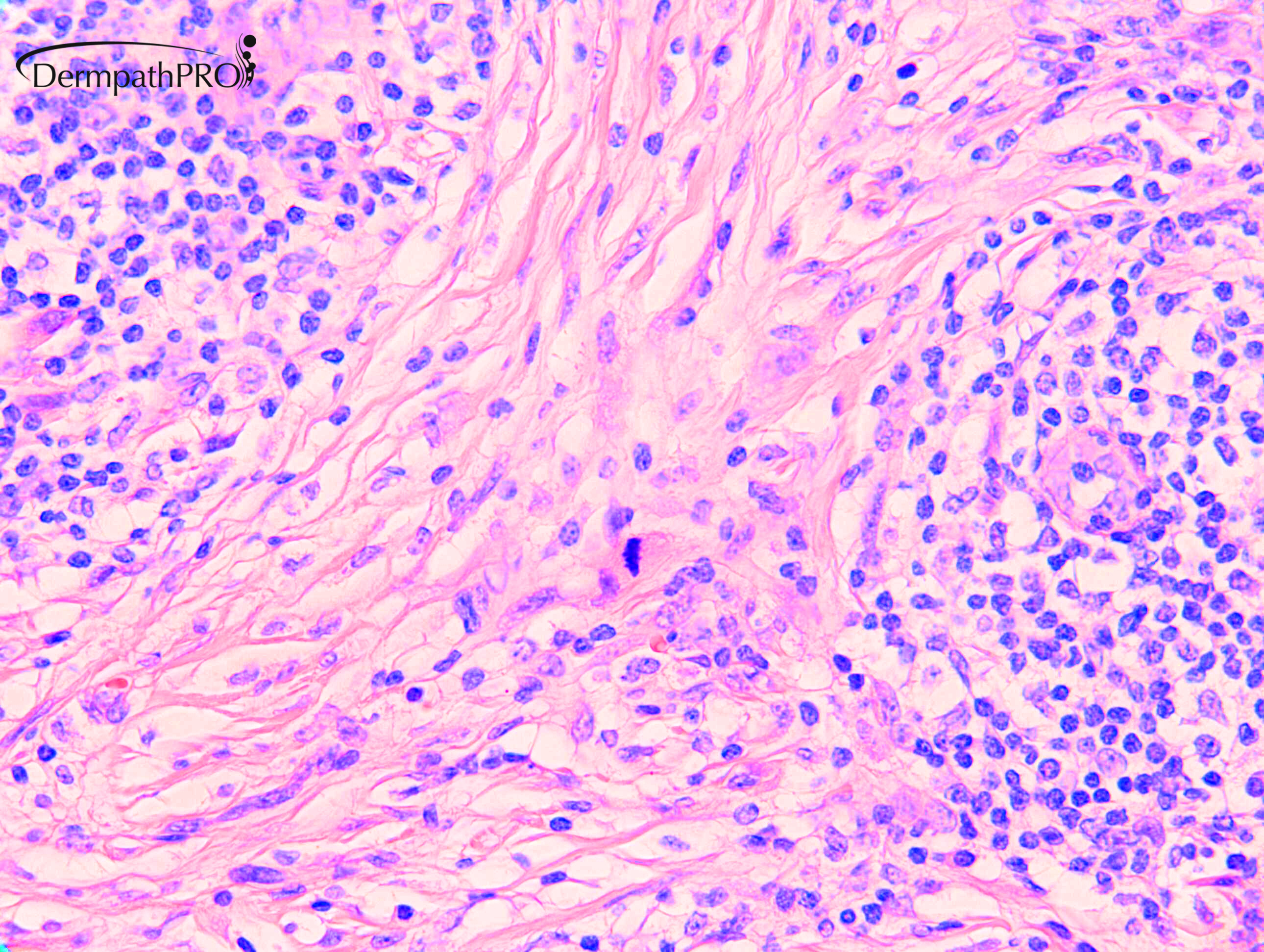
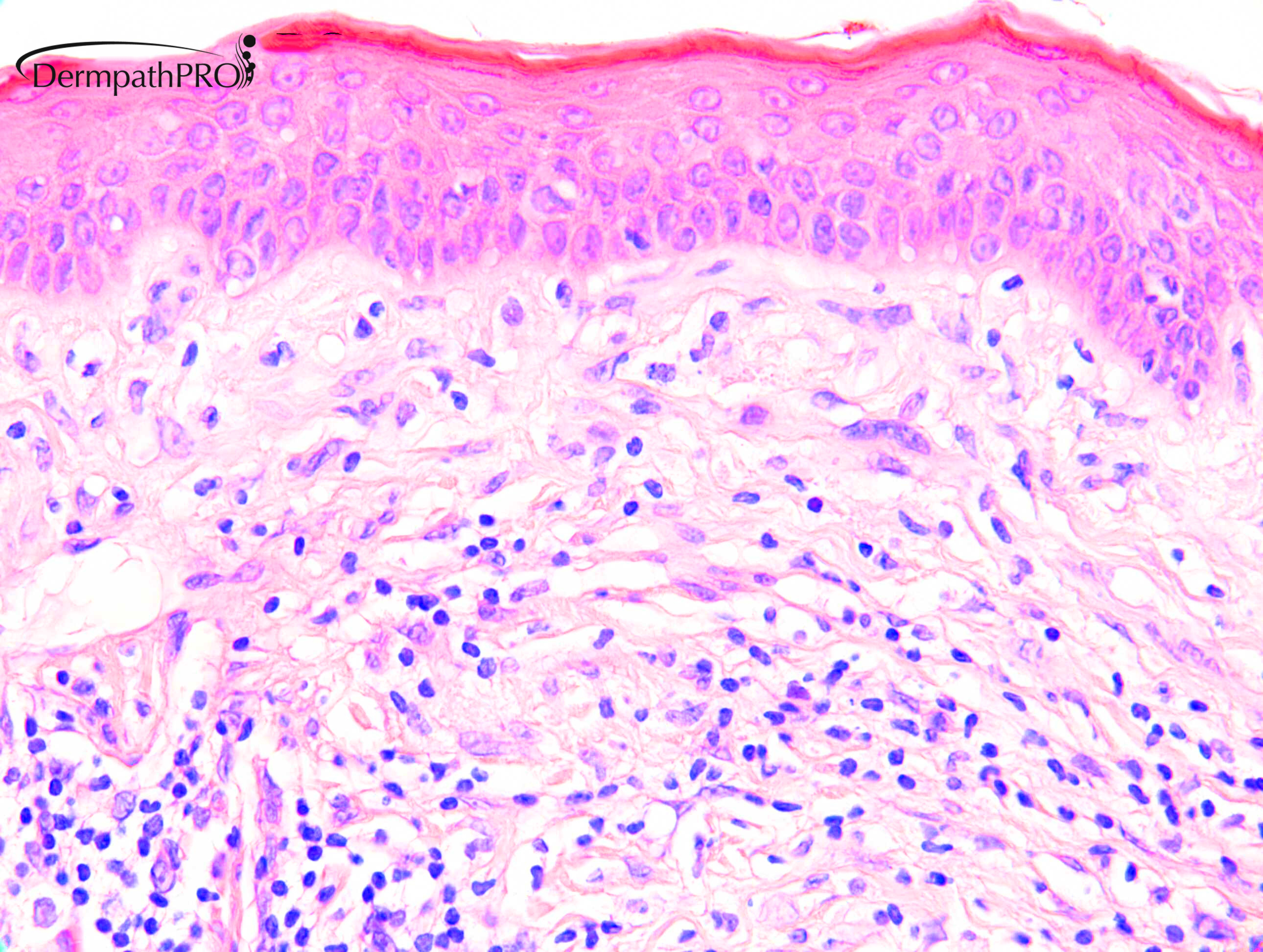
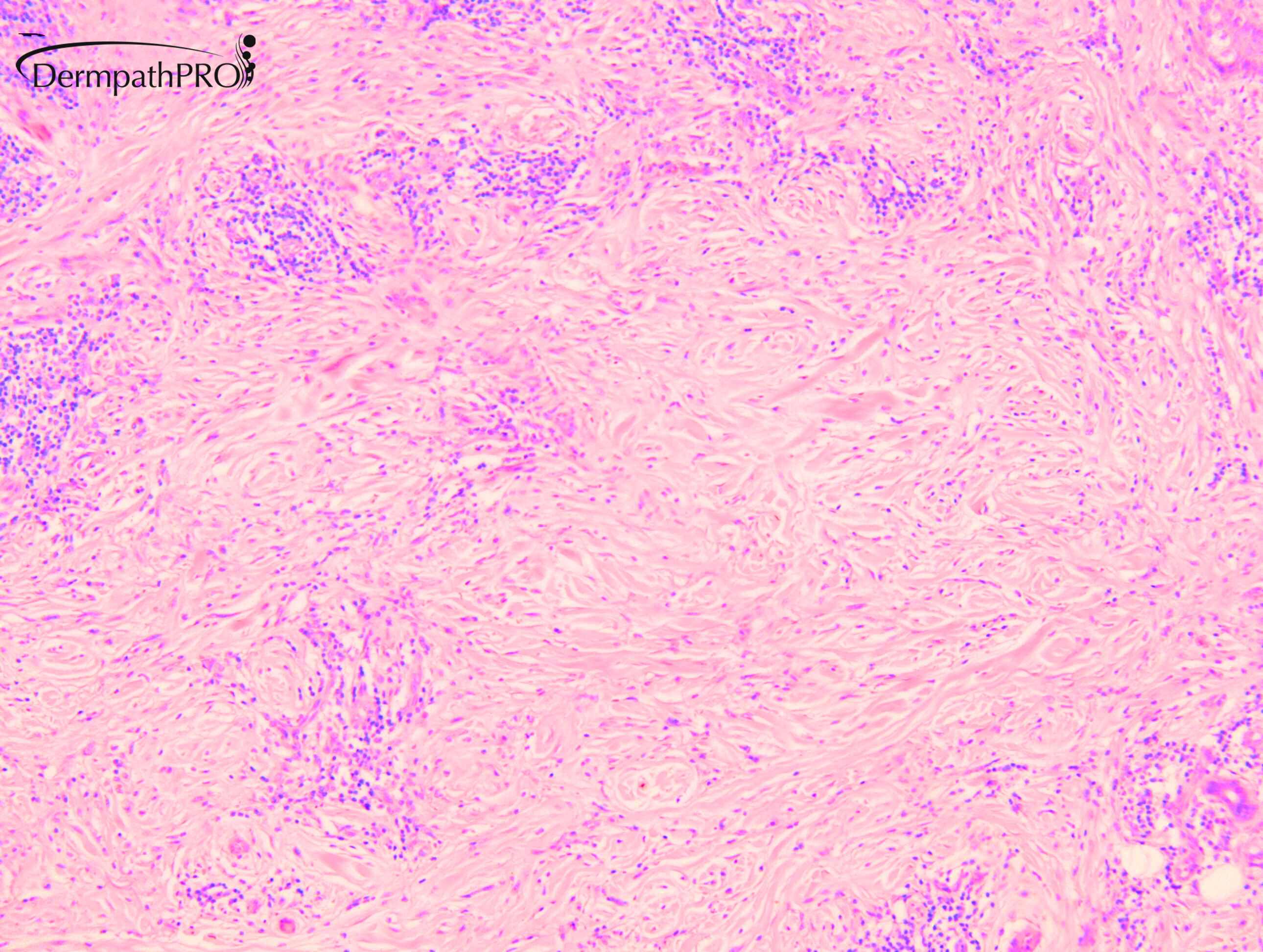
70-F. Erythematous plaque forehead.

  Report Record

User Feedback


Malignant spindle cell tumors for immunos. Ddx. Desmoplastic melanoma.

Share this comment


Link to comment
share_externally

Agree. Lymphoid nodular clusters in sclerotic dermis. There is something resembling Rosaj Dorfman Disease in pic 4, also.

S100 should be +ve in both, but SOX10 could be an appropriate immuno for ddx.


Share this comment


Link to comment
share_externally

Krishnakumar subramanian

Posted

slide 2,3 and 5 showing some collagenolytic changes could be granuloma multiforme, but a mitotic activity is seen in image 6

i dont know

Share this comment


Link to comment
share_externally

Meenakshi Batrani

Posted

On 17/04/2020 at 19:55, Hamad said:

Malignant spindle cell tumors for immunos. Ddx. Desmoplastic melanoma.

Agree, suspicious for spindle cell melanoma.

Share this comment


Link to comment
share_externally

Dr. Richard Carr

Posted

This case serves to support Phillip Mckee's adage that before every sign out say to yourself "desmoplastic melanoma" (DM). It is a good idea to perform S100 on any odd fibrohistiocytic / inflammatory looking lesions on the head & neck of an older individual. This case actually dates from several years before and there had been a previous lentigo maligna near this lesion. Despite this an otherwise good, but non-dermato-, pathologist possibly unaware of Phillip's adage signed it out as in keeping with lupus tumidus. We received a recent specimen (some 6 years later) that led us to review this biopsy. I'd like to thank Dr Domenico Mesiano for sharing this learning case. I think it also serves to illustrate the DM in it's pure form can be rather indolent. When I looked at the case "blind" I was thinking of morphoea which serves hopefully to illustrate the difficulty and that it's very easy to slip-up with this diagnosis so that clinical information is highly important. The case is quite similar to another one we illustrated on the leg (granuloma annulare-like).


See also Case 2460


Share this comment


Link to comment
share_externally

Dr. Mona Abdel-Halim

Posted

Nice! my thought was desmoplastic melanoma too. 

Share this comment


Link to comment
share_externally


Join the conversation

You can post now and register later. If you have an account, sign in now to post with your account.

Guest
Add a comment...

×   Pasted as rich text.   Paste as plain text instead

  Only 75 emoji are allowed.

×   Your link has been automatically embedded.   Display as a link instead

×   Your previous content has been restored.   Clear editor

×   You cannot paste images directly. Upload or insert images from URL.

Loading...

×
×
  • Create New...