Jump to content
In this section we have spot diagnoses posted on a daily basis since June 2010, now over 4000! You can review the archived cases and read the suggested diagnoses by users and the final comment by the contributors.
Case are uploaded each week day by 10 am UK time with the correct diagnosis will generally be posted at 8 pm UK time. Why not view the most recent spot diagnosis and proffer a diagnosis?

Case Number : Case 2556 - 23 April 2020 Posted By: Saleem Taibjee

Please read the clinical history and view the images by clicking on them before you proffer your diagnosis.
Submitted Date :
   (0 reviews)

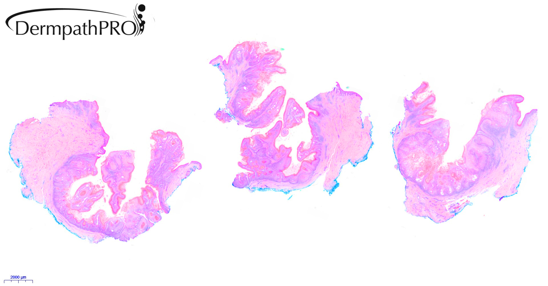
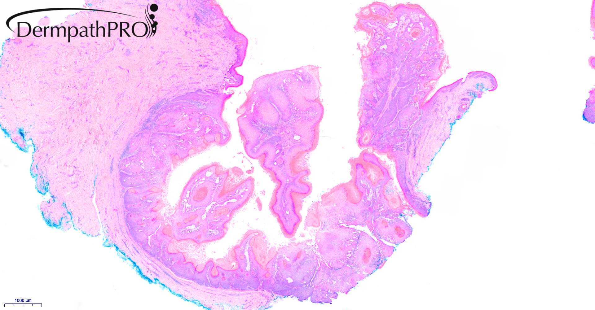
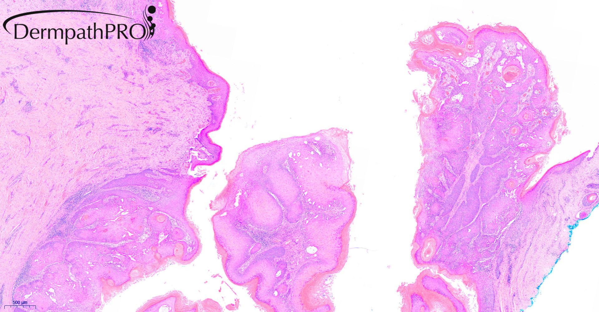
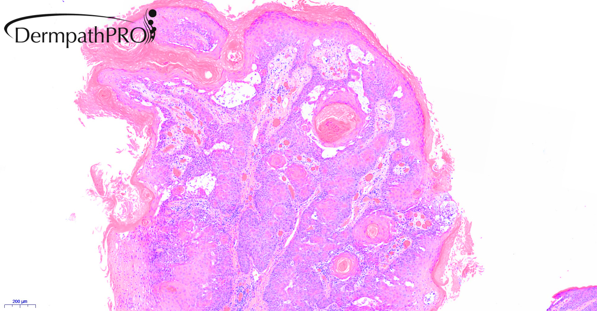
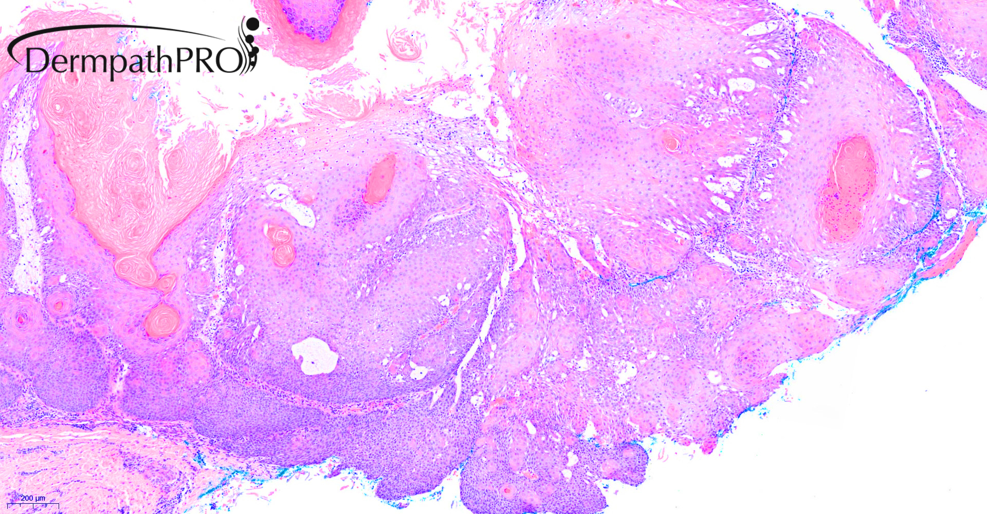
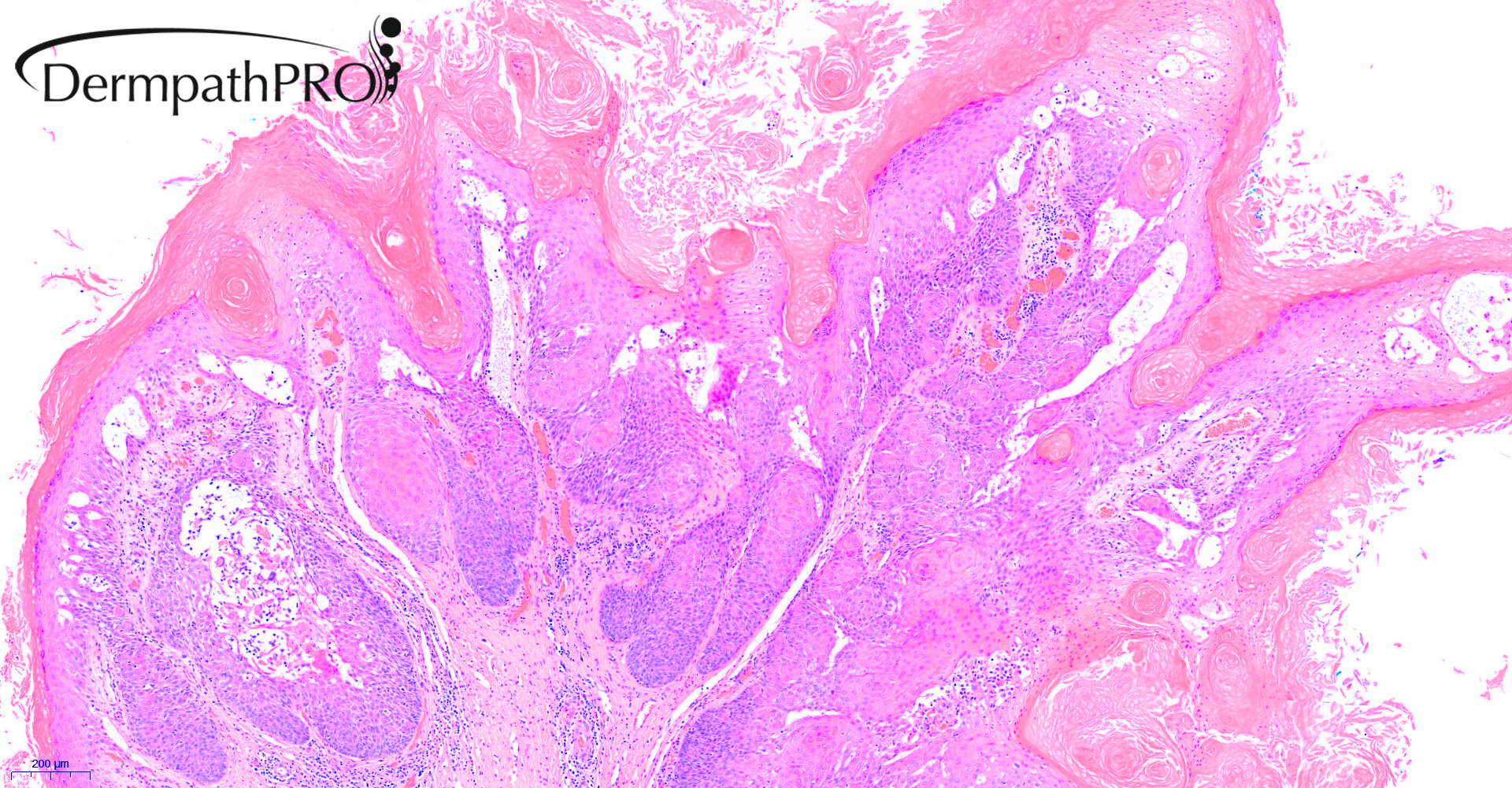
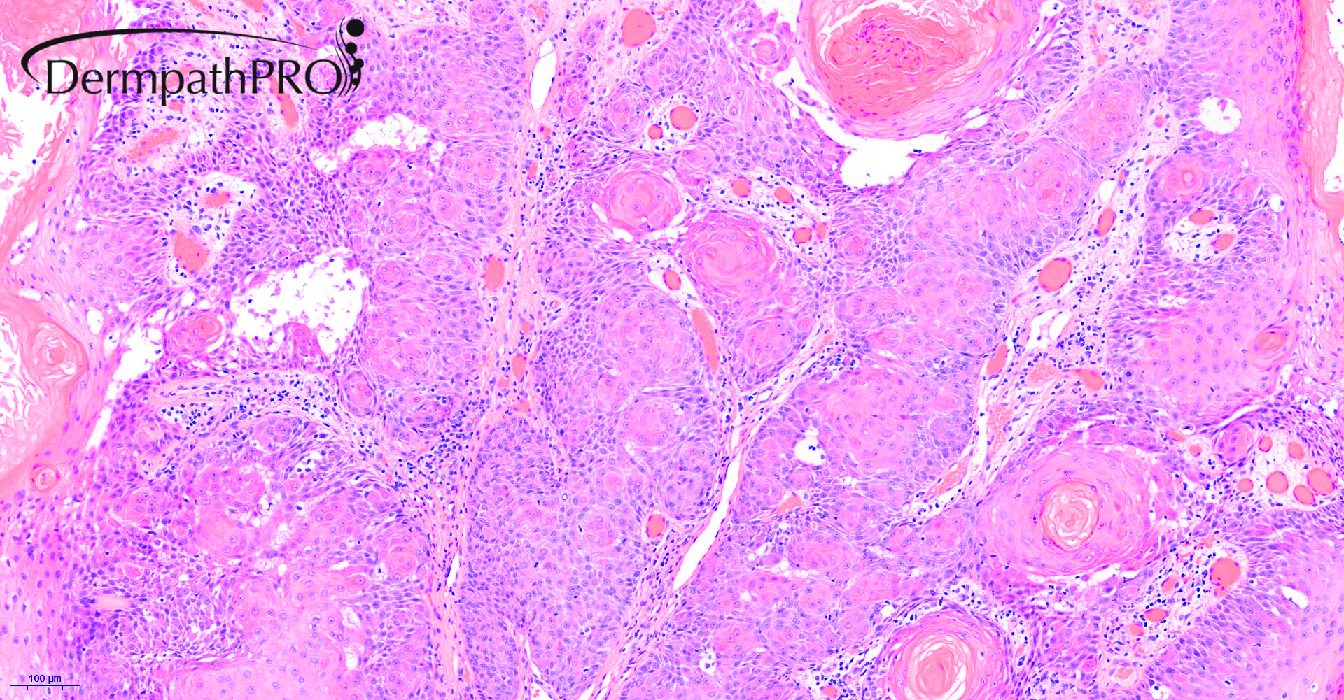
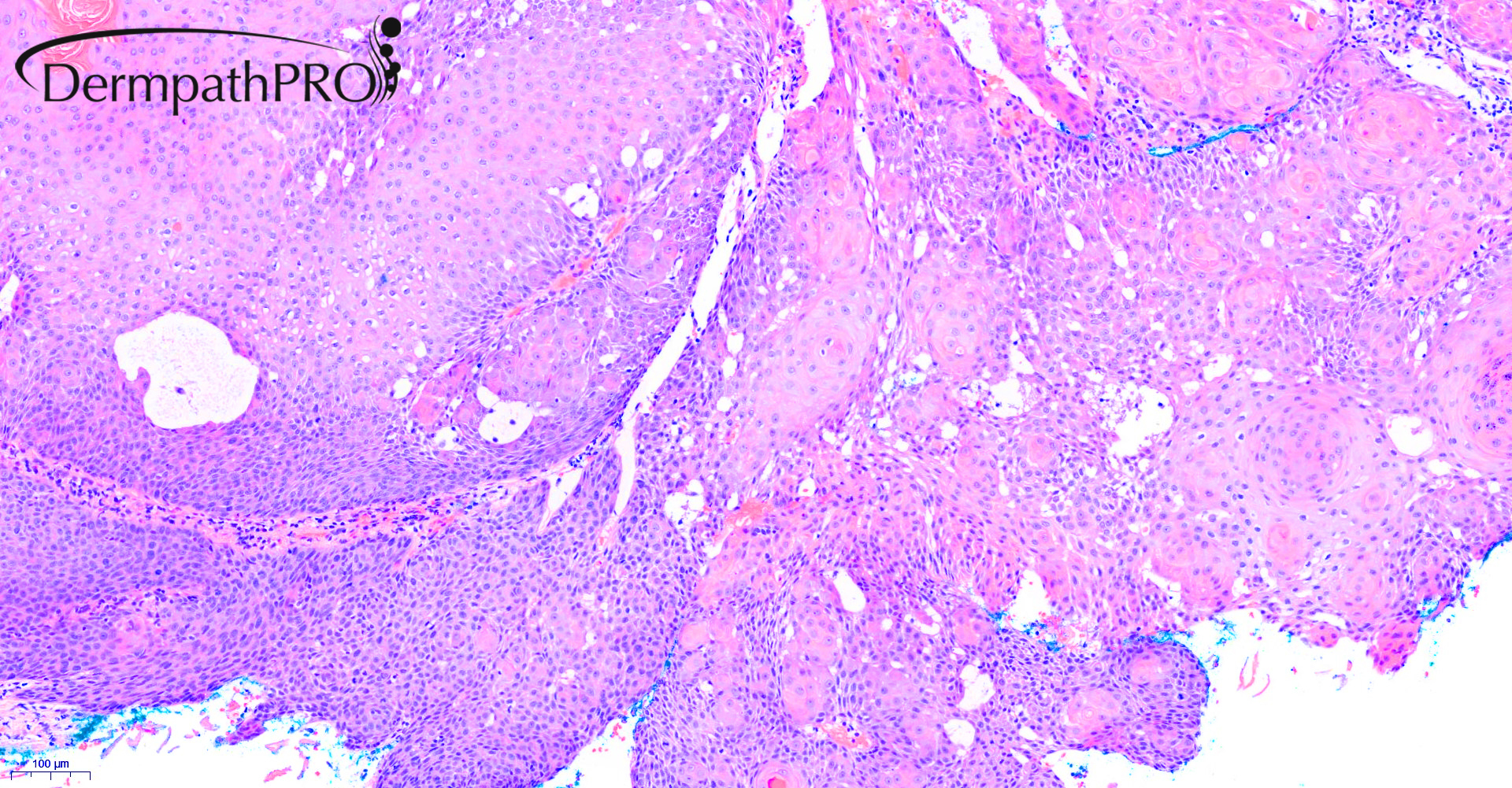
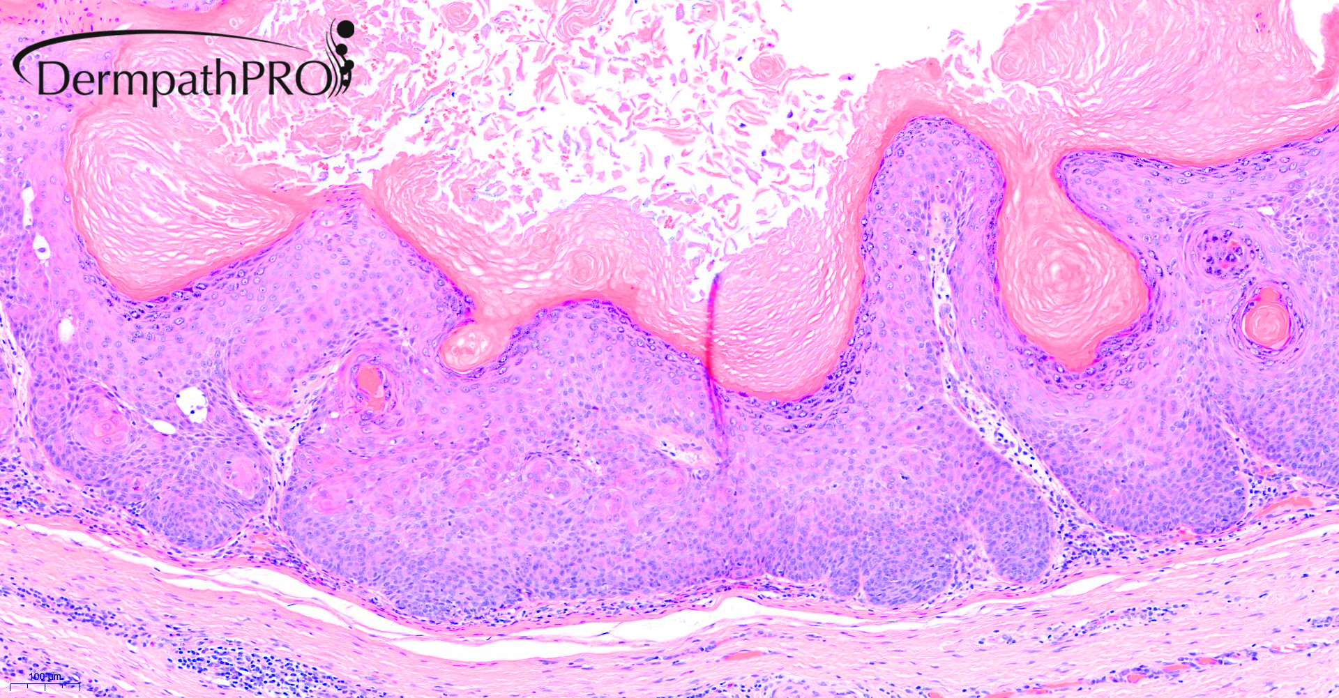
20M, Left pre-auricular region, 3cm subcutaneous lump with discharge ?epidermoid cyst


  • Like 1
  Report Record

User Feedback


The lining of the cystic lesion has a granular layer so I think it's not outer root sheath differentiation (proliferating trichilemmal cysts usual display outer roots sheath differentiation according to the newest WHO blue book , meaning abrupt keratinization without granular layer). Some might just call the lesion a well-differentiated keratinizing SCC and be done with it. I think if someone wants to call this proliferating variant of epidermal (infundibular) cyst (so-called proliferating epithelial cyst) because of the presence of the granular layer, I would agree too. 

Share this comment


Link to comment
share_externally

AmerSM

Posted (edited)

Inverted follicular keratosis


Edited by AmerSM

Share this comment


Link to comment
share_externally

Dr. Mona Abdel-Halim

Posted

I also consider this inverted follicular keratosis.

Share this comment


Link to comment
share_externally

Krishnakumar subramanian

Posted

agree Inverted follicular keratosis of helwig

Share this comment


Link to comment
share_externally

John Zhang

Posted

Interesting case. While features such as squamous eddies are compatible with IFK, the overall crateriform architecture makes me wonder for keratoacanthoma. A keratoacanthoma in a young patient raises the consideration for genetic syndromes such as Muir-Torre.

Share this comment


Link to comment
share_externally

Saleem Taibjee

Posted

Thanks for the interesting comments. I concluded that this is an example of proliferating epidermoid cyst. McKee's Pathology of the Skin states that this is rare, more common in males, anatomical sites of predilection including pelvic area, scalp and trunk. In some instances it is longstanding suggesting that it may arise within a pre-existing epidermoid cyst. Recurrence rate is 20%, but there are no reports of metastasis.

I could not ignore the cystic architecture in this case. Regarding histology, the cyst wall shows a granular layer with epidermal/infundibular keratinisation. Interestingly, the proliferative component may vary in this entity, but notably may show conspicuous squamous eddies (as in this case) reminiscent of inverted follicular keratosis. In other examples, the proliferative component may be multicystic, keratotic or verrucous.

I have alerted the clinician of the risk of local recurrence in this case.

Share this comment


Link to comment
share_externally

Dr. Richard Carr

Posted

I'm a bit late to the party but I'd go for an unusual cystic IFK with infundibular and tricholemmal differentiation. Nice mucin giving rise to pseudoacantholysis. I think I've seen a similar one in the recent past - not a common manifestation. 

Share this comment


Link to comment
share_externally


Join the conversation

You can post now and register later. If you have an account, sign in now to post with your account.

Guest
Add a comment...

×   Pasted as rich text.   Paste as plain text instead

  Only 75 emoji are allowed.

×   Your link has been automatically embedded.   Display as a link instead

×   Your previous content has been restored.   Clear editor

×   You cannot paste images directly. Upload or insert images from URL.

Loading...

×
×
  • Create New...