Jump to content
In this section we have spot diagnoses posted on a daily basis since June 2010, now over 4000! You can review the archived cases and read the suggested diagnoses by users and the final comment by the contributors.
Case are uploaded each week day by 10 am UK time with the correct diagnosis will generally be posted at 8 pm UK time. Why not view the most recent spot diagnosis and proffer a diagnosis?

Case Number : Case 2632 - 07 August 2020 Posted By: Dr. Richard Carr

Please read the clinical history and view the images by clicking on them before you proffer your diagnosis.
Submitted Date :
   (0 reviews)

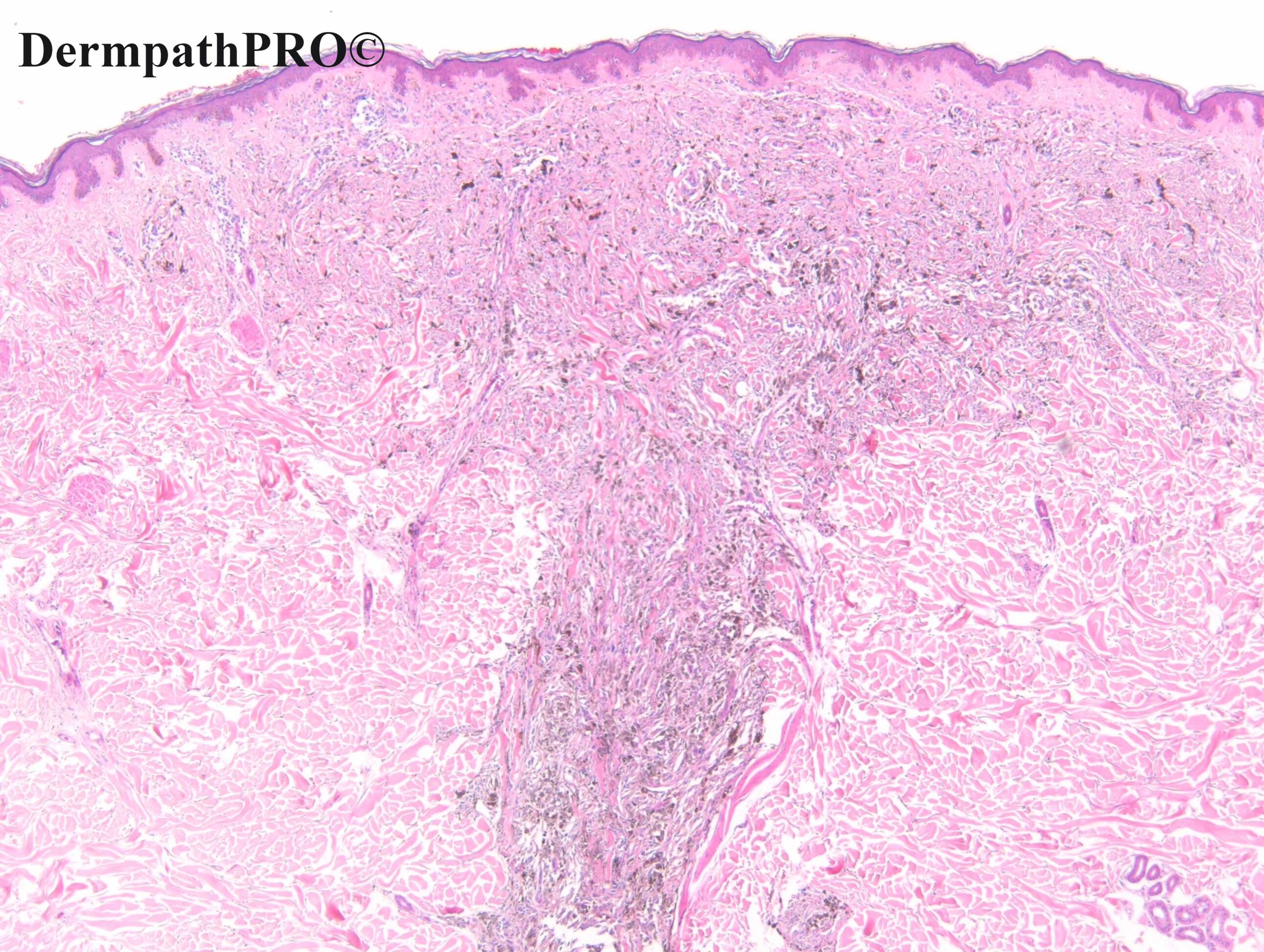
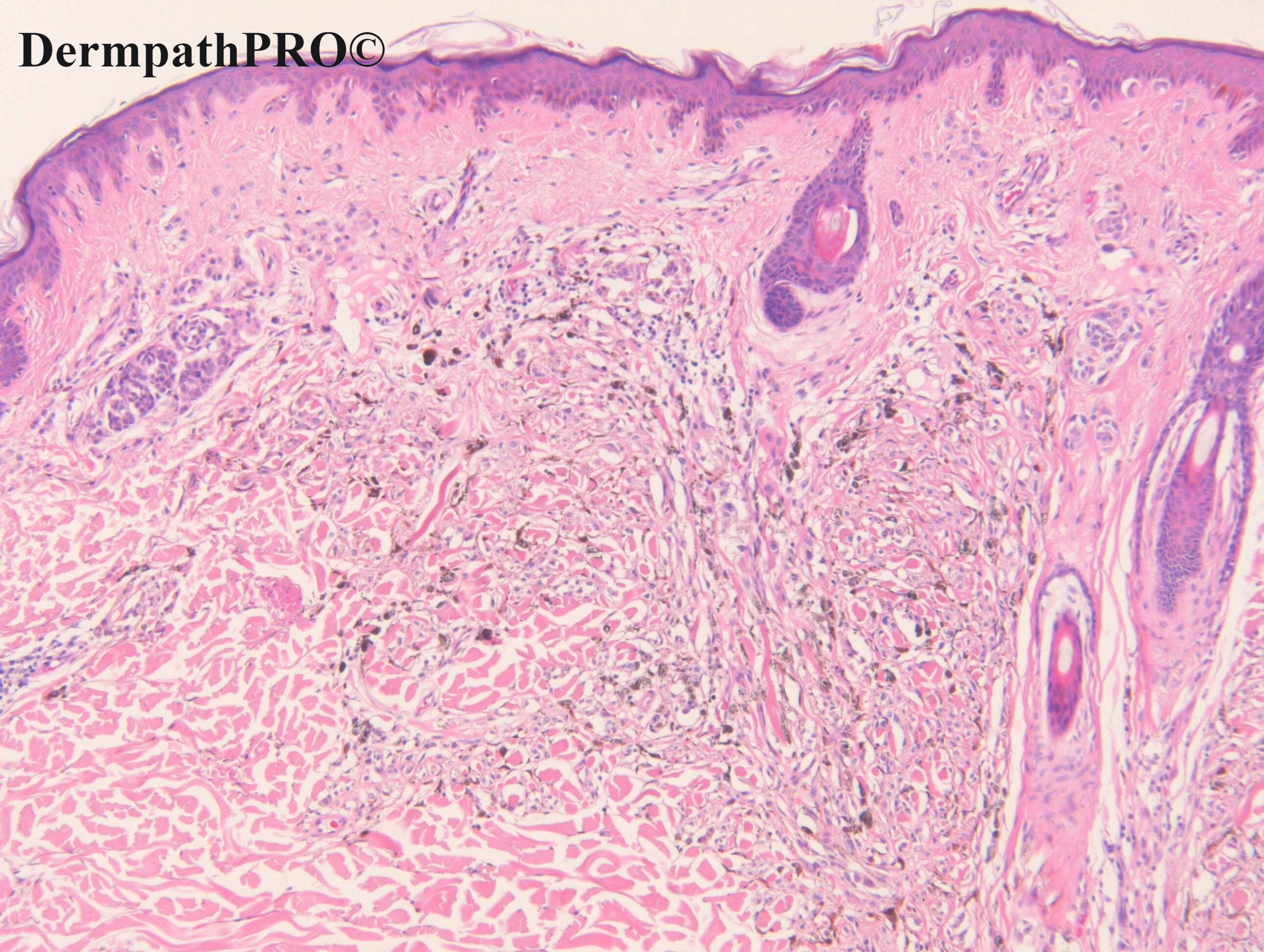
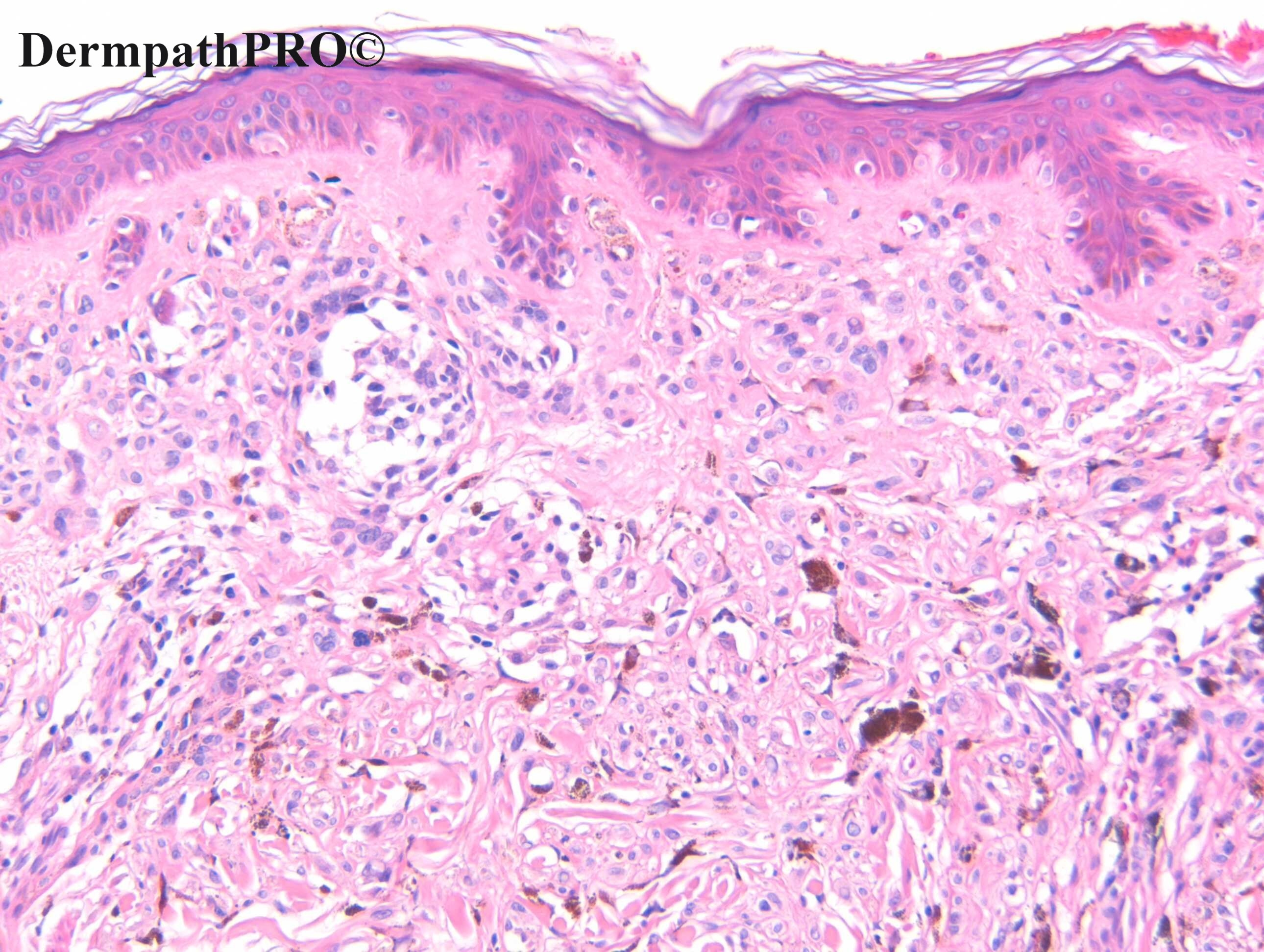
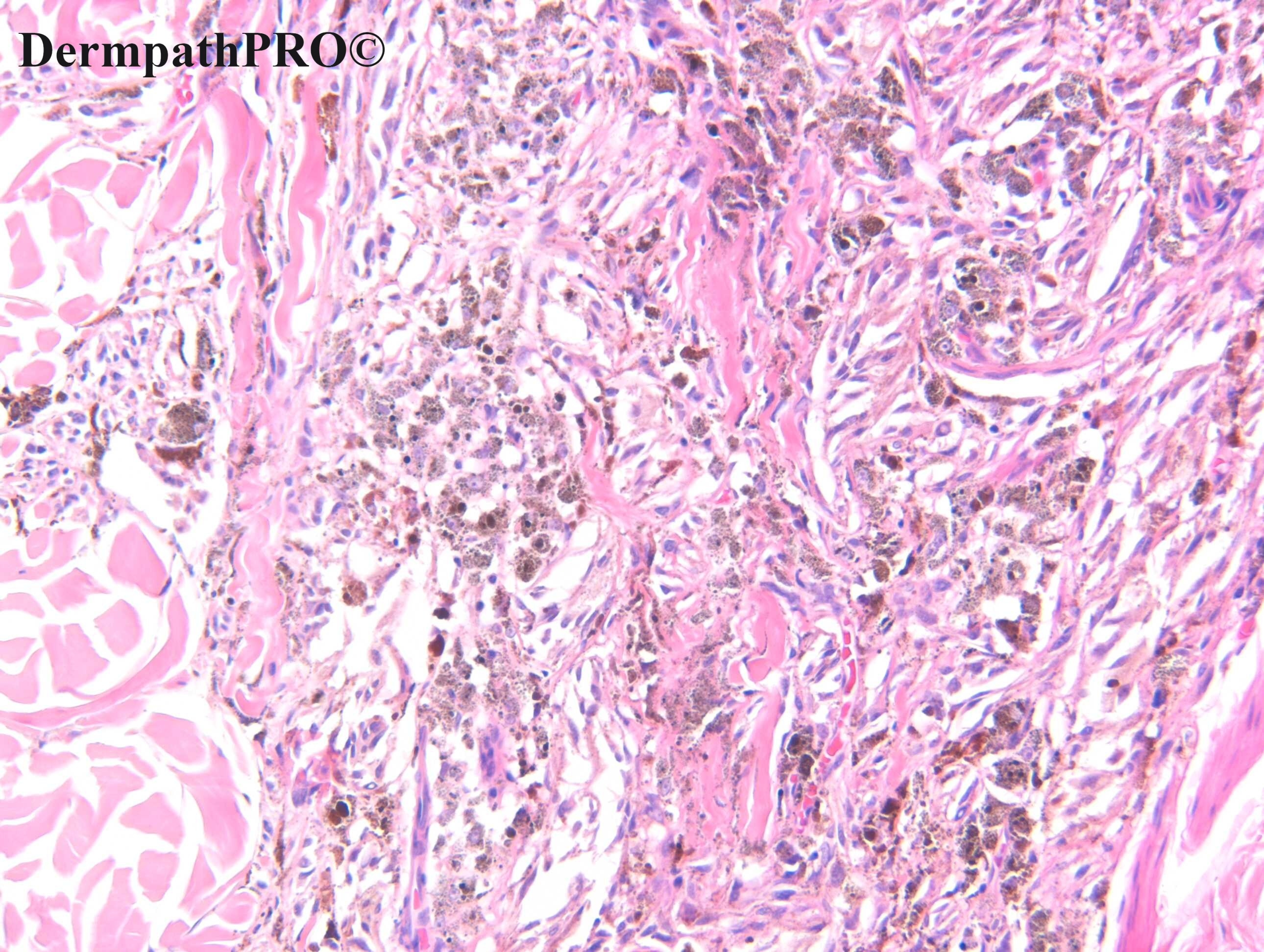
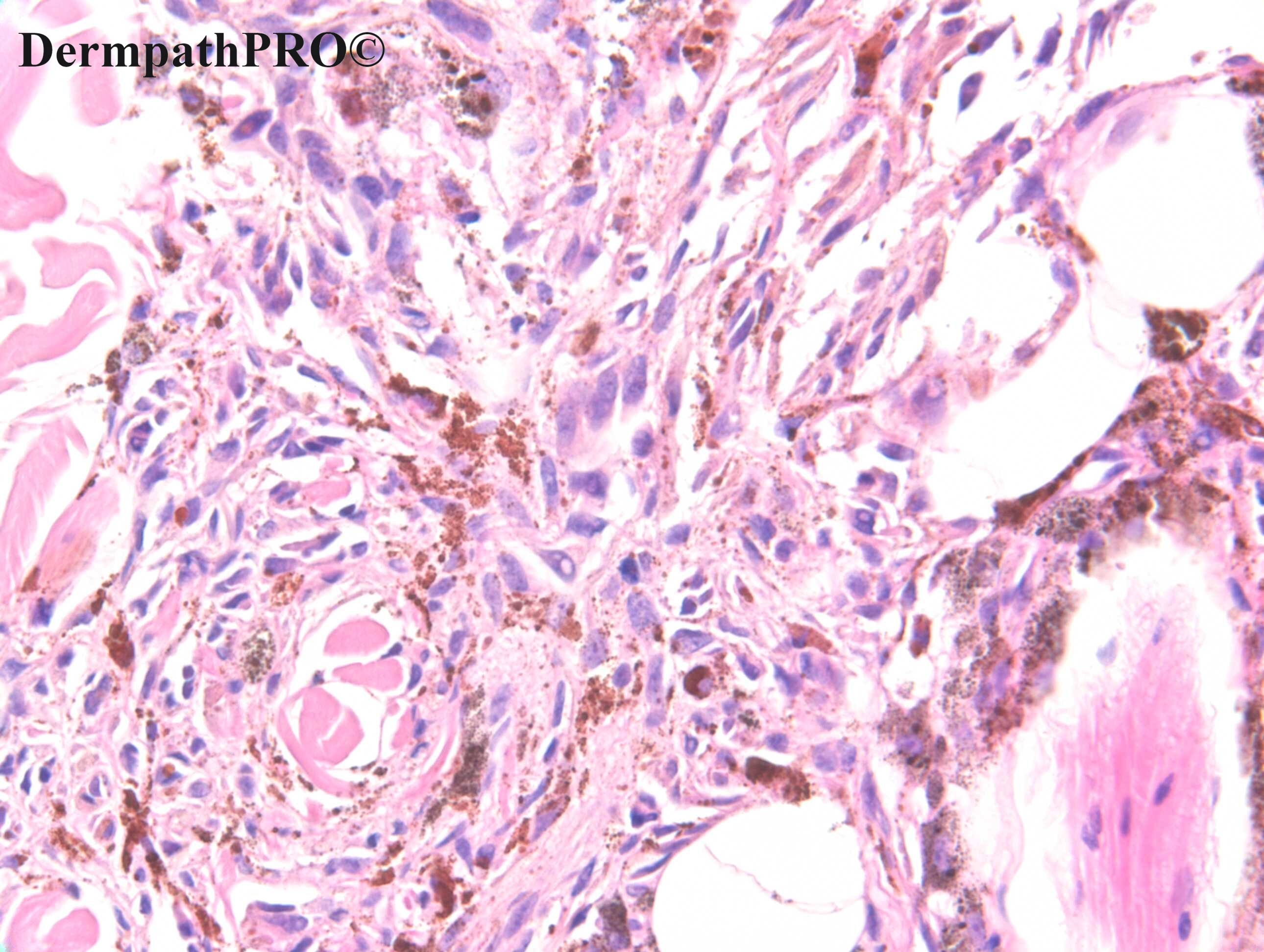
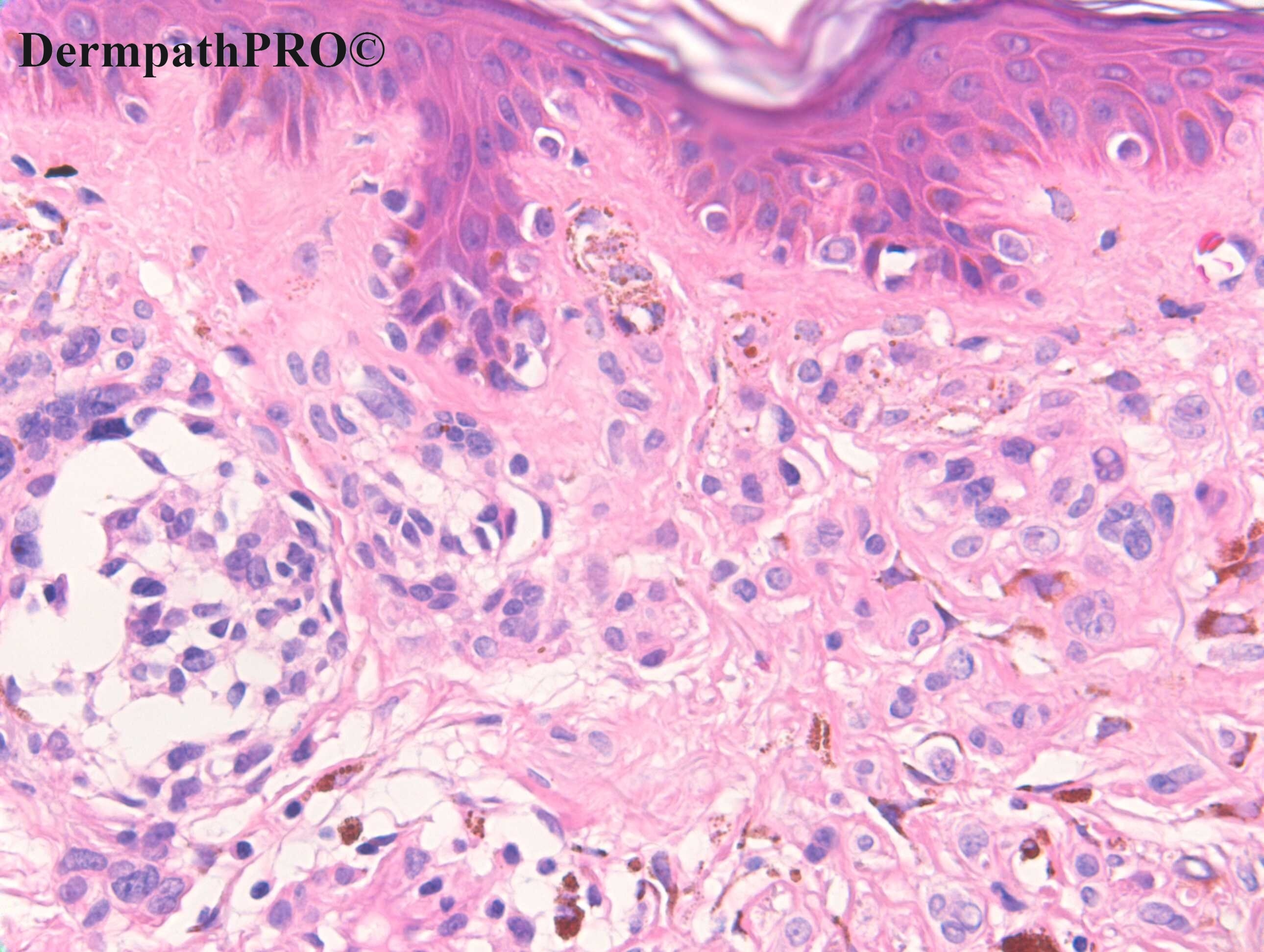
F-70. Oval black papule on back. (removed at the same time as a melanoma in situ from the arm).

Edited by Admin_Dermpath


  Report Record

User Feedback


Agree with DPN. Maybe there is a small component of conventional intradermal nevus(last fig),but not sure...

Share this comment


Link to comment
share_externally

Dr MONA MOHSEN

Posted

I dont feel the symmetry of cell nests distribution that is usually seen in nevi.. Plus atypical melanocytes with hyperchromatism, multinucleatiin and increase of N/C  seen in superfacial levels and in epidermis.. Suggesting melanoma

Share this comment


Link to comment
share_externally

Krishnakumar subramanian

Posted

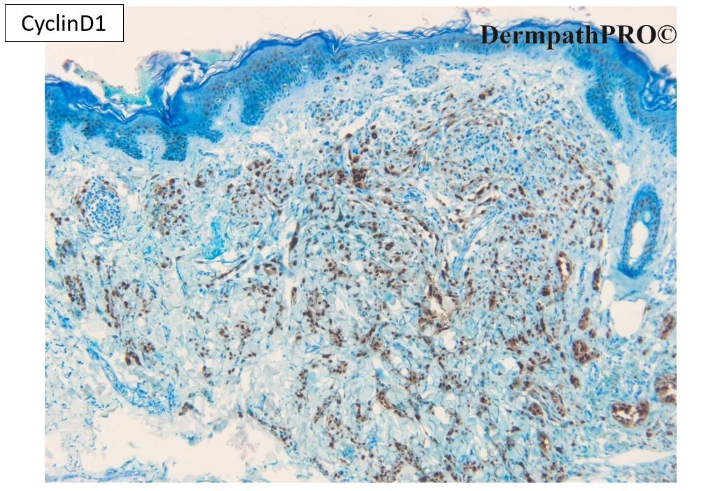
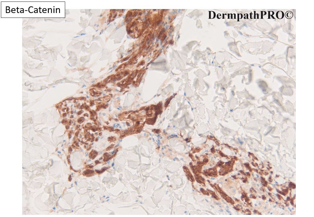
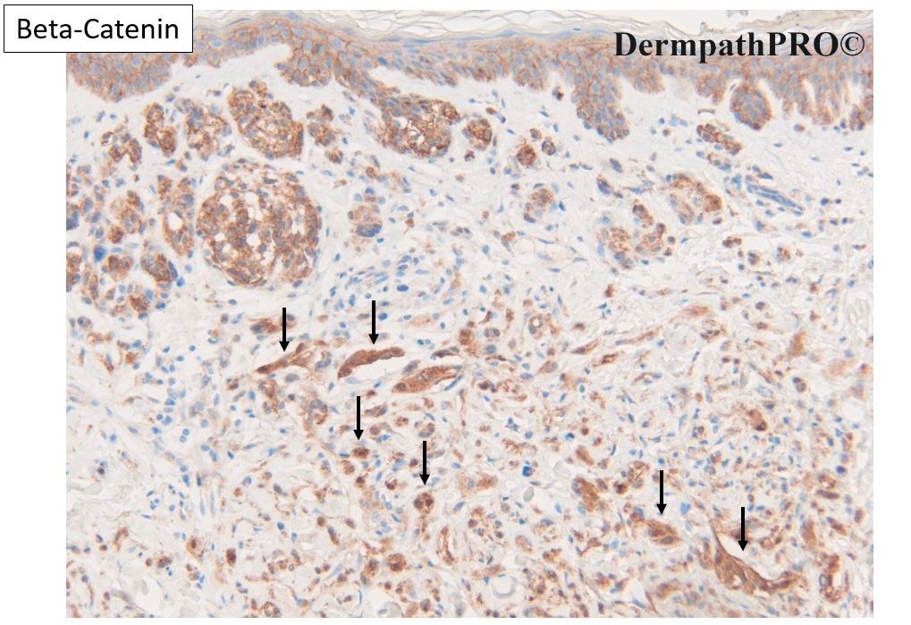
with cyclin D1 and beta catenin positivity -could it be a melanoma

Share this comment


Link to comment
share_externally

Krishnakumar subramanian

Posted

Dear sir, morphology looks like a nevus but with cyclin d1 and  nuclear beta catenin positive can it go as a DPN 

i saw this commentary , is morphology enough do we need molecular biology or the gene profile expression tests to look for class 1 and class 2 type

kindly suggest

https://www.aad.org/dw/dw-insights-and-inquiries/dermatopathology/deep-penetrating-thoughts-about-deep-penetrating-nevi

DEEP PENETRATING THOUGHTS ABOUT DEEP PENETRATING NEVI

By Warren R. Heymann, MD
Jan. 14, 2019

Share this comment


Link to comment
share_externally

Dr. Richard Carr

Posted

This is a biphenotypic naevocellular (background) naevus that has developed a clone - in this case deep penetrating type (also known as one variant of a combined naevus). I did not see any worrying features for malignancy. Given that very rarely DPN can undergo malignant transformation and the fact the lesion now has a driver mutatation (BRAF usually) and a second hit (Beta-catenin) experts usually recommend complete excision with clear margins (MPathDx class II). They usually have low-grade mitotic activity, low-grade atypia but I'd expect them to keep growing and of course diagnosis can be challenging so it is well to ensure complete removal. The beta-catenin can be quite hard to read but the staining in the DPN clone is nuclear & cytoplasm as opposed to the negative nuclear pattern in the naevocellular naevus. Helpfully the CyclinD1 is also expressed at all levels of the DPN clone whereas in benign naevi it's limited the more active, type A, superficial melanocytes. Remember that naevocellular naevi usually have BRAF or NRAS mutations which are mutually exclusive with blue naevi (usually G-protein drivers). So you don't really get combined naevocellular-true blue naevi. It's a common misconception and I still occasionally see speakers refer to combined naevocellular-blue naevi (this may occur as a very rare "collision" but usually the pigmented clone in a pre-existing naevocellular (i.e. common nested naevus) is either DPN or more rarely a PEM clone or unspecified (but not a G-protein). No doubt there are other types of clone to be discovered. The BAP1 loss clones are composed of hypopigmented large epithelioid cells with admixed lymphocytes again it's important to look for the background naevocellular naevus. Again some consider true "Spitz" naevi are mutally exclusive with BRAF/NRAS naevocellular naevi so an Spitz-like clone in a pre-existing naevocellular naevus would not have an HRAS for example but may have a fusion protein I believe. So in melanocytic pathology it's important to look for the background naevus whether it's naevocellular or blue (when you're thinking about atypical & malignant blues) or pure Spitz morphology.   

I'd like to thank Dr Mickhaiel Barrow for sharing the case with me.

Share this comment


Link to comment
share_externally


Join the conversation

You can post now and register later. If you have an account, sign in now to post with your account.

Guest
Add a comment...

×   Pasted as rich text.   Paste as plain text instead

  Only 75 emoji are allowed.

×   Your link has been automatically embedded.   Display as a link instead

×   Your previous content has been restored.   Clear editor

×   You cannot paste images directly. Upload or insert images from URL.

Loading...

×
×
  • Create New...