Jump to content
In this section we have spot diagnoses posted on a daily basis since June 2010, now over 4000! You can review the archived cases and read the suggested diagnoses by users and the final comment by the contributors.
Case are uploaded each week day by 10 am UK time with the correct diagnosis will generally be posted at 8 pm UK time. Why not view the most recent spot diagnosis and proffer a diagnosis?

Case Number : Case 2637 - 14 August 2020 Posted By: Dr. Richard Carr

Please read the clinical history and view the images by clicking on them before you proffer your diagnosis.
Submitted Date :
   (0 reviews)

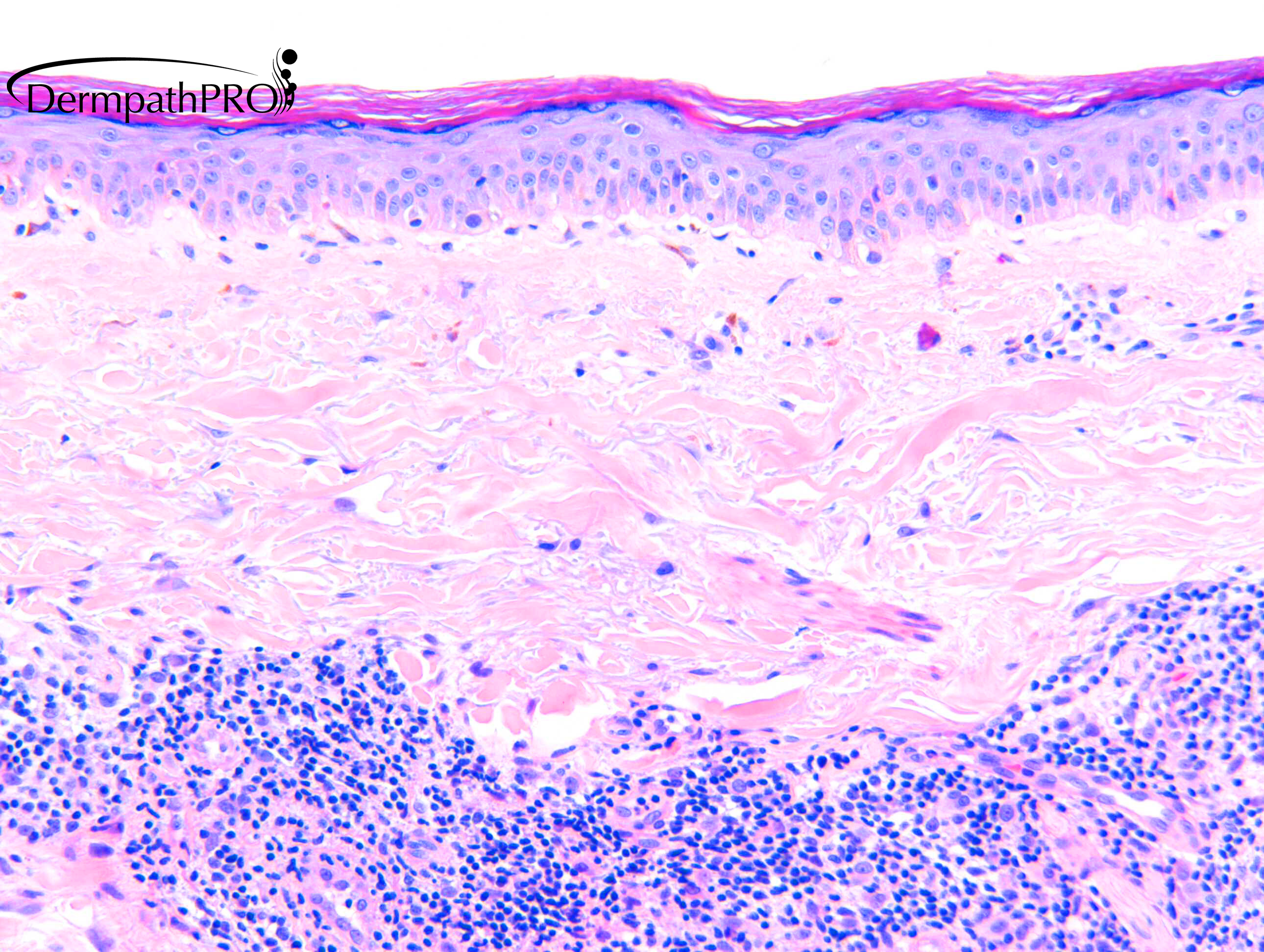
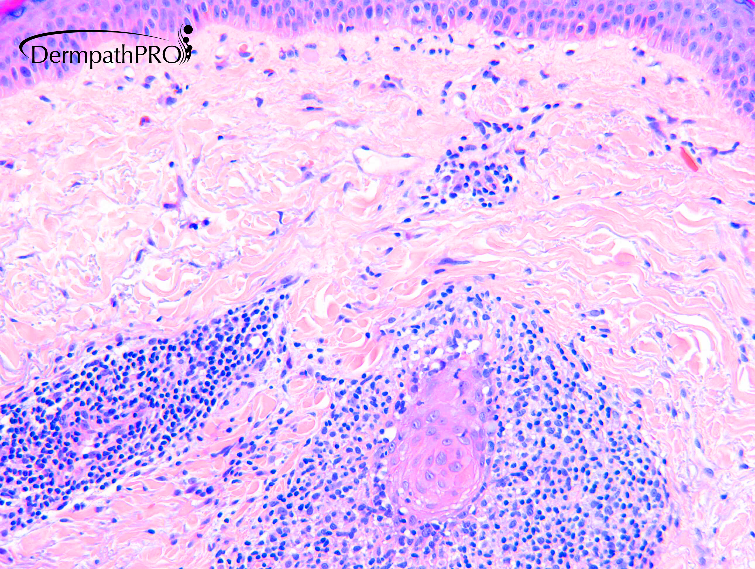
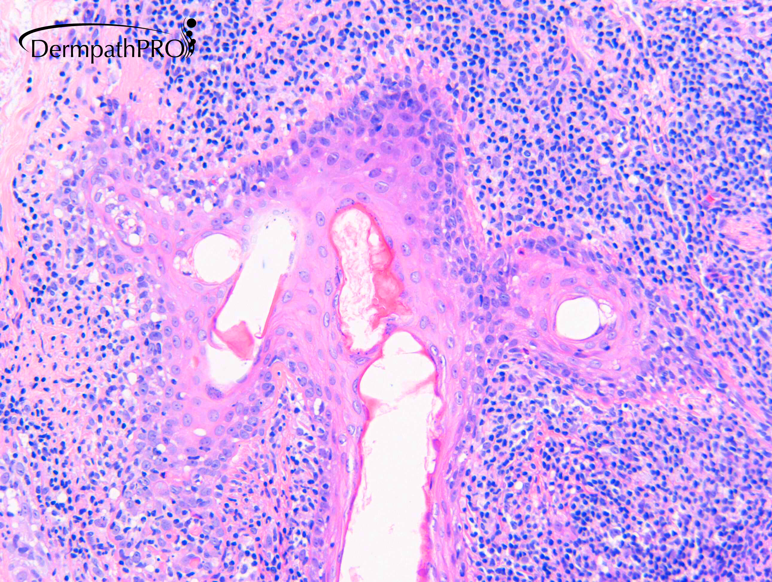
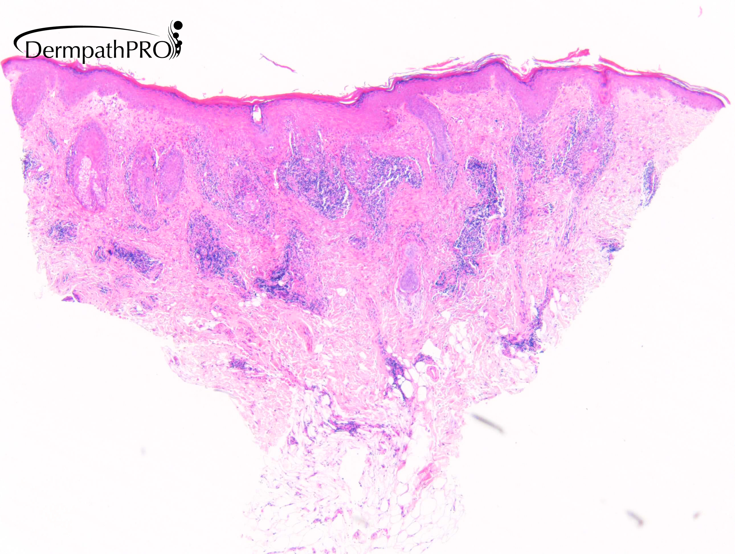
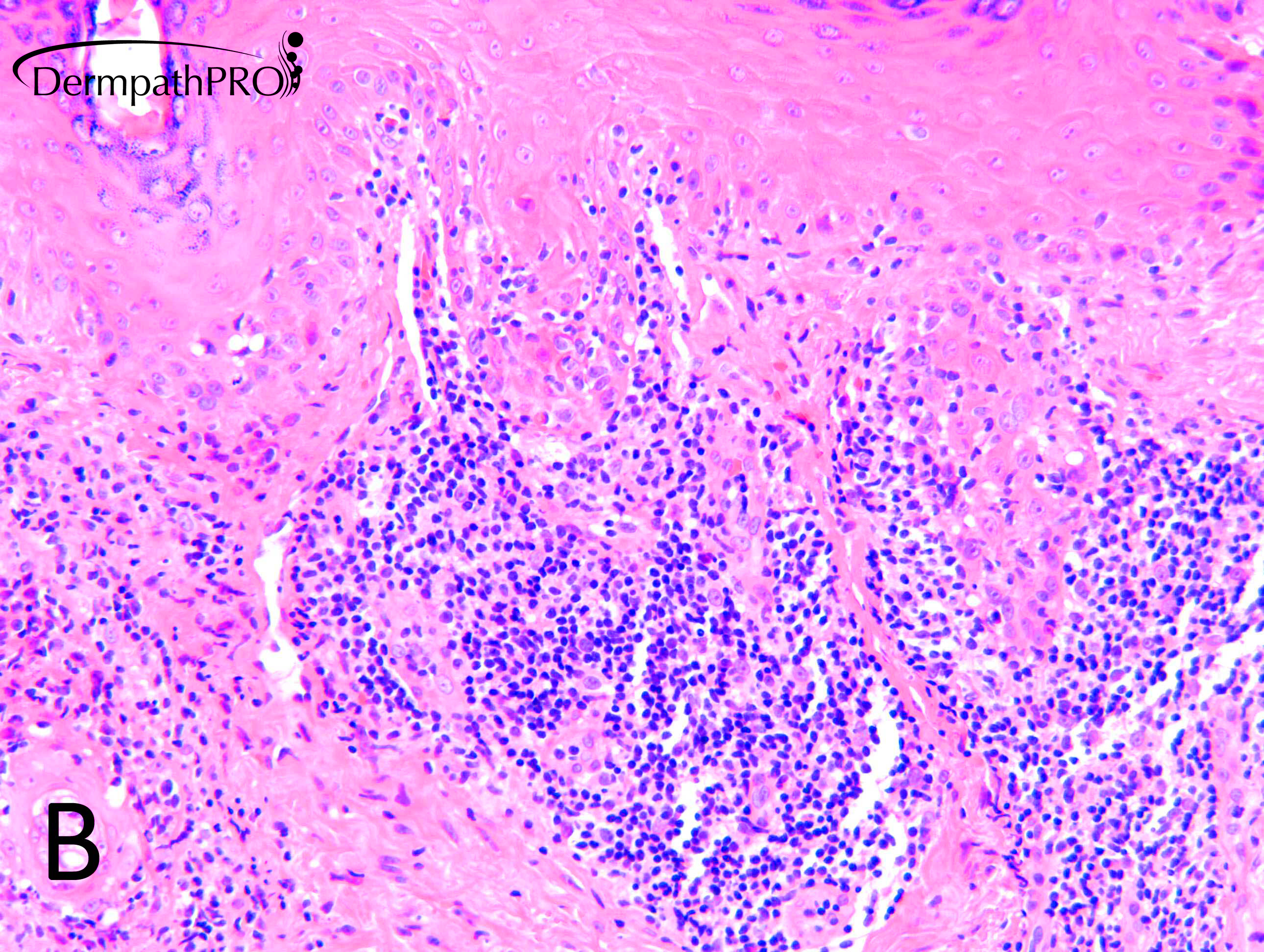
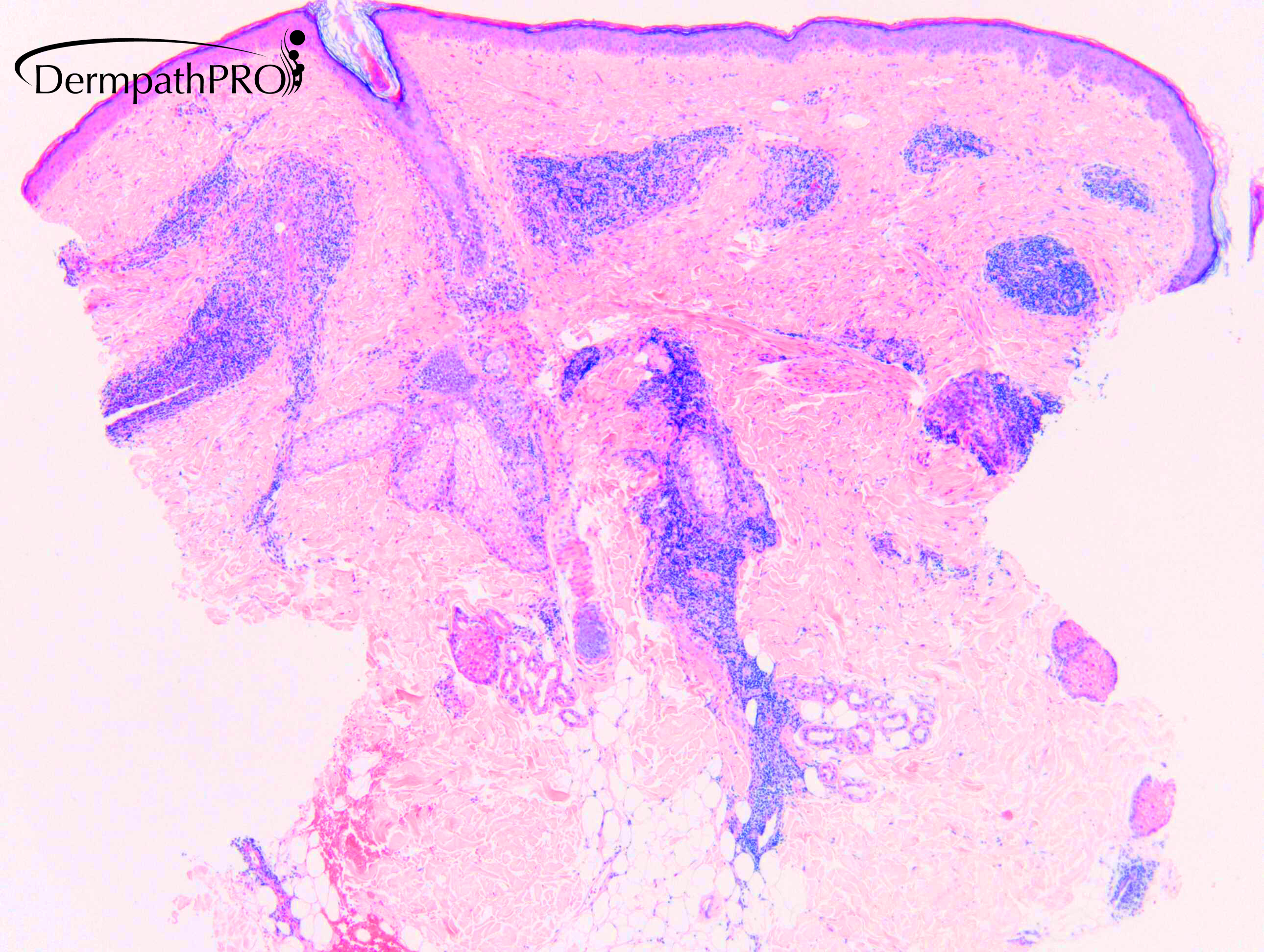
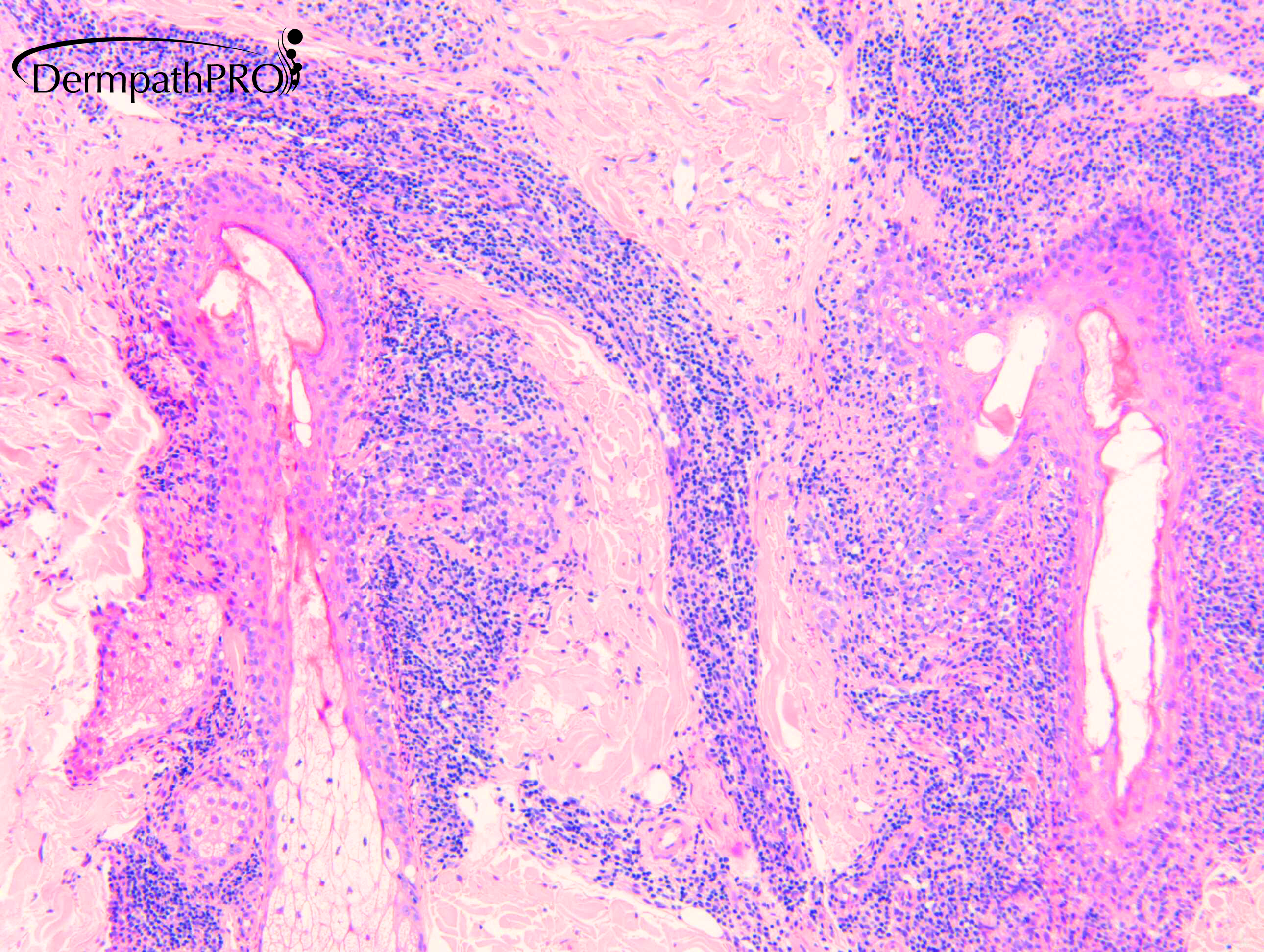
F50. A. 12 x 10 mm pink patch, fine telangiectasia ?Sup. BCC; B. 11 x 6mm indented telangiectactic papules ?BCC

  Report Record

User Feedback


Krishnakumar subramanian

Posted

perivascular and periadnexal lymphocytes, ? mucin-dermis

LE

Share this comment


Link to comment
share_externally

1 hour ago, Krishnakumar subramanian said:

perivascular and periadnexal lymphocytes, ? mucin-dermis

LE

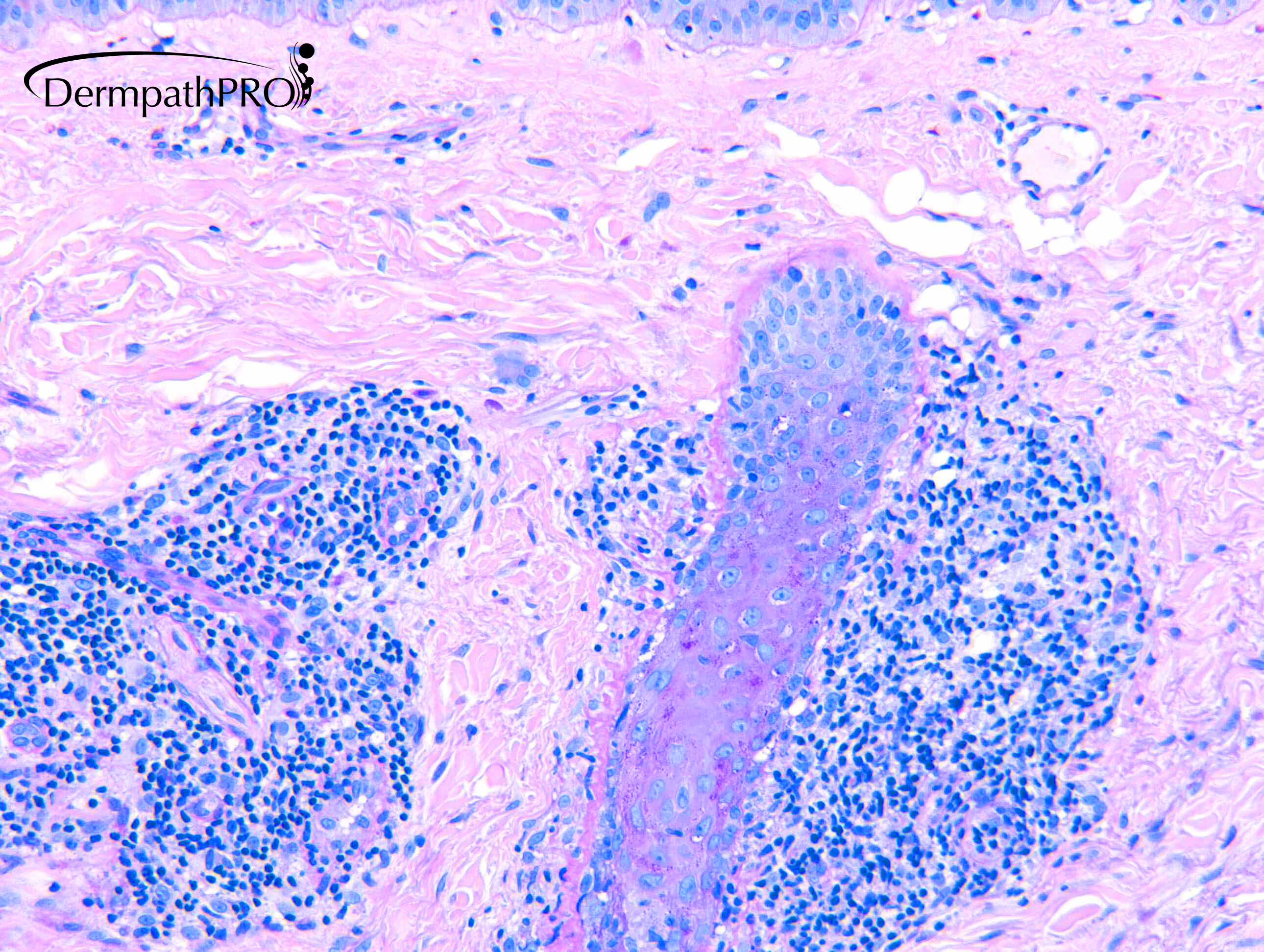
Agree, and there is also a periadnexal thickening of the basement membrane in fig 5.

Share this comment


Link to comment
share_externally

Eman El-Nabarawy

Posted

The B biopsy shows lichen planus like features. Thought of LE/LP overlap syndrome

Share this comment


Link to comment
share_externally

Dr. Richard Carr

Posted

Yes I reported this as suggesting discoid lupus erythematosus. We can see rather subtle basement membrane thickening on the PAS exclusive to the follicles. In this case mucin is inconspicuous and it's usually my experience that I "spot" the features of DLE at low power (fairly dense perivascular & periadnexal infiltrates with atrophic lichenoid pattern to the epidermis) and then notice mucin as a secondary phenomenon that rarely makes the diagnosis  On the other hand in cases like this where the disease is predominantly affecting the follicles and mimicking licheno planopilaris I do look closely for basement membrane thickening and will request a PAS if it's equivocal on H&E as in this instance.

Share this comment


Link to comment
share_externally

Krishnakumar subramanian

Posted

Thanks a lot sir for key points

Share this comment


Link to comment
share_externally

Dr. Mona Abdel-Halim

Posted

On 17/08/2020 at 10:53, Dr. Richard Carr said:

Yes I reported this as suggesting discoid lupus erythematosus. We can see rather subtle basement membrane thickening on the PAS exclusive to the follicles. In this case mucin is inconspicuous and it's usually my experience that I "spot" the features of DLE at low power (fairly dense perivascular & periadnexal infiltrates with atrophic lichenoid pattern to the epidermis) and then notice mucin as a secondary phenomenon that rarely makes the diagnosis  On the other hand in cases like this where the disease is predominantly affecting the follicles and mimicking licheno planopilaris I do look closely for basement membrane thickening and will request a PAS if it's equivocal on H&E as in this instance.

Beautiful! I follow the same approach!

Share this comment


Link to comment
share_externally


Join the conversation

You can post now and register later. If you have an account, sign in now to post with your account.

Guest
Add a comment...

×   Pasted as rich text.   Paste as plain text instead

  Only 75 emoji are allowed.

×   Your link has been automatically embedded.   Display as a link instead

×   Your previous content has been restored.   Clear editor

×   You cannot paste images directly. Upload or insert images from URL.

Loading...

×
×
  • Create New...