Jump to content
In this section we have spot diagnoses posted on a daily basis since June 2010, now over 4000! You can review the archived cases and read the suggested diagnoses by users and the final comment by the contributors.
Case are uploaded each week day by 10 am UK time with the correct diagnosis will generally be posted at 8 pm UK time. Why not view the most recent spot diagnosis and proffer a diagnosis?

Case Number : Case 2639 - 18 August 2020 Posted By: Uma Sundram

Please read the clinical history and view the images by clicking on them before you proffer your diagnosis.
Submitted Date :
   (0 reviews)

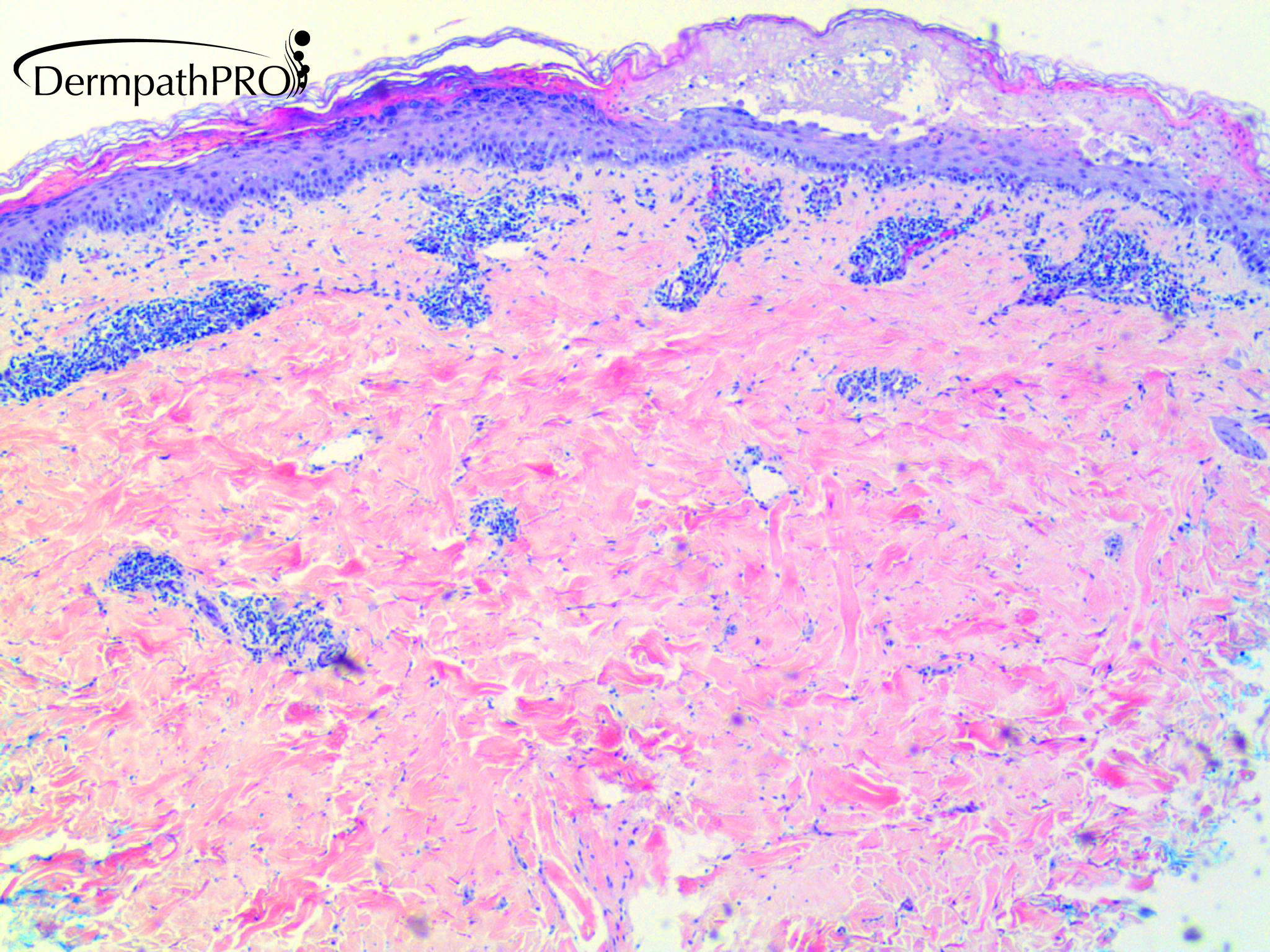
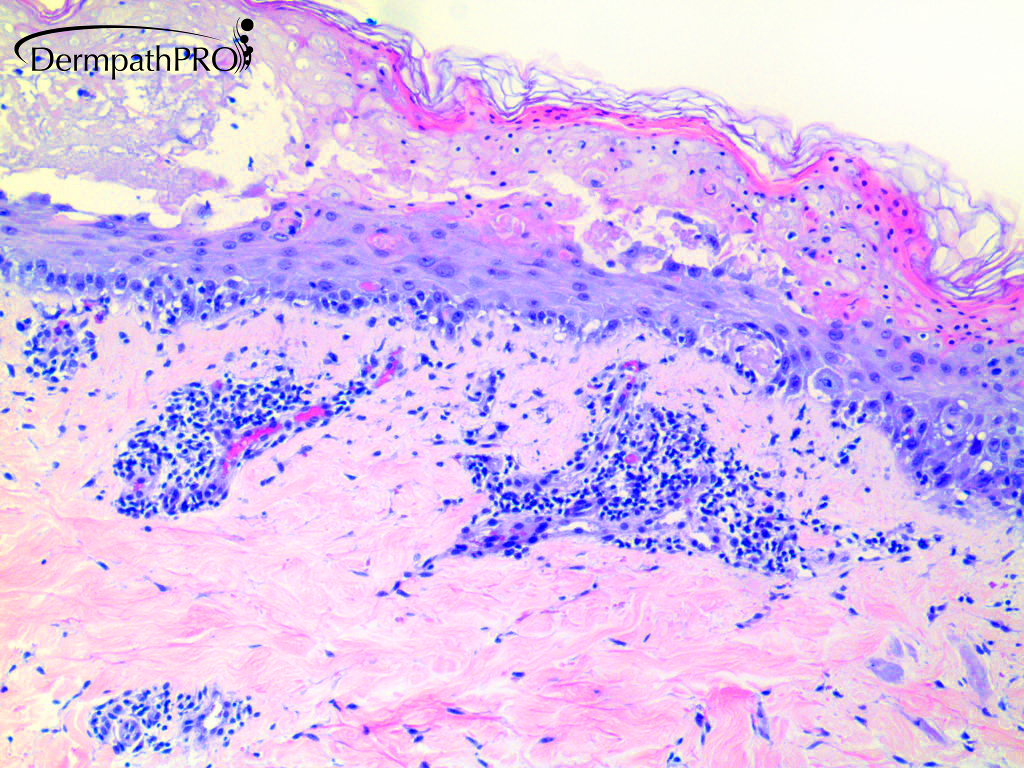
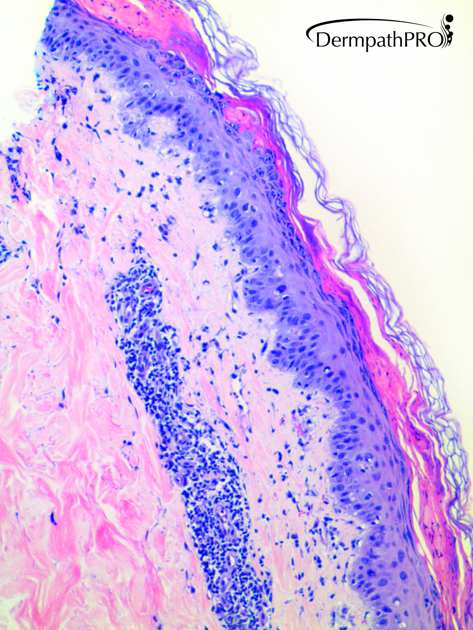
28 year old male with erythematous lesion on left arm.


  Report Record

User Feedback


Krishnakumar subramanian

Posted

epidermal necrosis in superficial portions with pallor and dermal perivascular lymphocytes

could be necrolytic erythema

Share this comment


Link to comment
share_externally

49 minutes ago, Krishnakumar subramanian said:

epidermal necrosis in superficial portions with pallor and dermal perivascular lymphocytes

could be necrolytic erythema

Necrolytic erythema could be a good differential.

This is an erythema multiforme-like single lesion (single area if I’m not mistaken ). ?FDE( but I don’t see any eosinophils nor melanophages). 

Difficult...


Share this comment


Link to comment
share_externally

Richard Logan

Posted

I see a fairly tight perivascular lymphocytic infiltrate in the upper and mid-dermis.  At one end of the biopsy (Fig 3) there is parakeratosis with a disturbance of the cytology of the lower epidermis similar to a solar keratosis, but probably secondary to the inflammation.  In the centre there is vaculoar change with some colloid bodies and necrosis of the upper epidermis.  I don't see any dyskeratotic cells.


Although the overall appearance is suggestive of necrolytic migratory erythema, this would be an unlikely diagnosis at this age.  More plausible would be erythema multiforme.  I would also consider the possibility of nutritional deficiency possibly complicating liver disease or HIV.

Share this comment


Link to comment
share_externally

Meenakshi Batrani

Posted

Apart from EM, FDE and nutritional,  I would add D/D of Pityriasis lichenoides in view of parakeratotic mounds underneath basket weave stratum corneum, interface pathology and EM like necrosis. 

Share this comment


Link to comment
share_externally

Dr. Mona Abdel-Halim

Posted

The clinical points to a solitary lesion mostly, so early FDE is my first thought.. it needs CPC

Share this comment


Link to comment
share_externally

Uma Sundram

Posted

A solitary lesion. Fixed drug eruption is a good thought; this is what we suggested and fit with the clinical. We also thought about irritant contact dermatitis. The patient is otherwise healthy. 

Share this comment


Link to comment
share_externally


Join the conversation

You can post now and register later. If you have an account, sign in now to post with your account.

Guest
Add a comment...

×   Pasted as rich text.   Paste as plain text instead

  Only 75 emoji are allowed.

×   Your link has been automatically embedded.   Display as a link instead

×   Your previous content has been restored.   Clear editor

×   You cannot paste images directly. Upload or insert images from URL.

Loading...

×
×
  • Create New...