Jump to content
In this section we have spot diagnoses posted on a daily basis since June 2010, now over 4000! You can review the archived cases and read the suggested diagnoses by users and the final comment by the contributors.
Case are uploaded each week day by 10 am UK time with the correct diagnosis will generally be posted at 8 pm UK time. Why not view the most recent spot diagnosis and proffer a diagnosis?

Case Number : Case 2646 - 27 August 2020 Posted By: Saleem Taibjee

Please read the clinical history and view the images by clicking on them before you proffer your diagnosis.
Submitted Date :
   (0 reviews)

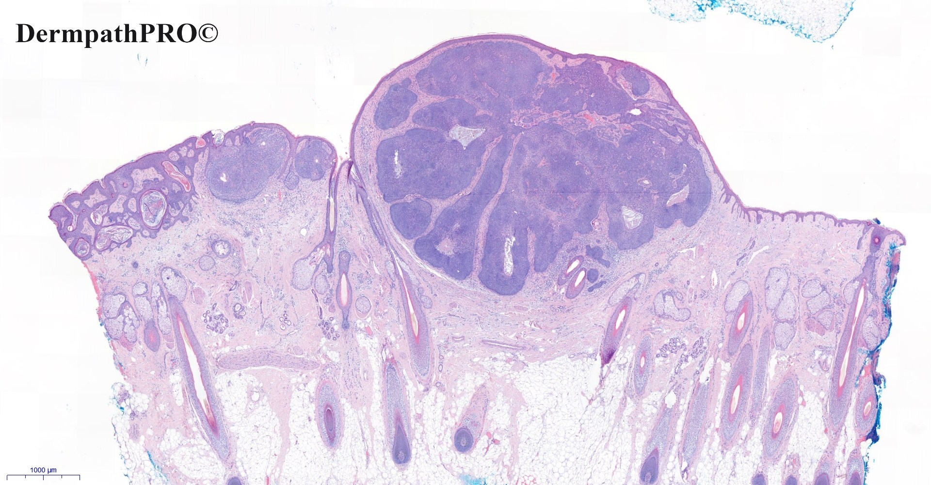
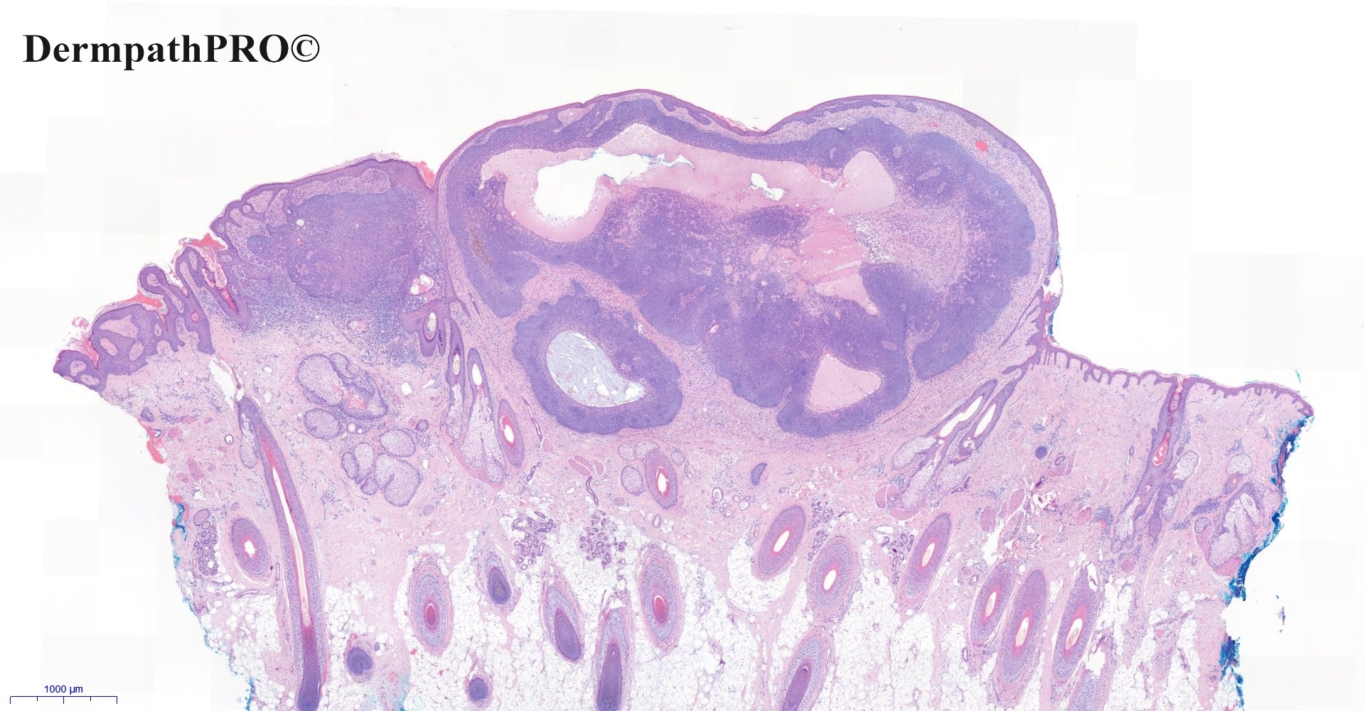
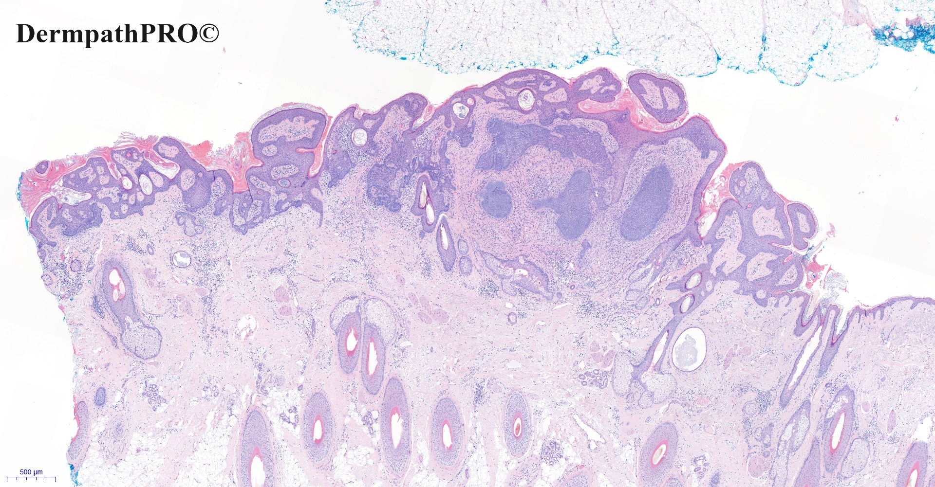
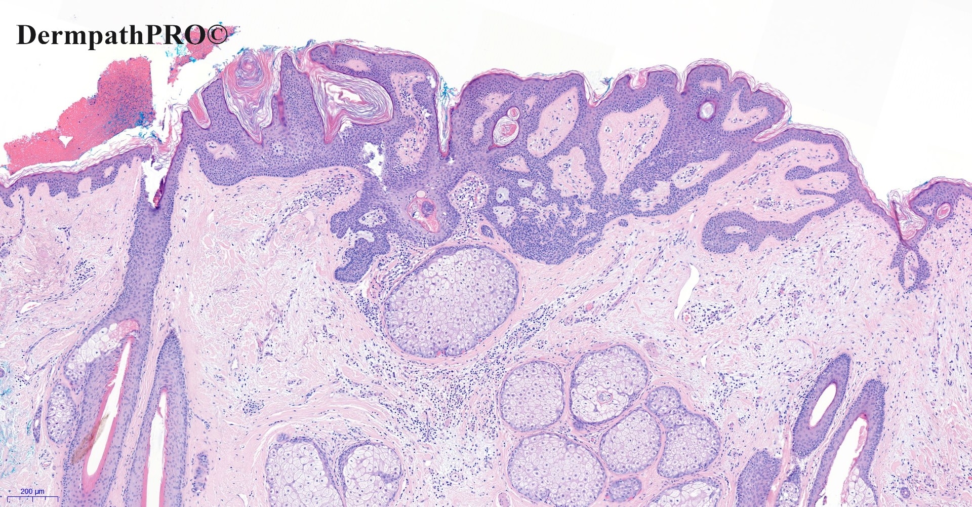
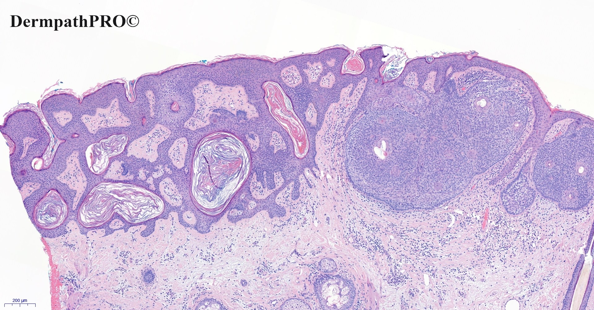
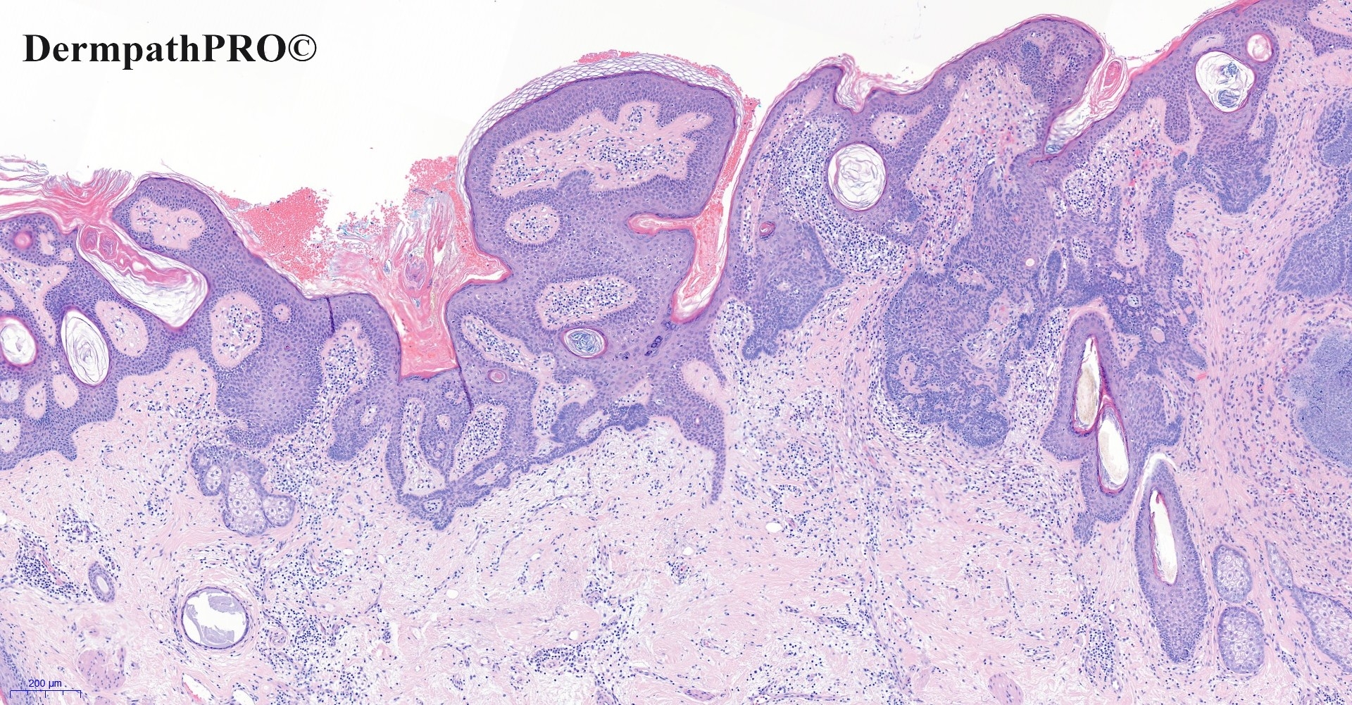
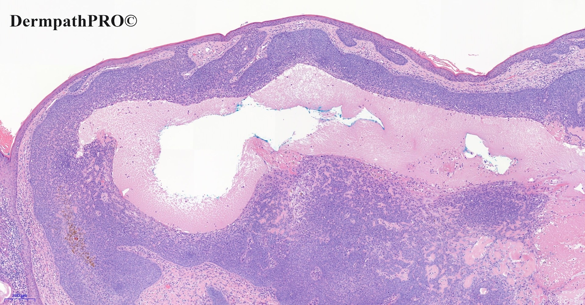
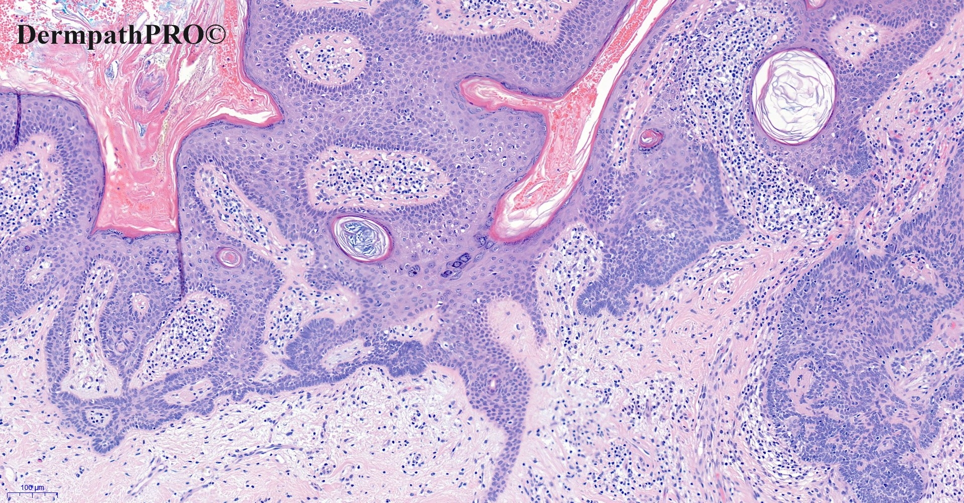
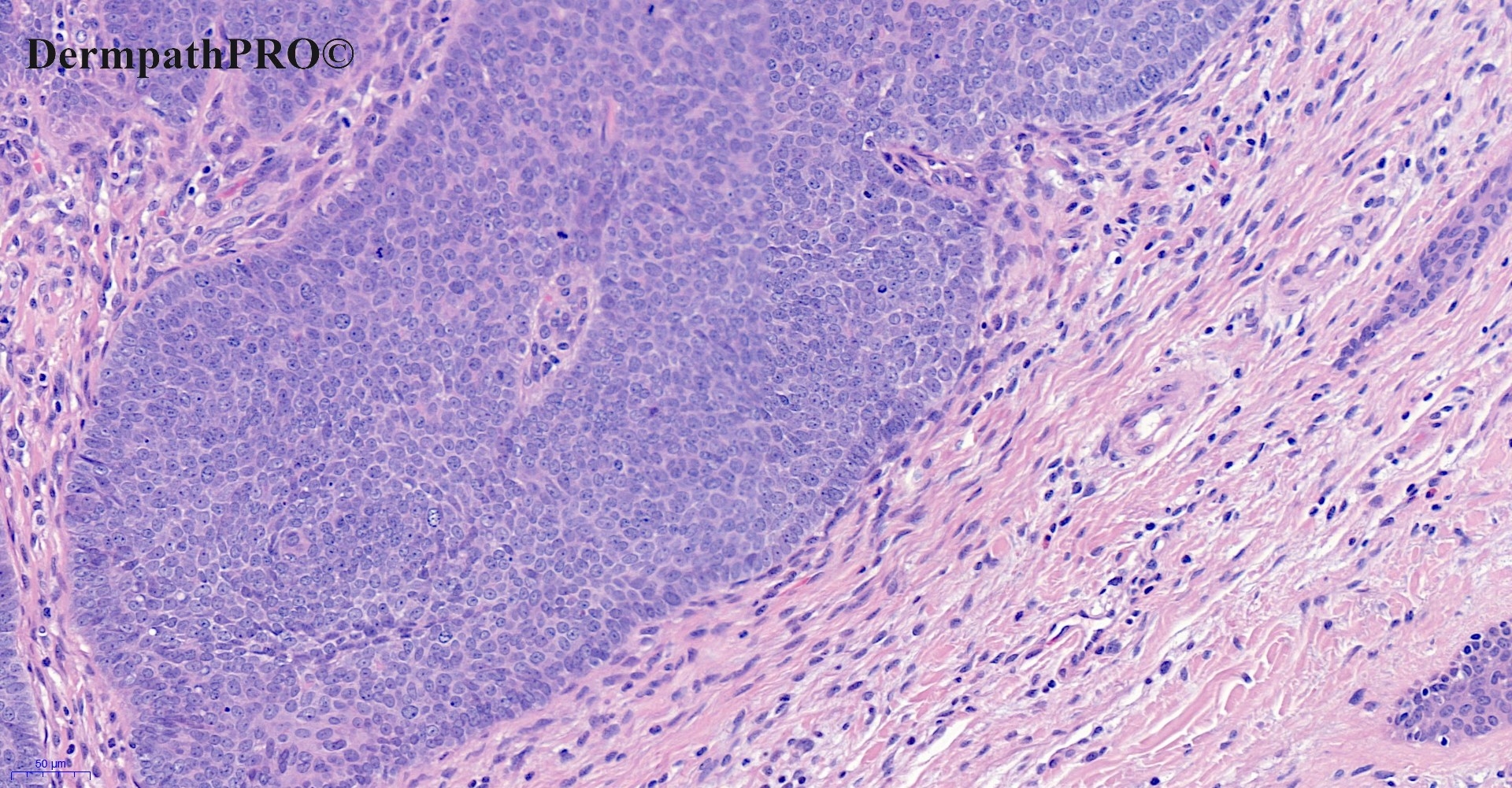
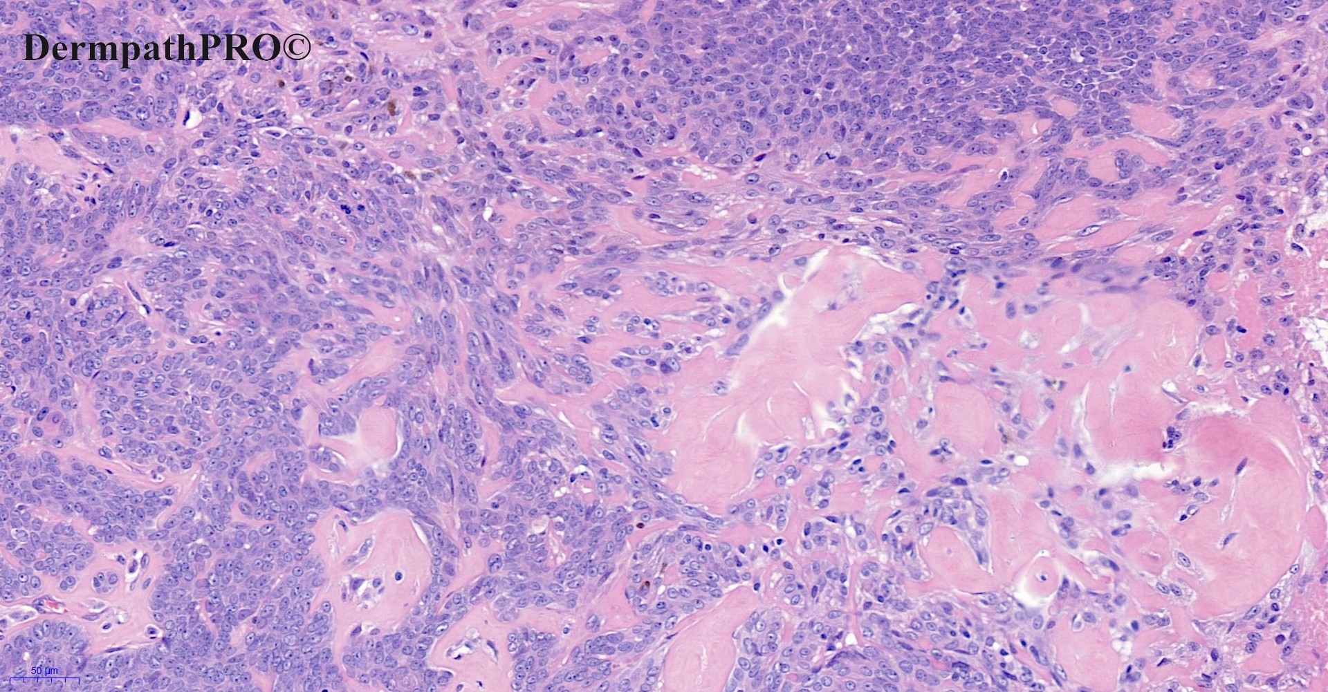
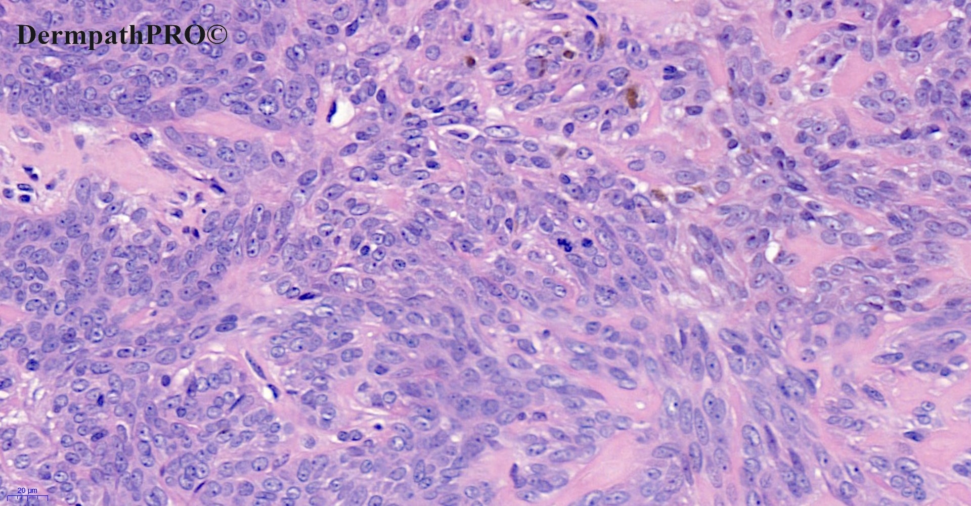
53M, Excision right scalp.

  Report Record

User Feedback


Krishnakumar subramanian

Posted

Epidermal hyperplasia with horn cysts and sebaceous glands looking like sebaceous nevus now showing basaloid proliferation and something like a trichelemmoma with mitosis


Share this comment


Link to comment
share_externally

Coexistent Nevus sebaceous-associated and focally desmoplastic, trichilemmoma, trichiepithelioma and pigmented trichoblastoma. 

Share this comment


Link to comment
share_externally

Eman El-Nabarawy

Posted

Nevus sebacous with basaloid proliferation, desmoplastic trichilemmoma and trichoblastoma.

Share this comment


Link to comment
share_externally

Saleem Taibjee

Posted

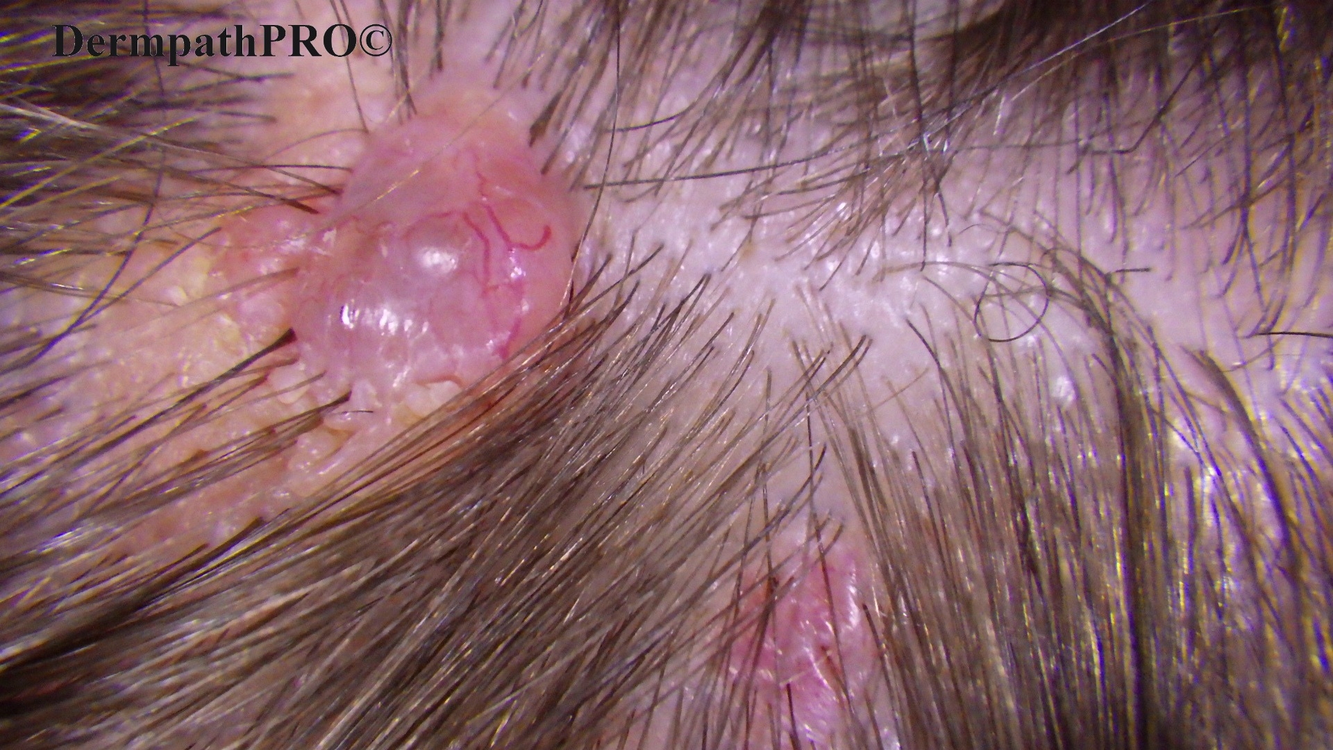
Please note the clinical photo. The main nodule is pearly with surface telangiectasia. There is a more subtle surrounding yellowish warty plaque within the scalp.

Here are the immunostains (I requested a limited panel only):

CD10 showed staining of the tumour epithelium (mainly peripheral staining pattern) within the larger basaloid nodule, and focal accentuation of staining in the stroma only. No evidence of Merkel cells within the main nodule (noting positive control of Merkel cells in the adjacent follicular infundibula and epidermis).04904_5.0x CD10 labelled.jpg

04904_5.0x CK20 labelled.jpg


04904_10.0x CK20 labelled.jpg

Share this comment


Link to comment
share_externally

Richard Logan

Posted

What's wrong with a BCC arising in a sebaceous naevus?  Thay's what it looks like clinically, and the CD10 staining is compatible.

Share this comment


Link to comment
share_externally

Dr. Mona Abdel-Halim

Posted

4 hours ago, Richard Logan said:

What's wrong with a BCC arising in a sebaceous naevus?  Thay's what it looks like clinically, and the CD10 staining is compatible.

Agree... 

Share this comment


Link to comment
share_externally

Meenakshi Batrani

Posted

18 hours ago, Richard Logan said:

What's wrong with a BCC arising in a sebaceous naevus?  Thay's what it looks like clinically, and the CD10 staining is compatible.

Agree

Share this comment


Link to comment
share_externally

Krishnakumar subramanian

Posted

can we tell that nevus sebaceous with basal cell carcinoma and trichelemmoma [ ? malignant]

Share this comment


Link to comment
share_externally

Could it be a basaloid type of fSCC? ( ...I don’t know much about this pathology area ). 

Share this comment


Link to comment
share_externally

Saleem Taibjee

Posted

Hi. This is a recent patient on whom I operated. The clinical photo shows a pearly nodule with telangiectasia suggestive of BCC, but there is a subtle adjacent yellow warty change.

For me this is an uncommon example of Basal cell carcinoma arising in naevus sebaceous. In the majority of cases we see trichoblastoma arising in NS, rather than BCC. However,  this is an uncommon exception. Features favouring BCC include the multifocal connection to overlying epidermis, tumour-derived amyloid, relative absence of stromal cell aggregates adjacent to tumour. The immunostains also support BCC in showing peripheral tumour staining with CD10 (as opposed to absent tumour staining, but mesenchymal staining in  trichoblastoma), and absence of Merkel cells. However, it is not possible to fully exclude 'hybrid' features. I did not show, but BerEP4 is strongly positive, EMA completely negative, and CD34 essentially negative, also supporting BCC rather than any of the differentials such as trichilemmoma, follicular SCC, etc.

Share this comment


Link to comment
share_externally

Dr. Richard Carr

Posted

Trichoblastoma (superficial areas) with BCC-like nodule. I've yet to see a convincingly "infiltrative or deeply invasive nodular BCC" in NS. These BCC-like nodular lesions are hard to distinguish from BCC and it's probably academic although would be nice if there was a reliable marker.

Share this comment


Link to comment
share_externally


Join the conversation

You can post now and register later. If you have an account, sign in now to post with your account.

Guest
Add a comment...

×   Pasted as rich text.   Paste as plain text instead

  Only 75 emoji are allowed.

×   Your link has been automatically embedded.   Display as a link instead

×   Your previous content has been restored.   Clear editor

×   You cannot paste images directly. Upload or insert images from URL.

Loading...

×
×
  • Create New...