Jump to content
In this section we have spot diagnoses posted on a daily basis since June 2010, now over 4000! You can review the archived cases and read the suggested diagnoses by users and the final comment by the contributors.
Case are uploaded each week day by 10 am UK time with the correct diagnosis will generally be posted at 8 pm UK time. Why not view the most recent spot diagnosis and proffer a diagnosis?

Case Number : Case 2716 - 03 December 2020 Posted By: Saleem Taibjee

Please read the clinical history and view the images by clicking on them before you proffer your diagnosis.
Submitted Date :
   (0 reviews)

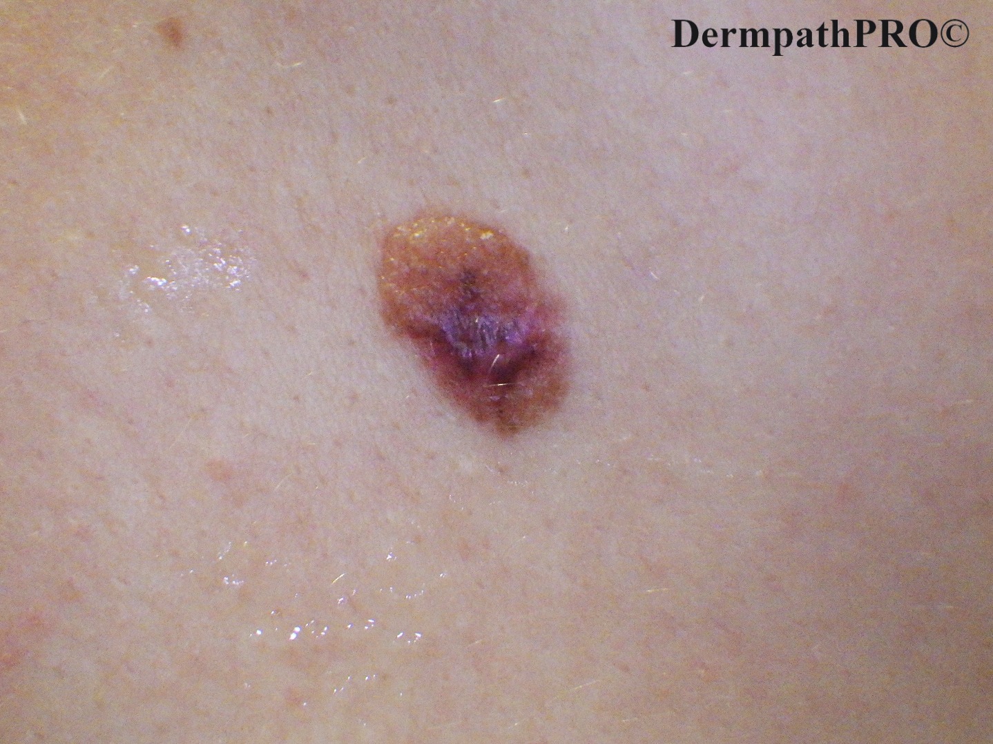
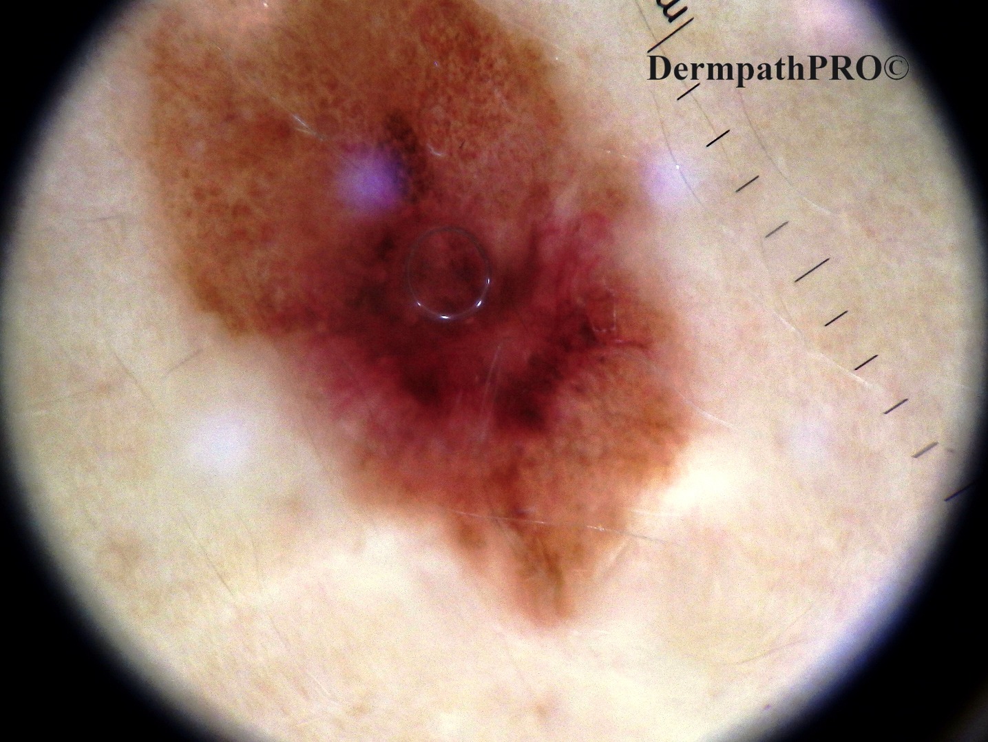
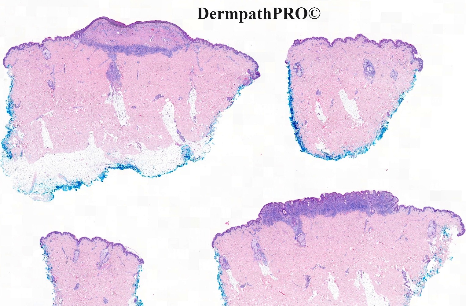
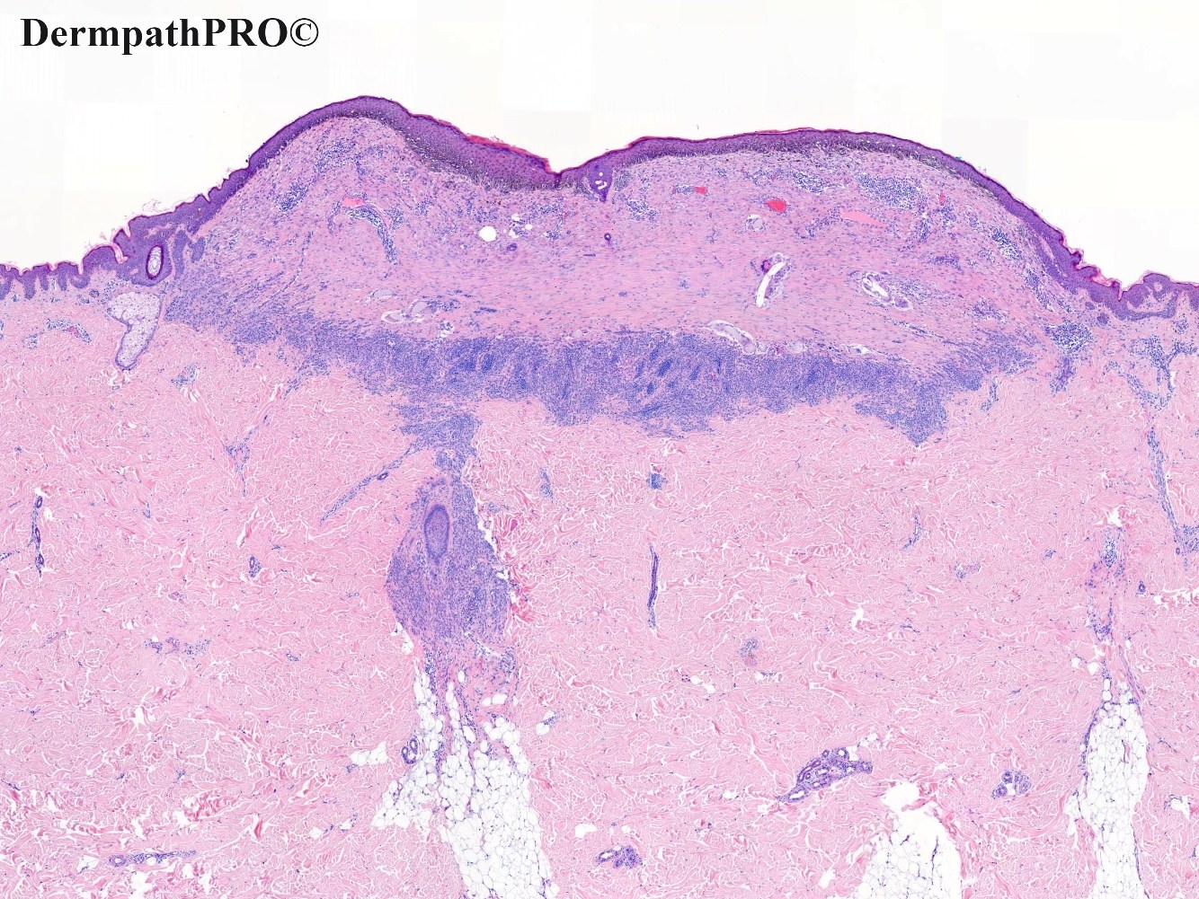
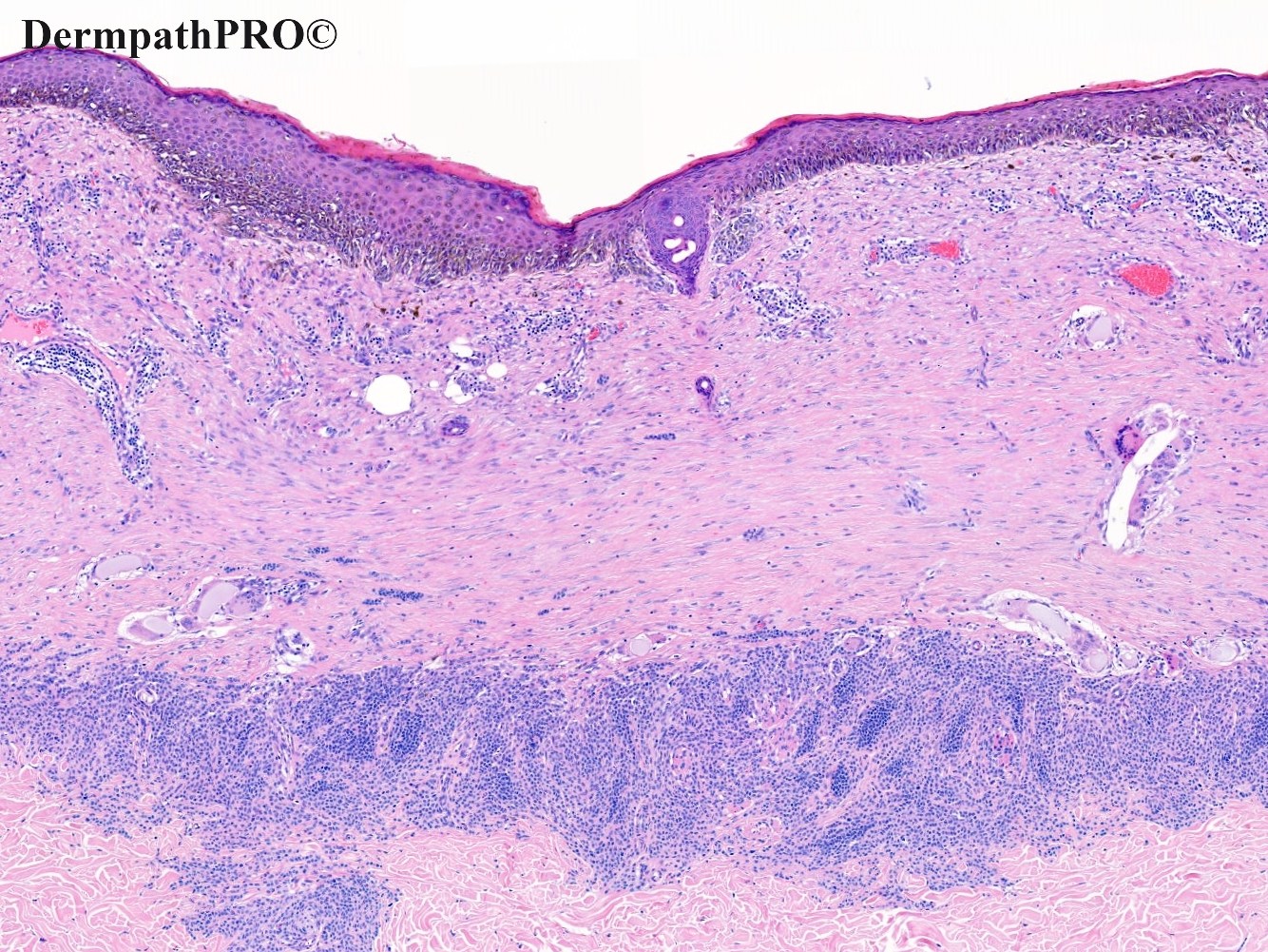
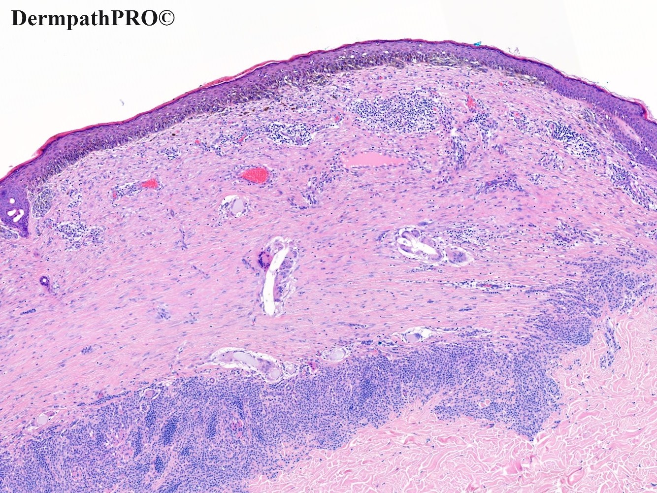
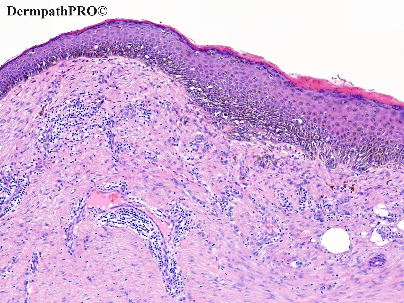
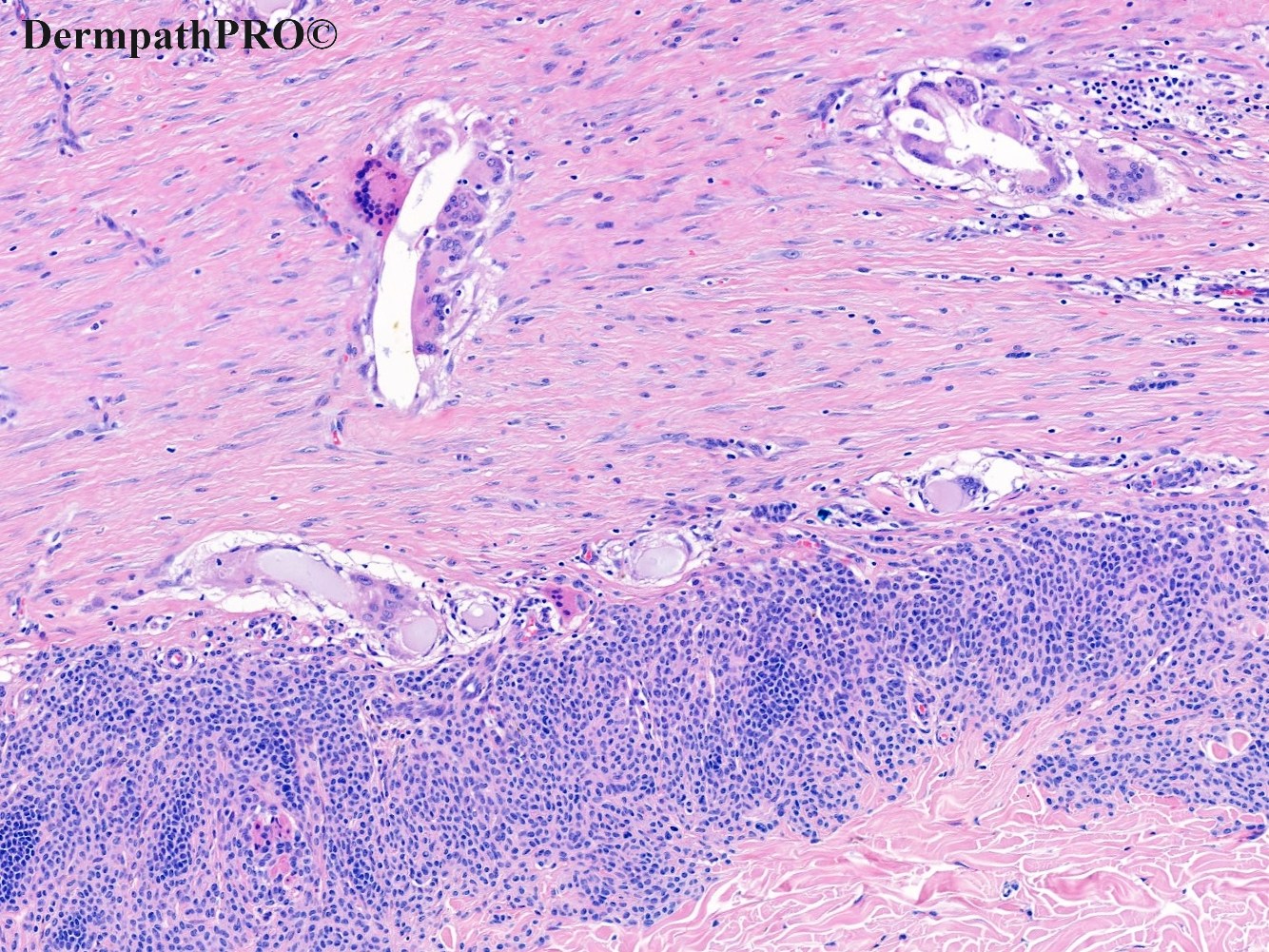
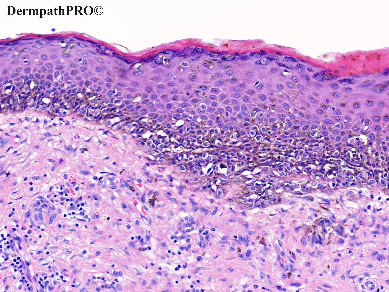
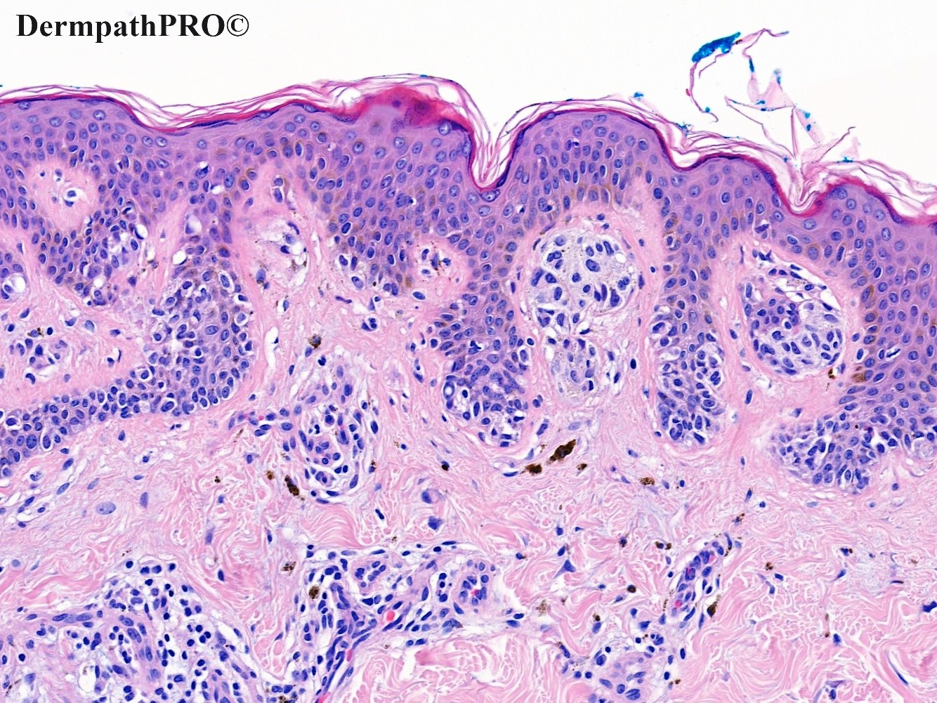
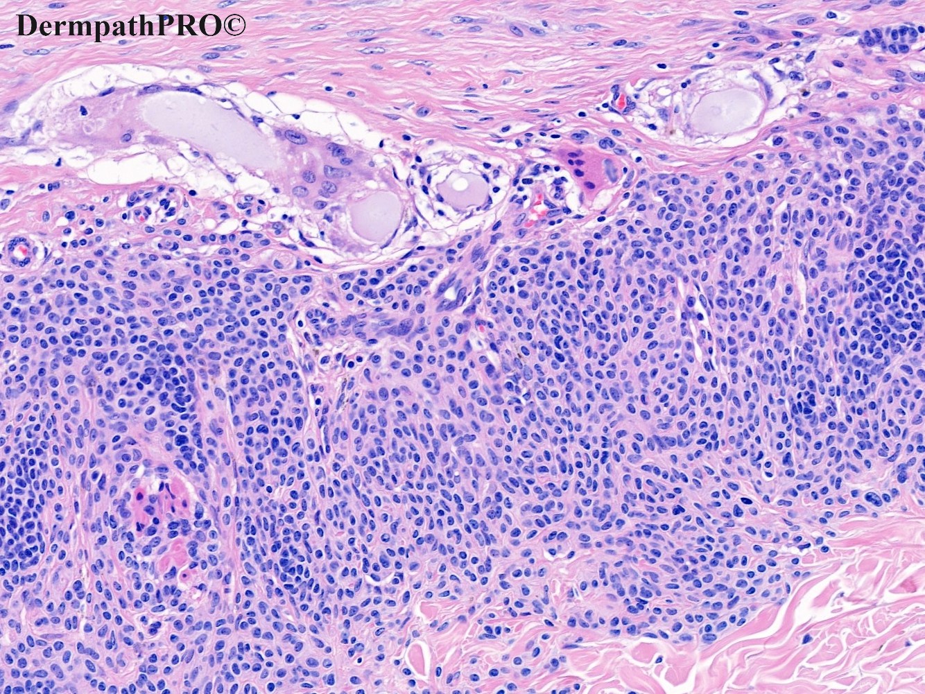
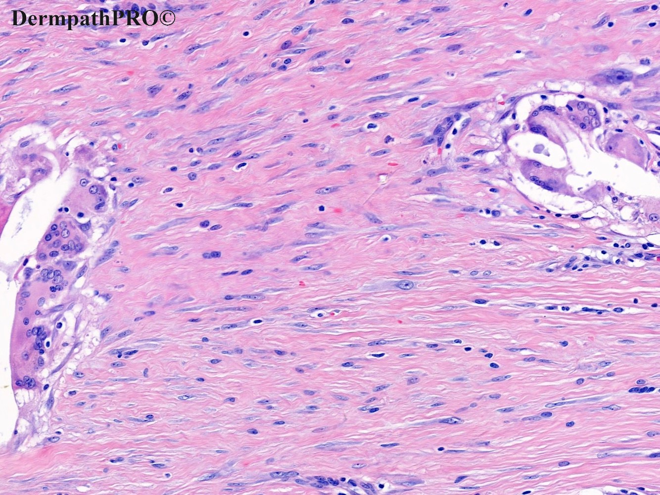
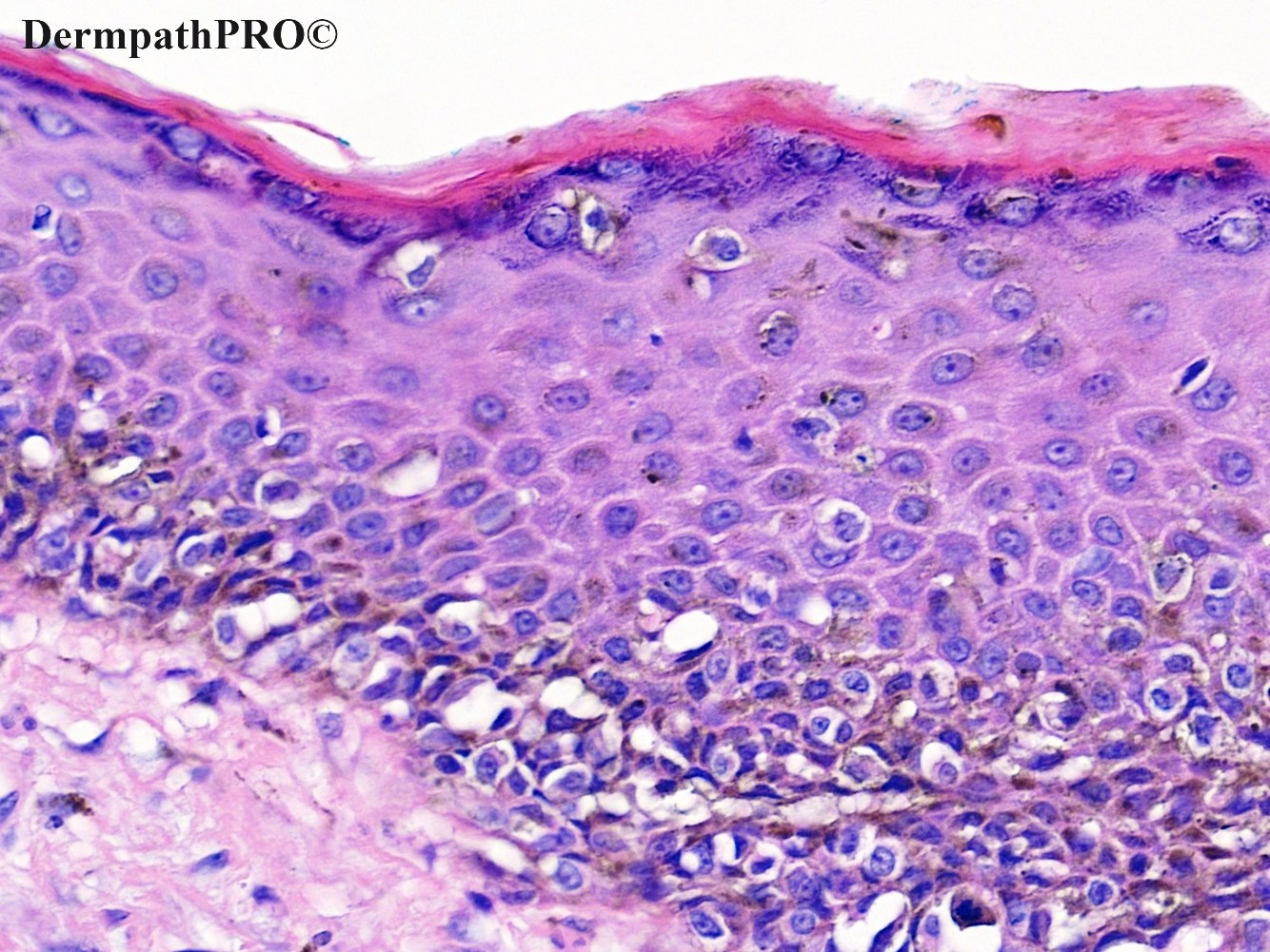
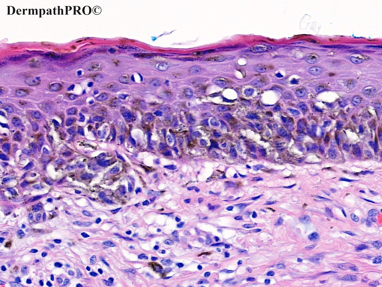
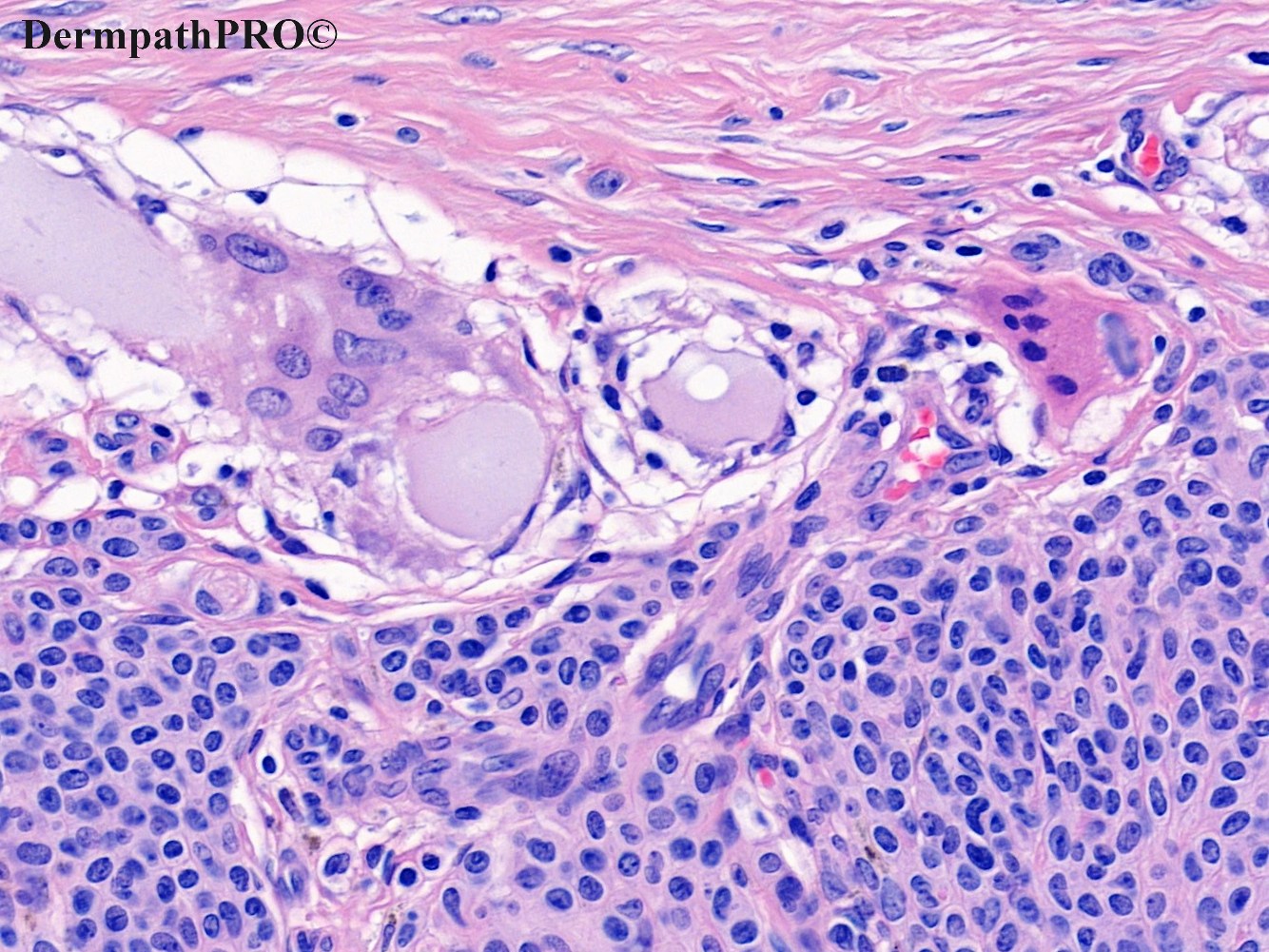
27F, excision right back, longstanding pigmented lesion, incidentally noticed by GP when the patient attended for an unrelated medical reason. No other history available.


  • Thanks 1
  Report Record

User Feedback


Richard Logan

Posted

This is a lovely example of pseudomelanoma.  There is a dermal scar from previous naevus surgery and also foci of foreign body reaction, possibly to suture material.  The lower part of the naevus is still present below the scar, and atypical melanocytic proliferation in the epidermis above it.  A good reason for leaving benign naevi alone!

Share this comment


Link to comment
share_externally

Cem Leblebici

Posted

Agree with Dr Logan.

Benign Nevus with Pseudomelanomatous Features. 

Share this comment


Link to comment
share_externally

There are two melanocytic lesions in the same specimen 

1): Favor "Recurrent nevus” over “scar/traumatized nevus”, because of body foreign granuloma, probably versus suture material. 

2): Question: is that a residual shoulder of previous nevus? Or is that another separate nevus from the previous one by the two nrormal skin specimens in fig 3?

I think the last not cicatricial appearing nevus is the residual adjacent half nevus. 

Share this comment


Link to comment
share_externally

Nice recurrent nevus. Shows all its typical characteristics

Share this comment


Link to comment
share_externally

Saleem Taibjee

Posted

Hi. Yes this is a nice recent example of recurrent naevus, with probably the most impressive pseudomelanoma phenomenon I have personally seen, including prominent upward spread. Amongst the diagnostic clues is the horizontal demarcation of the atypical junctional component to the area of underlying scarring. The dermoscopy is also quite interesting.

At the time of surgery, the patient provided no relevant history. However, when I telephoned her with the histology result, she then recalled scraping off the top of the mole on a wooden beam in the attic a few years previously!

Share this comment


Link to comment
share_externally

Very interesting case. So the foreign material was probably a wood splinter...

Share this comment


Link to comment
share_externally

Richard Logan

Posted

What do others think about this entity being called "recurrent naevus"?  This name implies that the naevus was removed and then regrew.  In fact, of course, this situation arises when only the top of a naevus (almost always pigmented) is removed, either surgically, by inappropriate cryotherapy or by accident as in this case.  More accurately it is a persistent naevus due to incomplete removal.  That portion beneath the scar is part of the original naevus.  The atypical melanocytic proliferation above the scar is not really naevus at all, but it is the part which causes all the concern because of its typical pigmentary pattern.

I prefer the designation of pseudomelanoma.

Share this comment


Link to comment
share_externally

I use recurrent/persistent nevus. very nice example.

Share this comment


Link to comment
share_externally


Join the conversation

You can post now and register later. If you have an account, sign in now to post with your account.

Guest
Add a comment...

×   Pasted as rich text.   Paste as plain text instead

  Only 75 emoji are allowed.

×   Your link has been automatically embedded.   Display as a link instead

×   Your previous content has been restored.   Clear editor

×   You cannot paste images directly. Upload or insert images from URL.

Loading...

×
×
  • Create New...