Jump to content
In this section we have spot diagnoses posted on a daily basis since June 2010, now over 4000! You can review the archived cases and read the suggested diagnoses by users and the final comment by the contributors.
Case are uploaded each week day by 10 am UK time with the correct diagnosis will generally be posted at 8 pm UK time. Why not view the most recent spot diagnosis and proffer a diagnosis?

Case Number : Case 2721 - 10 December 2020 Posted By: Saleem Taibjee

Please read the clinical history and view the images by clicking on them before you proffer your diagnosis.
Submitted Date :
   (0 reviews)

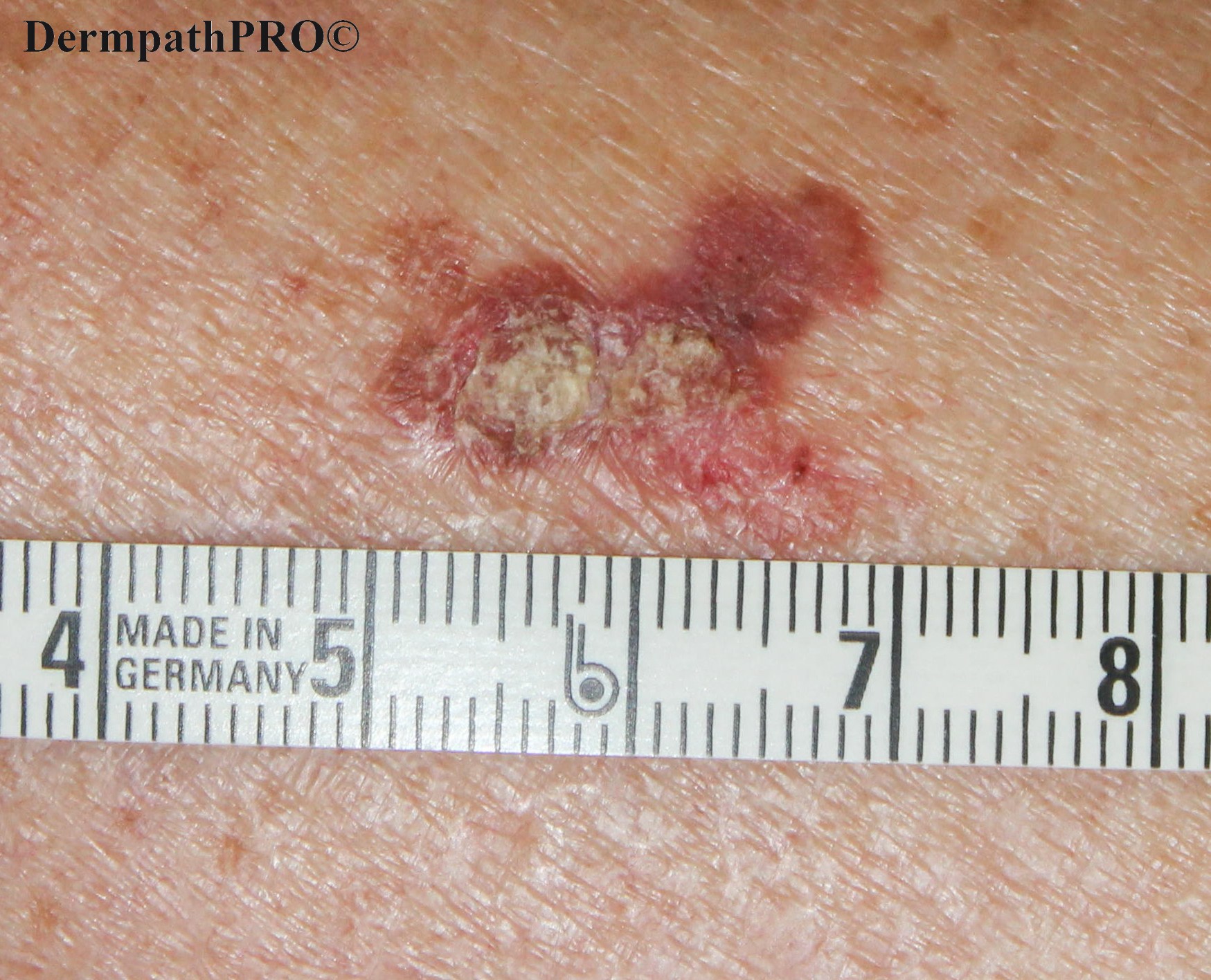
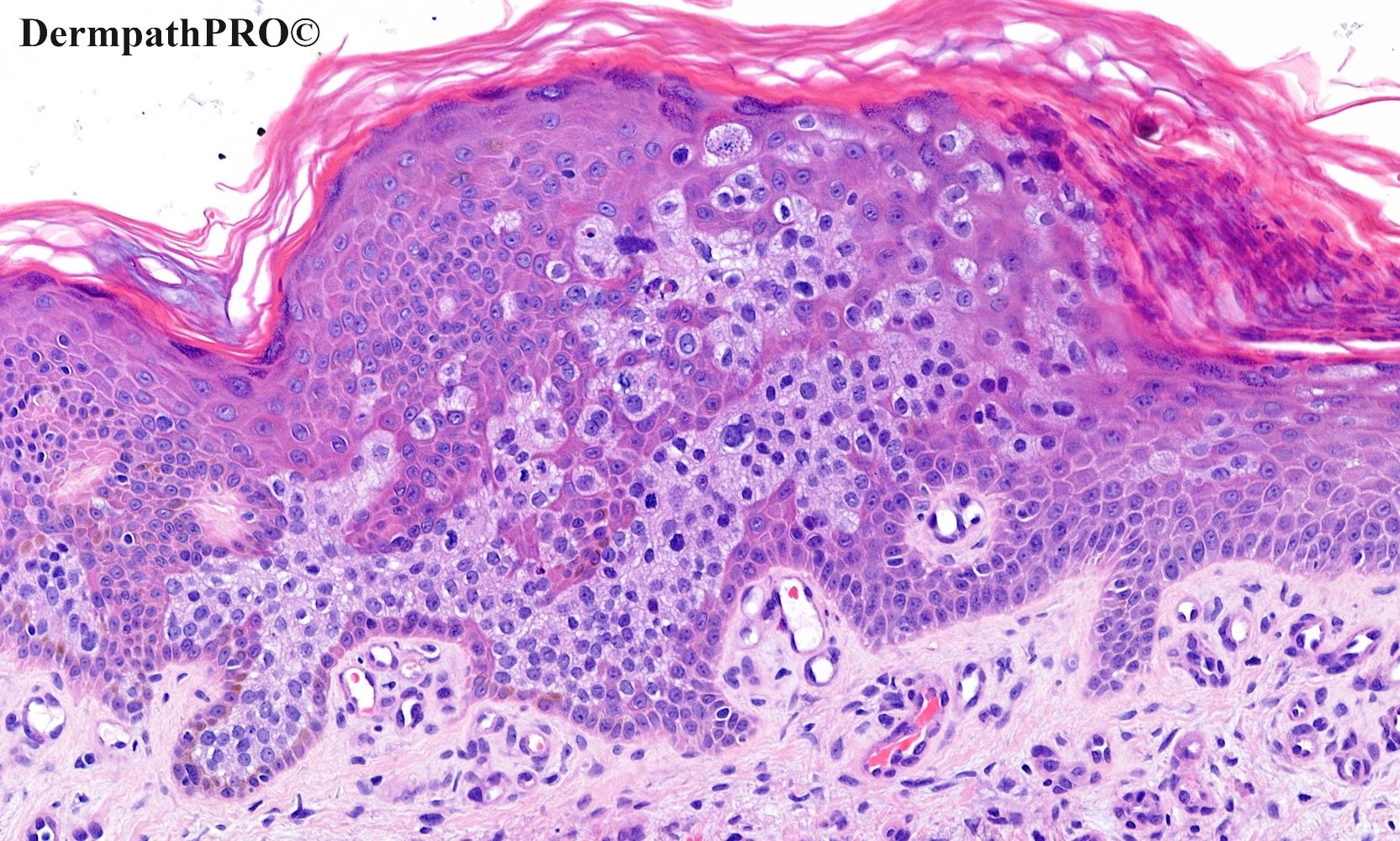
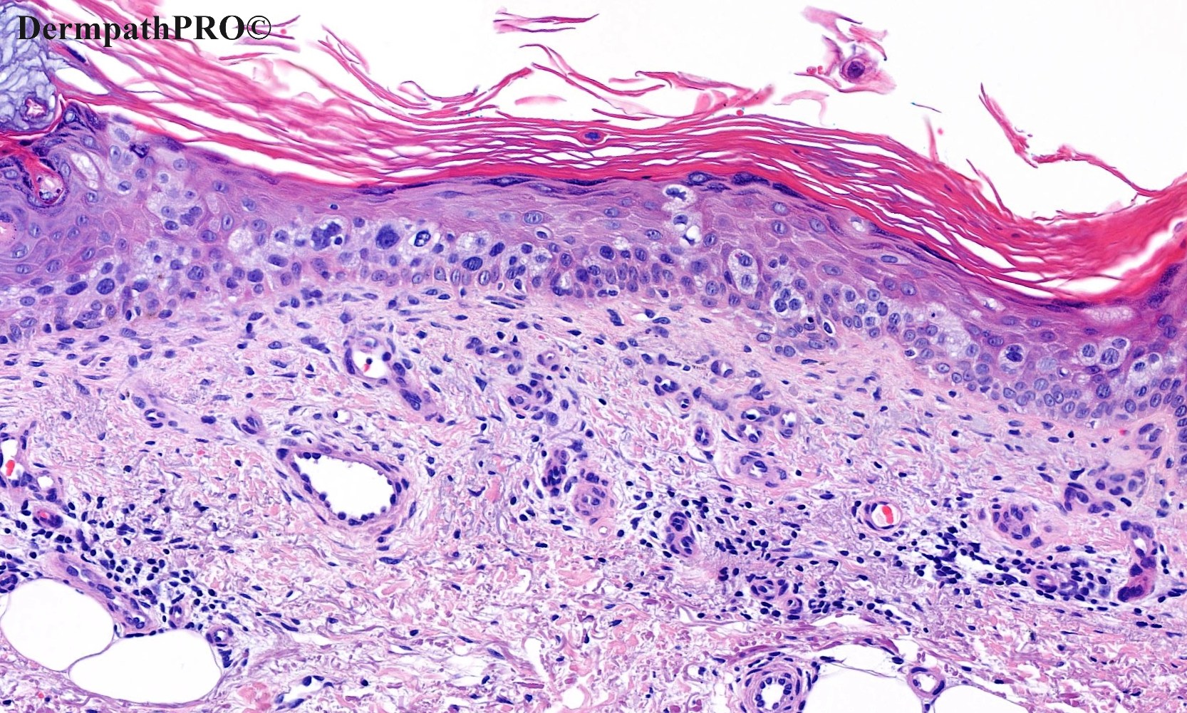
78F, Pink brown scaly plaque on the lower leg.


  • Like 2
  Report Record

User Feedback


David Llorca

Posted

Pigmented Bowen disease. clinical and histological 

Share this comment


Link to comment
share_externally

Richard Logan

Posted

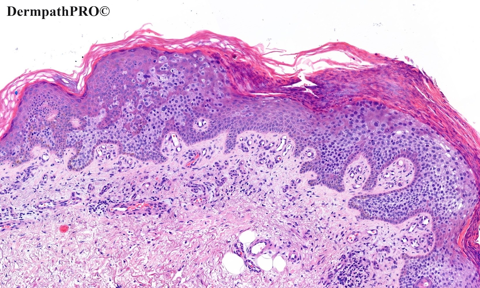
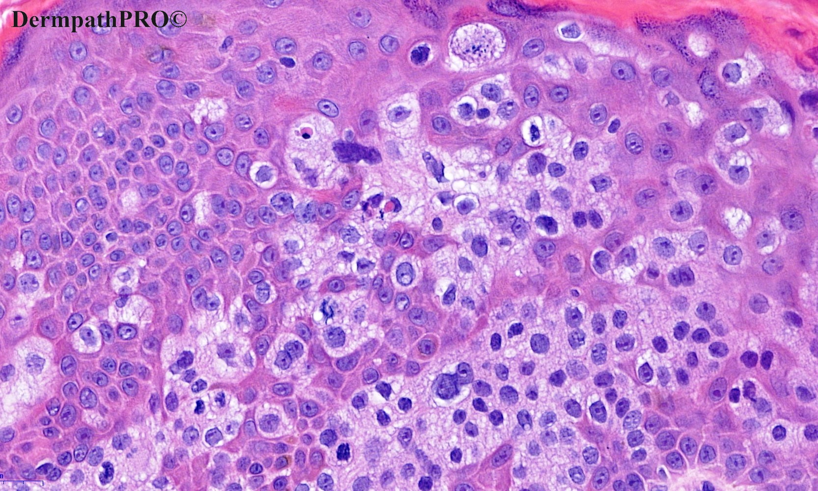
The cells involved in the Pagetoid spread have a rather bubbly cytoplasm.  I wonder if this is In-situ sebaceous carcinoma, but of course at this anatomical site pigmented Bowen's disease would be far more likely.  IHC needed.

Share this comment


Link to comment
share_externally

Meenakshi Batrani

Posted

Pagetoid Bowen's disease. Also, extamammary Paget's disease has rarely been described on unusual location other that perineal region. IHC will help. 

Share this comment


Link to comment
share_externally

Sebaceus carcinoma x pagetoid Bowen´s x extramammary Paget disease. IHQ

Share this comment


Link to comment
share_externally

Saleem Taibjee

Posted

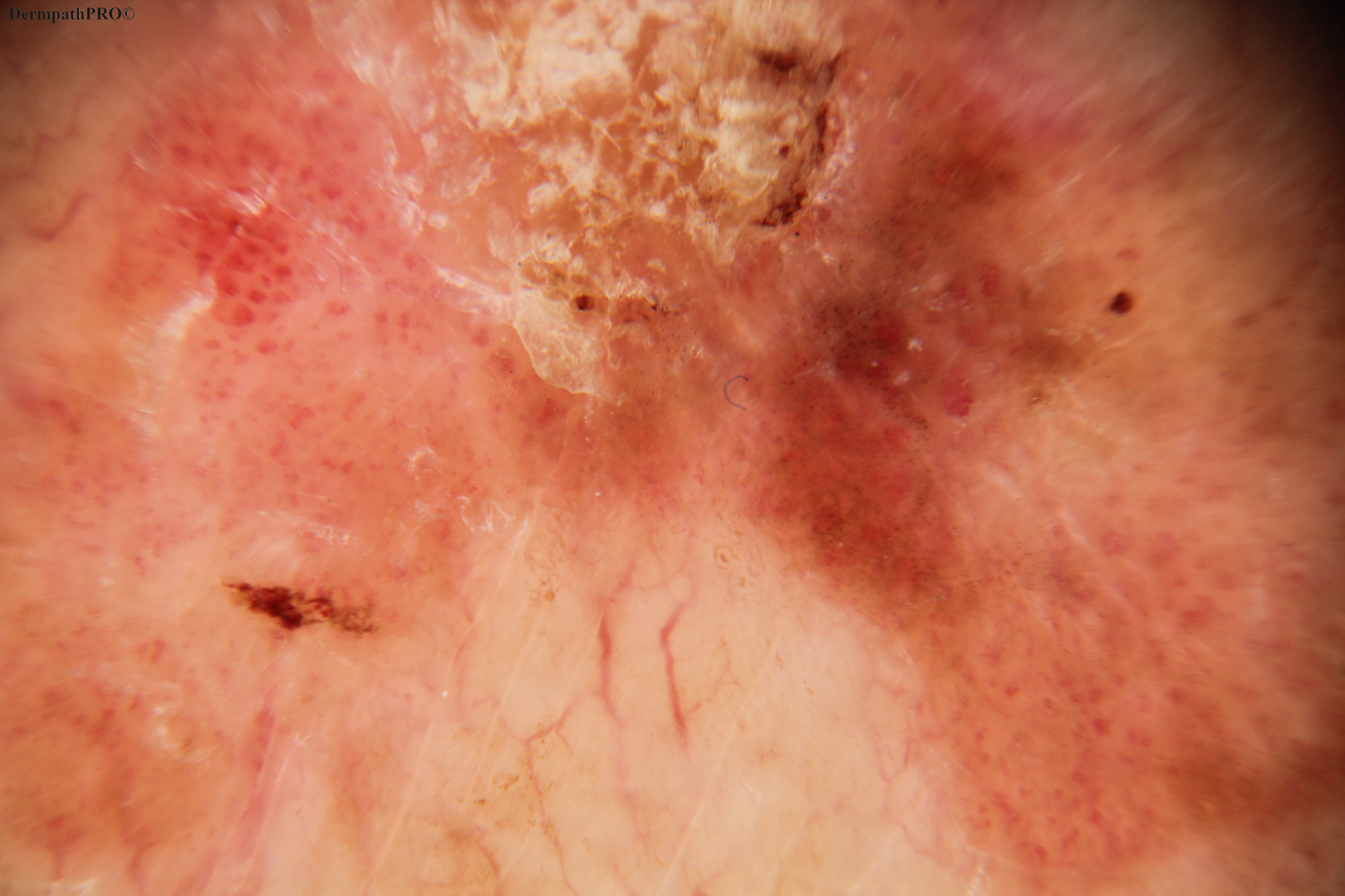
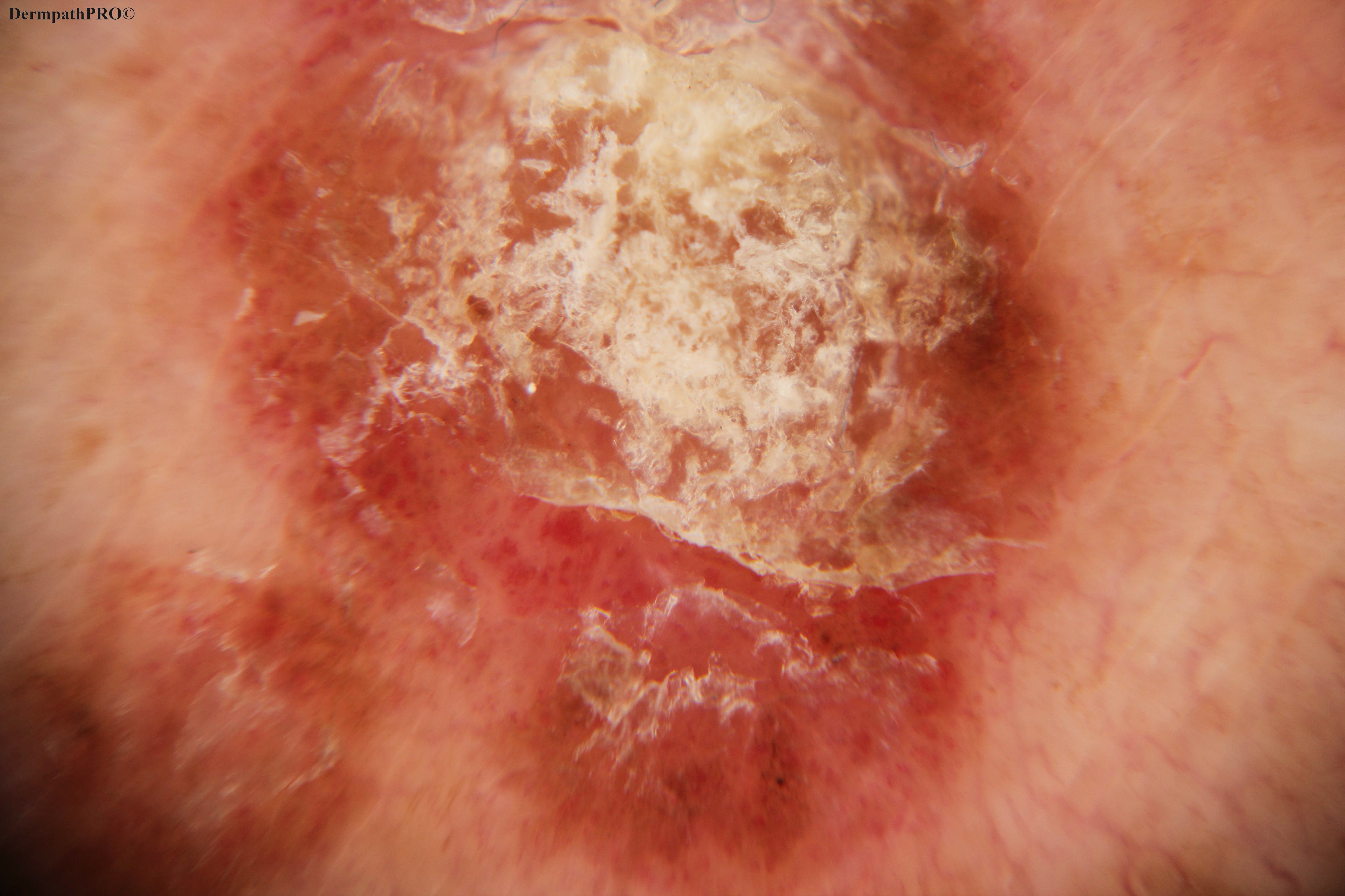
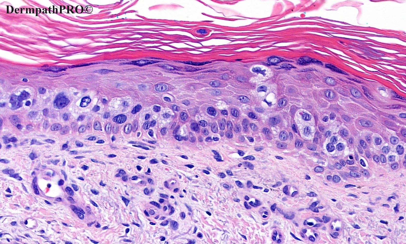
Yes, well done, this is pigmented and pagetoid Bowen's. I picked out the most pagetoid areas for the Spot Diagnosis, but there were more conventional Bowen's areas also (see below). It is a recent case where it was nice to have the clinical and dermoscopic images to correlate with the histology. The pigment is quite striking on the dermoscopy, but notably there is no true pigment network, and the vessels are arguably 'glomeruloid', all in keeping with Bowen's. Of course, a histological clue that this is Bowen's and not melanoma with pagetoid spread is that there is preservation of the basal layer which rims the pleomorphic cells, and that any apparent nesting is not actually junctional. At a higher power, one is able to discern the desmosomal attachments. I agree that the cells are a bit bubbly, but the cells lack the indented/crenalated nuclei for true sebaceous differentiation, and this would be an odd anatomical site for sebaceous carcinoma. Hence I did not run immunos, and indeed it might be difficult to conceive an immunopanel which would reliably distinguish Bowen's from sebaceous carcinoma. I do not have access to adipophilin at our lab. I have wondered about using PRAME as a surrogate for sebaceous differentiation, but have been discouraged by others' more extensive experience (e.g. Klaus Busam).

BW, Saleem

08380_10.0x.jpg

08380_20.0x.jpg


Share this comment


Link to comment
share_externally

Victor Delgado

Posted

Beautiful case, tks for sharing. SCC in situ

Share this comment


Link to comment
share_externally


Join the conversation

You can post now and register later. If you have an account, sign in now to post with your account.

Guest
Add a comment...

×   Pasted as rich text.   Paste as plain text instead

  Only 75 emoji are allowed.

×   Your link has been automatically embedded.   Display as a link instead

×   Your previous content has been restored.   Clear editor

×   You cannot paste images directly. Upload or insert images from URL.

Loading...

×
×
  • Create New...