Jump to content
In this section we have spot diagnoses posted on a daily basis since June 2010, now over 4000! You can review the archived cases and read the suggested diagnoses by users and the final comment by the contributors.
Case are uploaded each week day by 10 am UK time with the correct diagnosis will generally be posted at 8 pm UK time. Why not view the most recent spot diagnosis and proffer a diagnosis?

Case Number : Case 2722 - 11 December 2020 Posted By: Dr. Richard Carr

Please read the clinical history and view the images by clicking on them before you proffer your diagnosis.
Submitted Date :
   (0 reviews)

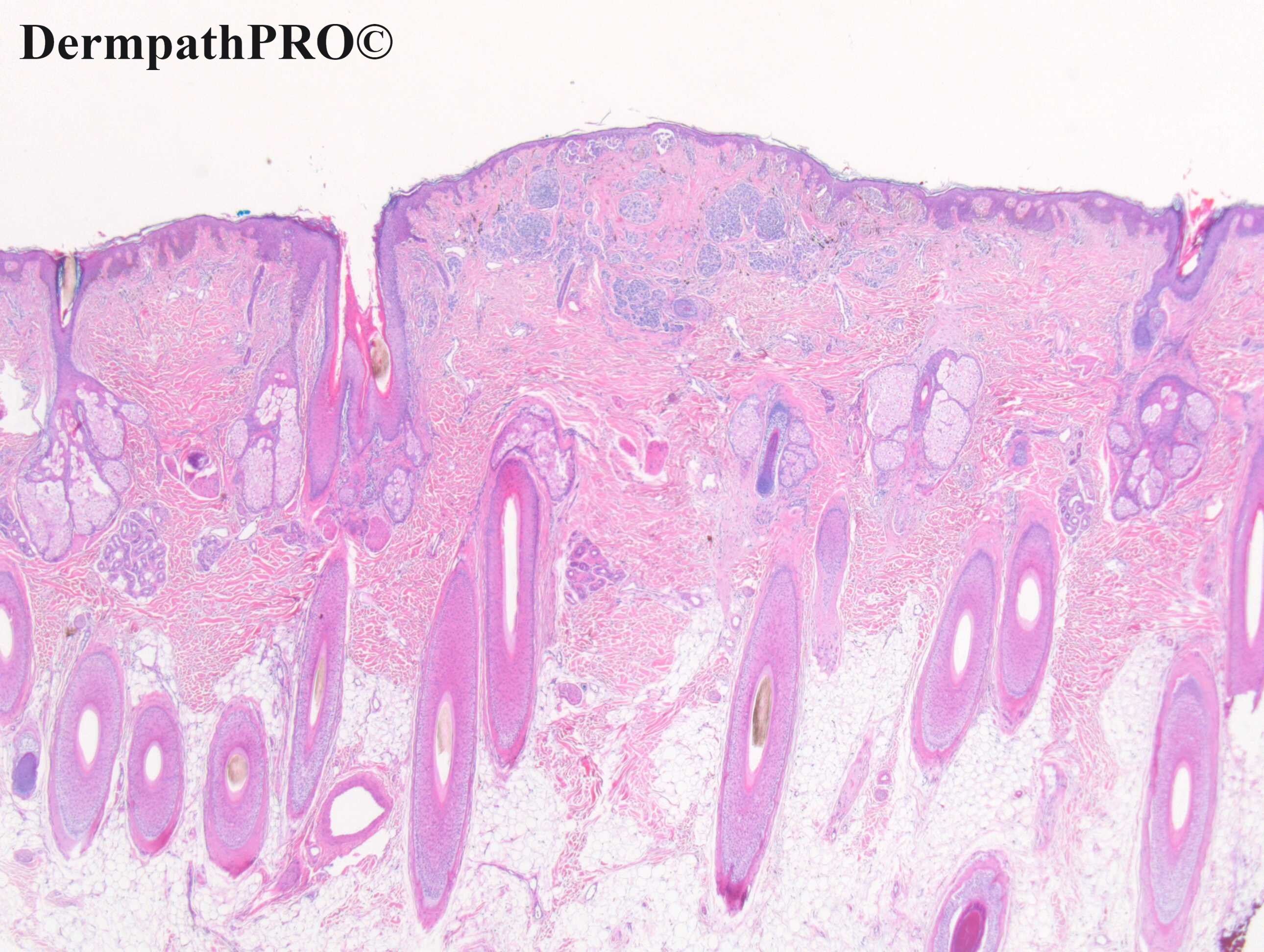
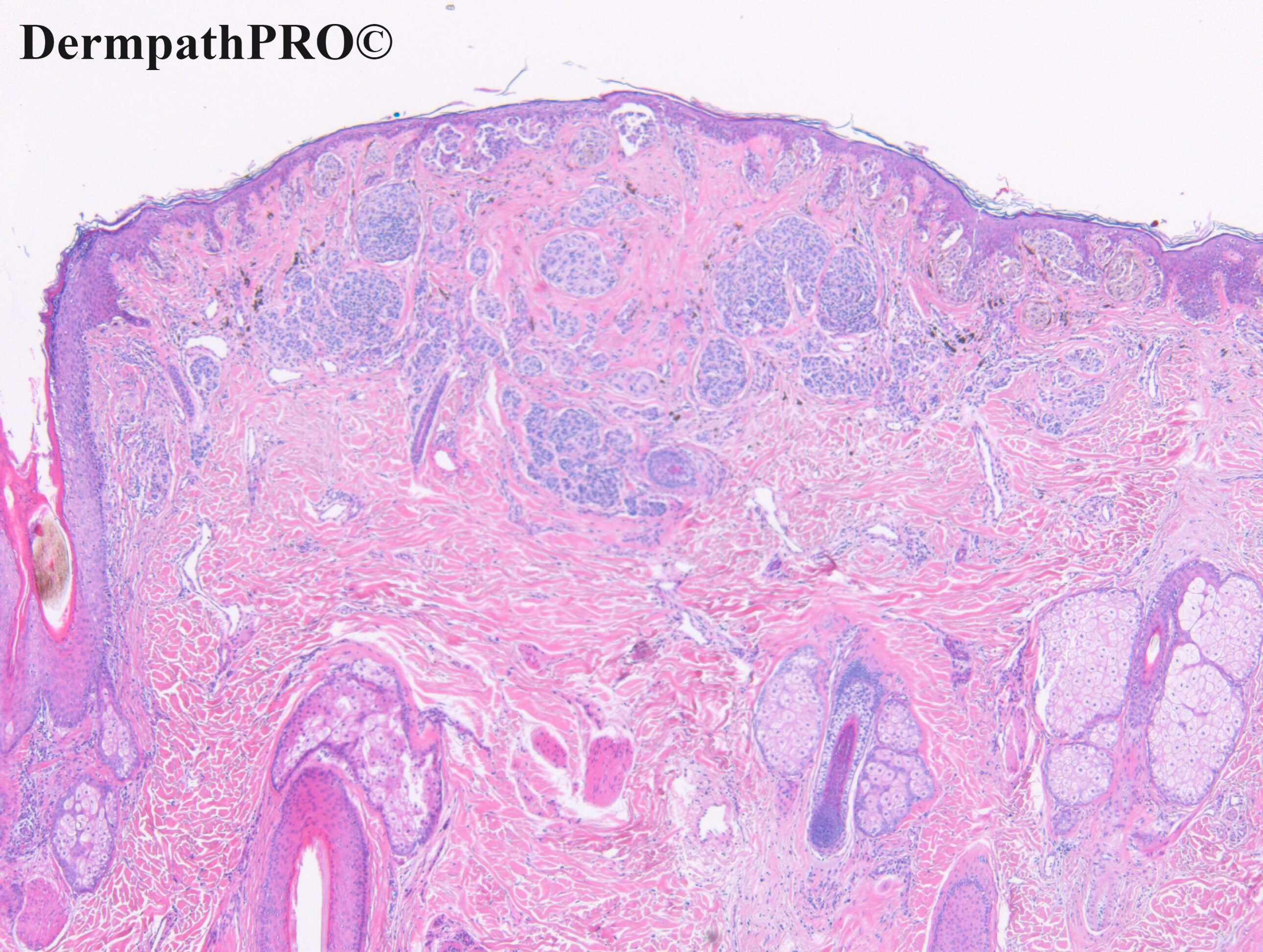
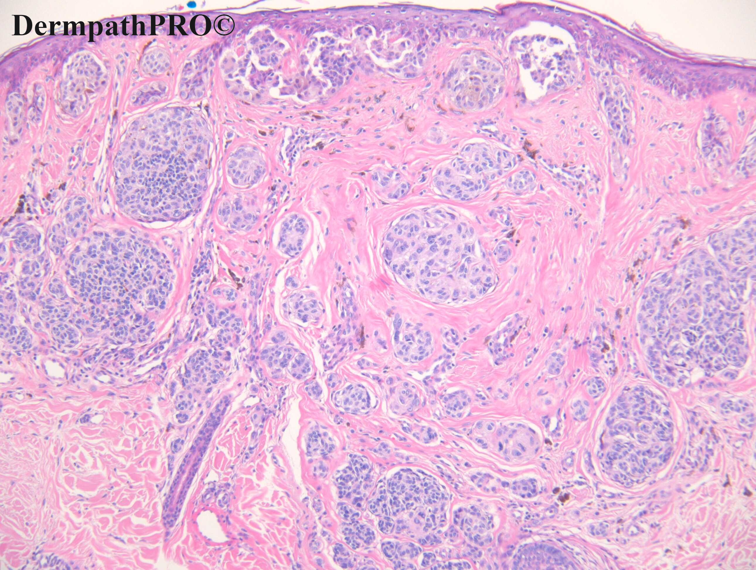
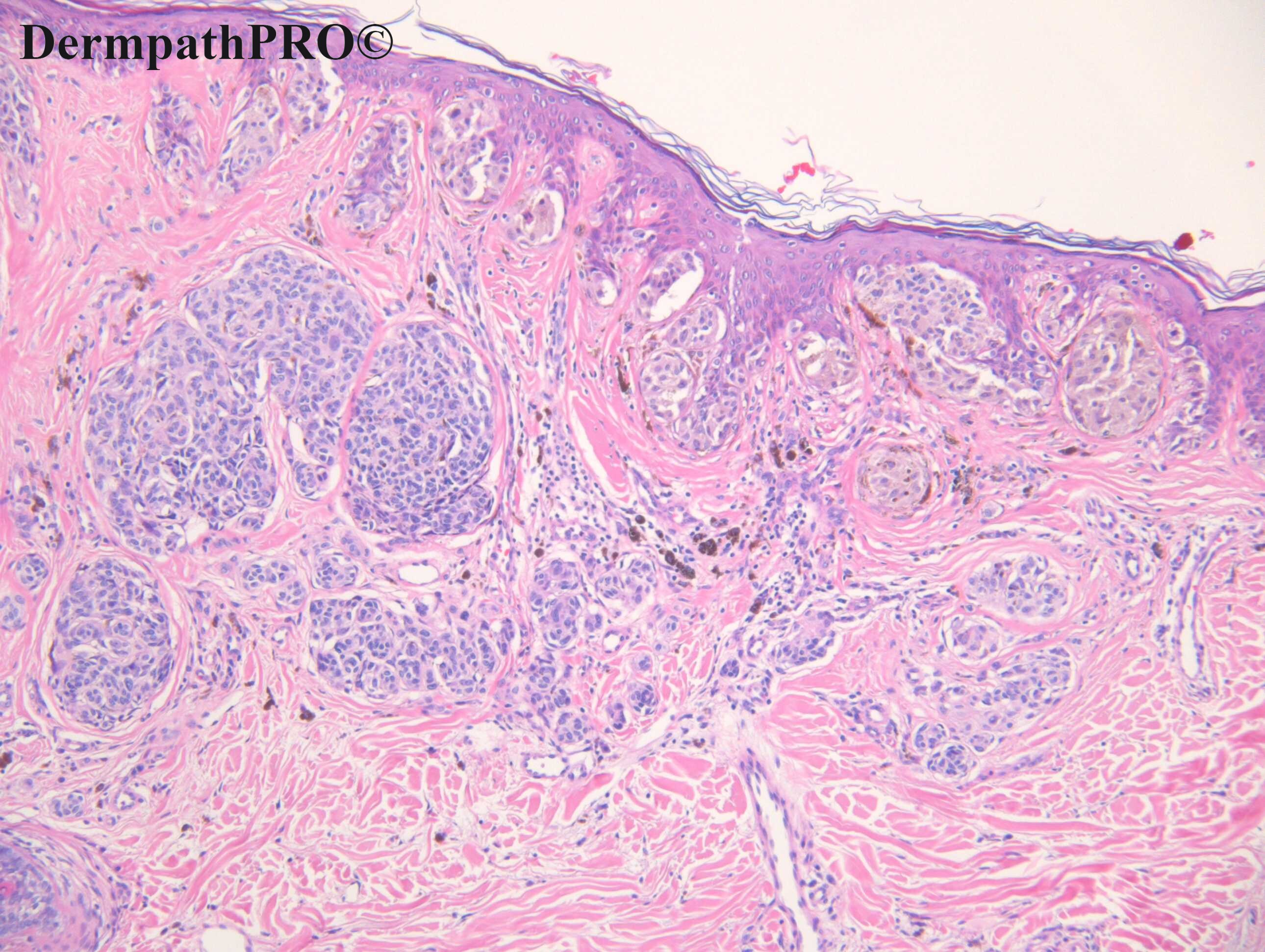
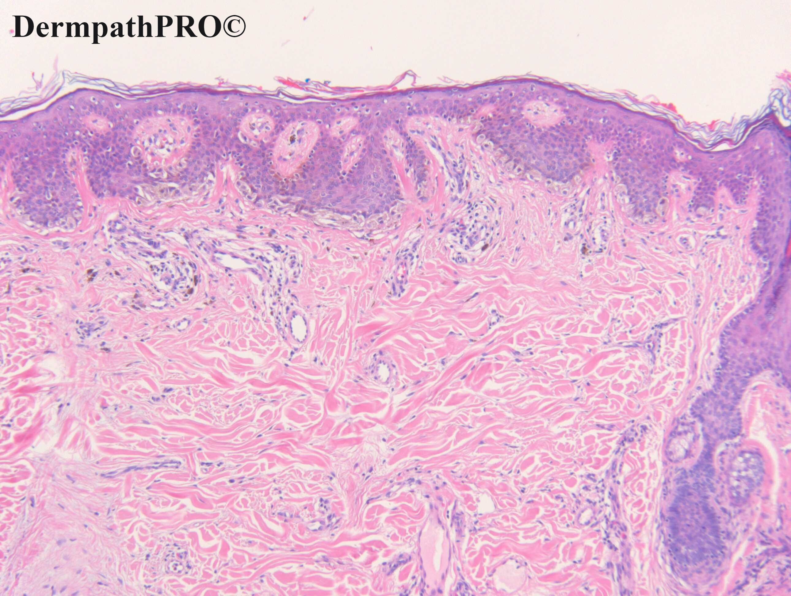
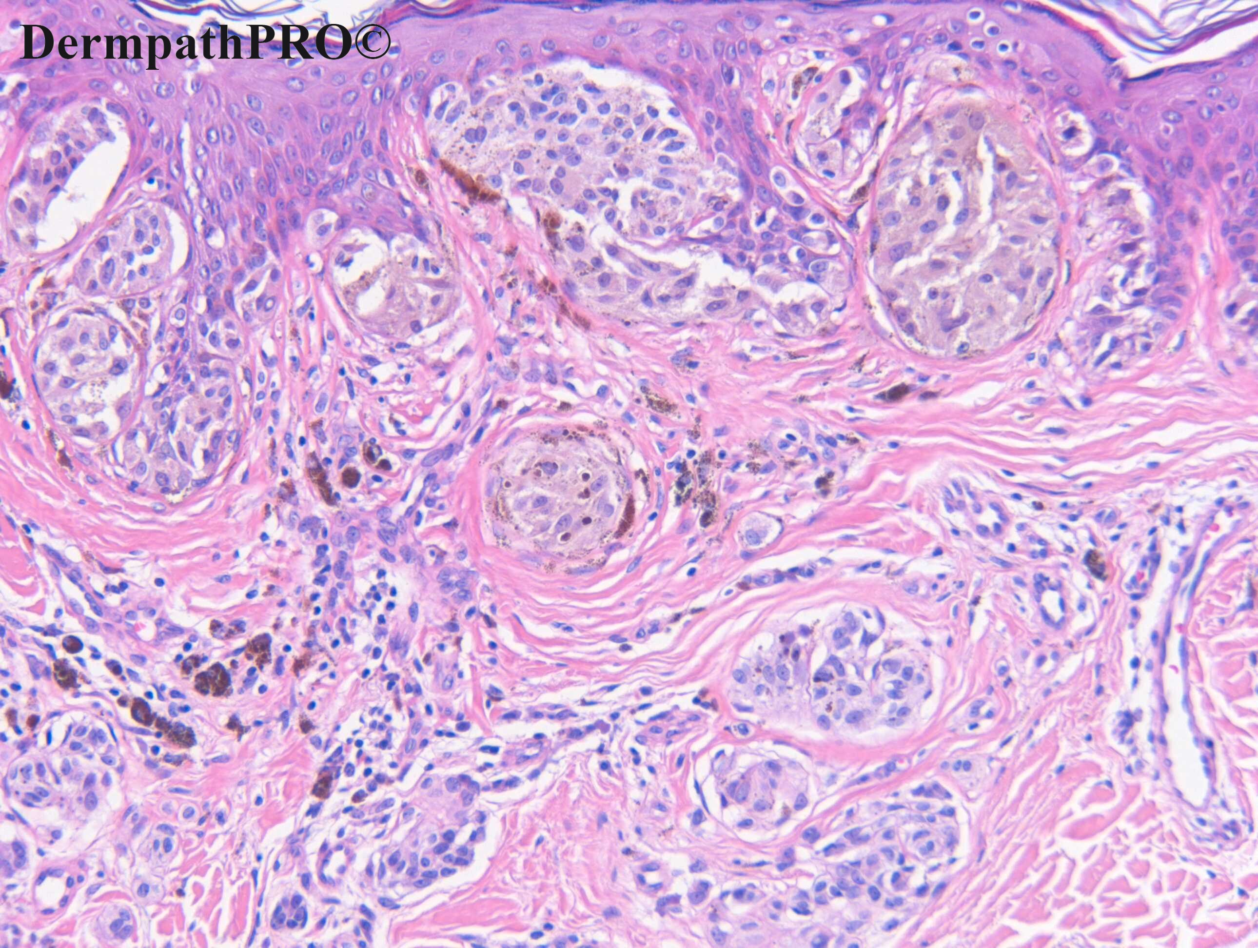
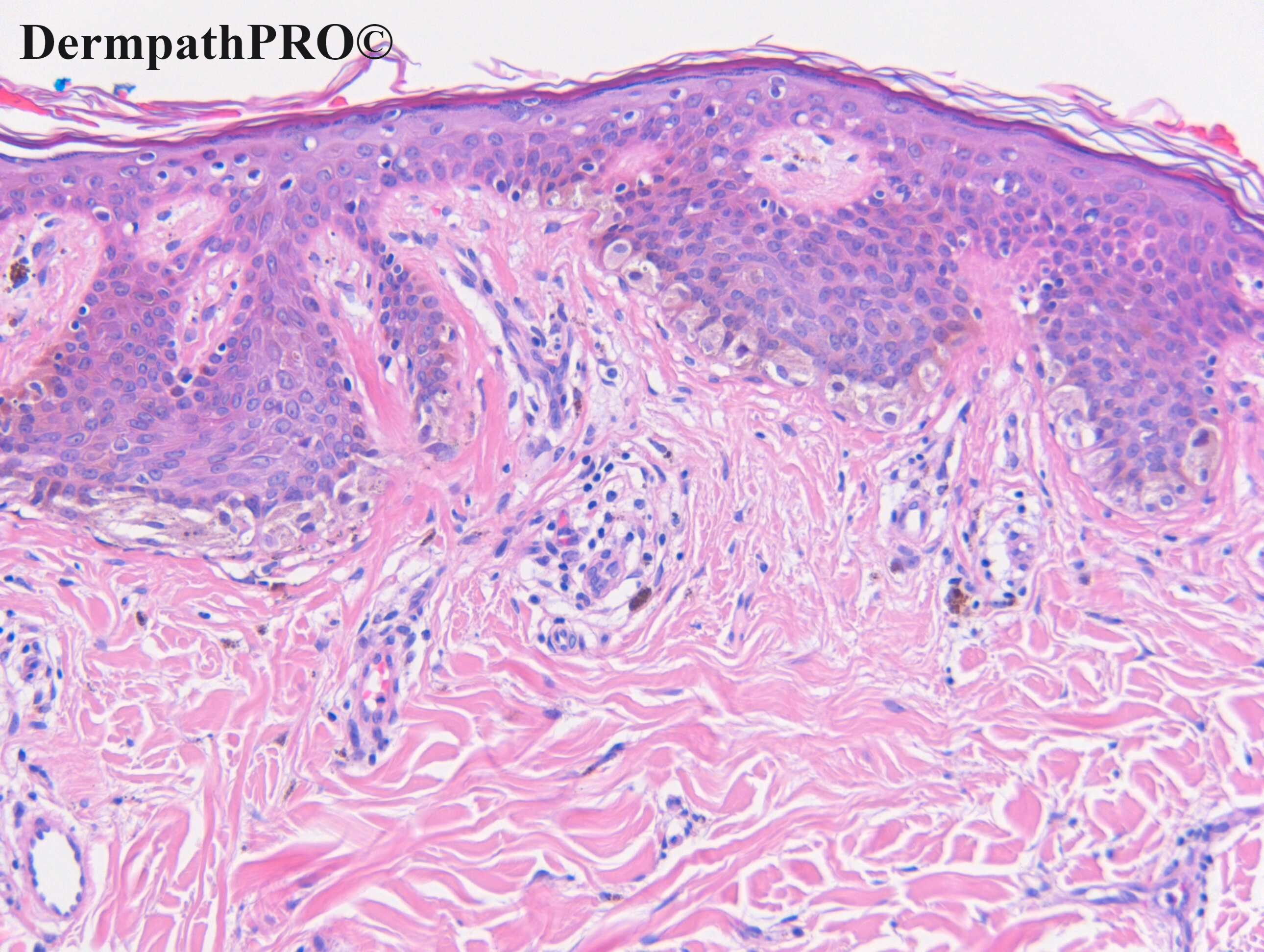
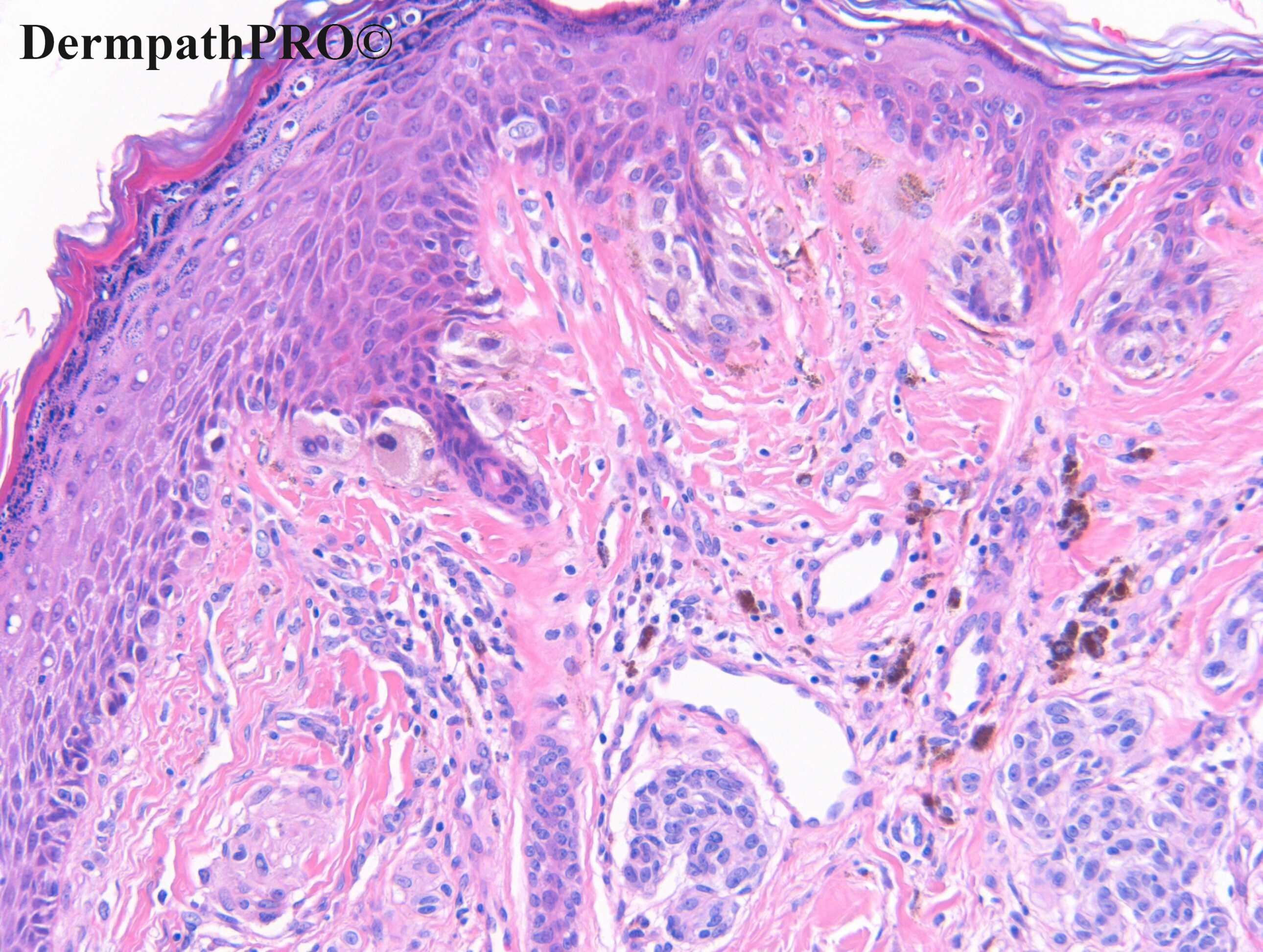
M19, Scalp. 7mm changing pigmented lesion ?Naevus ?MM


  Report Record

User Feedback


Dr. Richard Carr

Posted

20 views and no comments (apart from this one)

Share this comment


Link to comment
share_externally

I think there is maturation in this composite melanocytic lesion. The intraepidermal growth shows some basal spreading, but not Pagetoid or epidermal destructive consumption. So my spot is Atypical/Dysplastic Nevus

Share this comment


Link to comment
share_externally

I think this is a combined nevus, maybe a BAP1-inactivated nevus. It would be nice to see BAP1 and BRAF ihq in this lesion.

Share this comment


Link to comment
share_externally

58 minutes ago, IgorSC said:

I think this is a combined nevus, maybe a BAP1-inactivated nevus. It would be nice to see BAP1 and BRAF ihq in this lesion.

Usually the Bap1 negative cells, as I know, are large epithelioid cells, and intradermal nodule forming. But I’m happy to share the same benign suggestion with Igor. 

Share this comment


Link to comment
share_externally

Meenakshi Batrani

Posted

I am also not in favour of melanoma. Atypical/dysplastic nevus or may be BAP1 inactivated. 

Share this comment


Link to comment
share_externally

Dr. Richard Carr

Posted

Case c/o Dr Brinder "Buzz" Chohan.

My interpretation: Many thanks for the opportunity to review this case. This is a highly junctionally active compound lesion
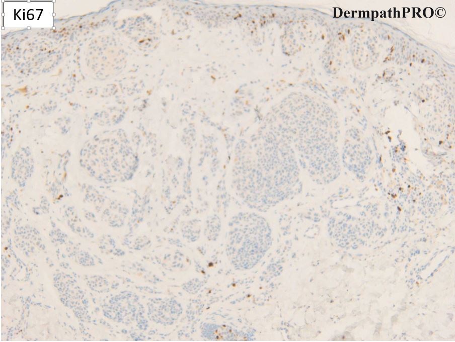
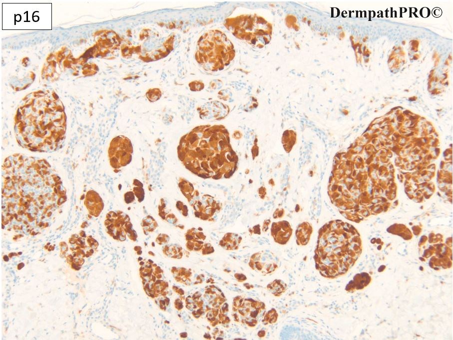
with slightly enlarged type A melanocytes. Overall relatively good symmetry. Nice chequer board p16 throughout and minimal Ki67 nuclear proliferation. This is a special site naevus with some architectural atypical features as described in younger individuals on the scalp.
Excision appears complete with appropriate margins.

Share this comment


Link to comment
share_externally

On 14/12/2020 at 07:58, Dr. Richard Carr said:

Case c/o Dr Brinder "Buzz" Chohan.

My interpretation: Many thanks for the opportunity to review this case. This is a highly junctionally active compound lesion
with slightly enlarged type A melanocytes. Overall relatively good symmetry. Nice chequer board p16 throughout and minimal Ki67 nuclear proliferation. This is a special site naevus with some architectural atypical features as described in younger individuals on the scalp.
Excision appears complete with appropriate margins.

Agree. Thanks Dr Carr.

Share this comment


Link to comment
share_externally

Scalp nevus can be as "striking" as though "ear" nevus.

Share this comment


Link to comment
share_externally


Join the conversation

You can post now and register later. If you have an account, sign in now to post with your account.

Guest
Add a comment...

×   Pasted as rich text.   Paste as plain text instead

  Only 75 emoji are allowed.

×   Your link has been automatically embedded.   Display as a link instead

×   Your previous content has been restored.   Clear editor

×   You cannot paste images directly. Upload or insert images from URL.

Loading...

×
×
  • Create New...