Jump to content
In this section we have spot diagnoses posted on a daily basis since June 2010, now over 4000! You can review the archived cases and read the suggested diagnoses by users and the final comment by the contributors.
Case are uploaded each week day by 10 am UK time with the correct diagnosis will generally be posted at 8 pm UK time. Why not view the most recent spot diagnosis and proffer a diagnosis?

Case Number : Case 2730 - 23 December 2020 Posted By: Iskander H. Chaudhry

Please read the clinical history and view the images by clicking on them before you proffer your diagnosis.
Submitted Date :
   (0 reviews)

68M
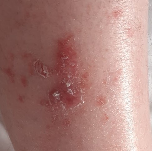
Right leg 6mm punch. Broad papulosquamous rash.


  Report Record

User Feedback


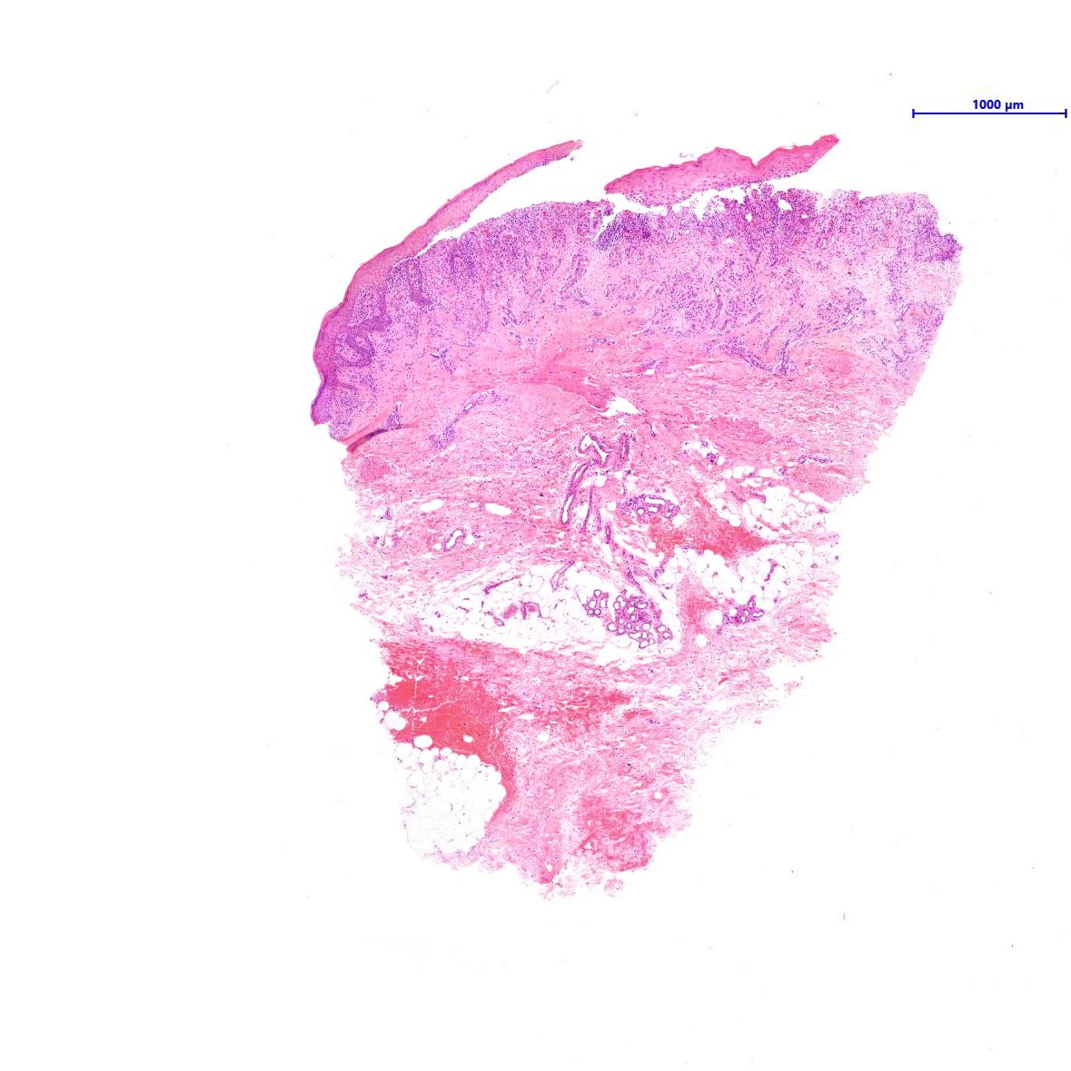
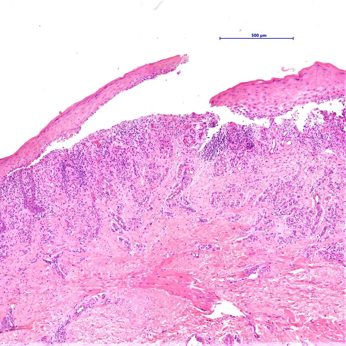
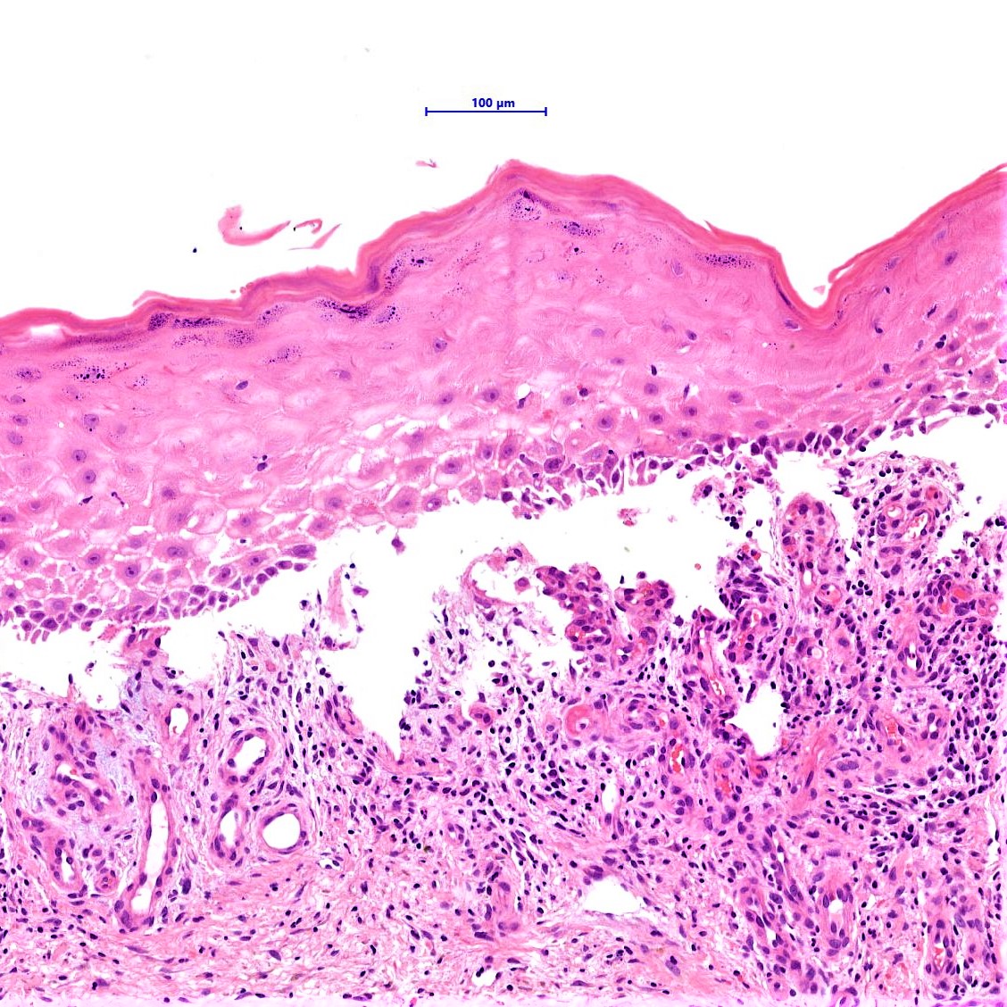
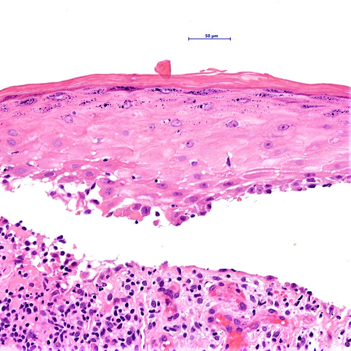
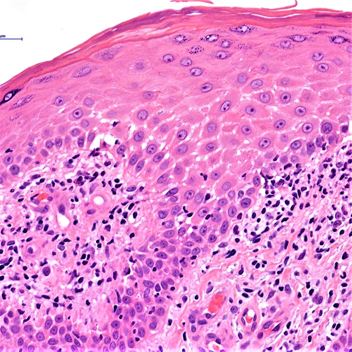
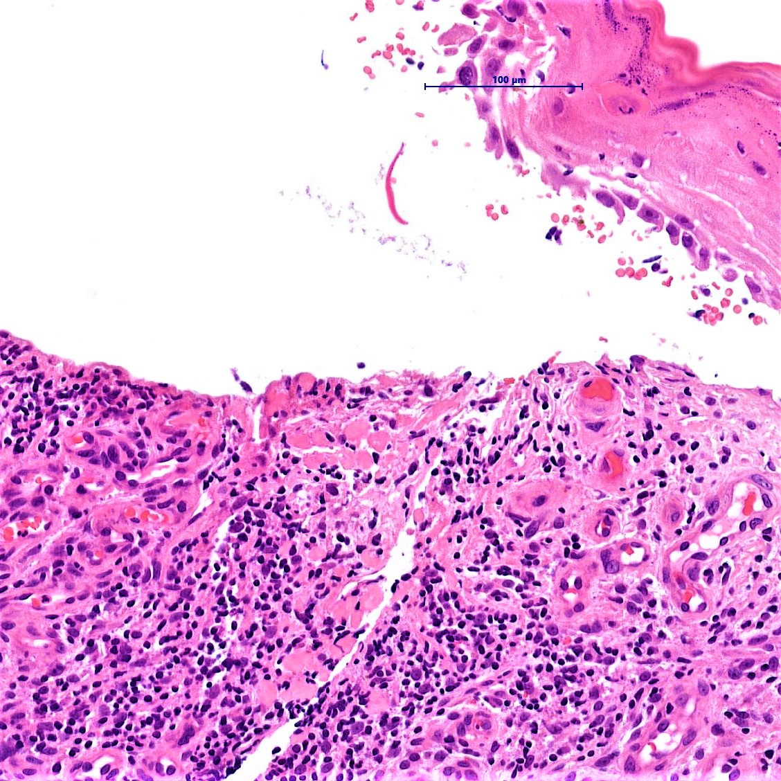
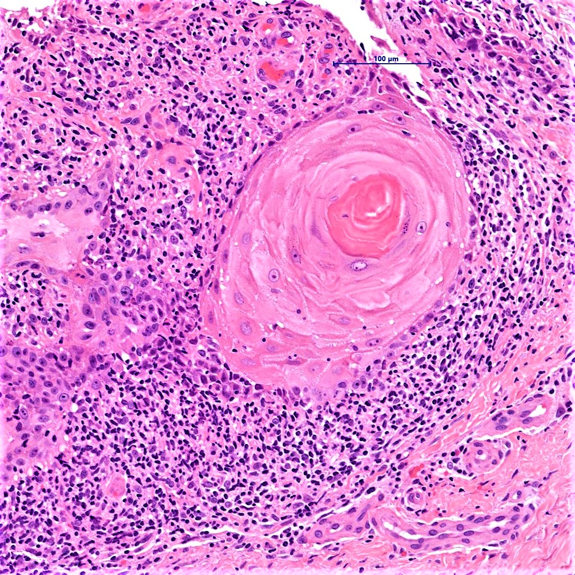
Dermoepidermal blister, dermal colloid bodies, hypergranulosis and orthokeratosis, a quasi-sawtooth acanthosis, make me think of Bullous Lichen Planus. 

Share this comment


Link to comment
share_externally

Richard Logan

Posted

Something about this case doesn't quite fit. 

I don't think the clinical photograph is representative of the sort of lesion that was biopsied.  The photograph shows a very superficial scaly erythematous problem which clinically looks like asteatotic eczema (eczema craquelee).  However, histologically there is a sub-epidermal blister with a lichenoid reaction, spongiosis and mild hypergranulosis rather than parakeratosis.  Were it not for the blistering, histologically I would be wondering about pityriasis lichenoides.  I would certainly do immunofluorescence in case this is lichen plano-pemphigoides.  I would enquire about the drug history.

Share this comment


Link to comment
share_externally

Iskander H. Chaudhry

Posted

Dear all thank you for your insightful comments. Richard I was on the same lines as you! 

Skin with spongiosis and interface change. There is a sub-epidermal split which may have occurred at the time of biopsy. No eosinophils are seen. No fungal hyphae are seen on specials. The features are those of a combined interface and spongiotic reaction pattern and the favoured differential is Lichen Planus. Spongiosis can be seen at this site particularly in the hypertrophic variant. The differential includes a bullous drug eruption.

COMMENT:

The clinician felt it would fit with Hypertrophic LP but was going to exclude the other differentials. 


Share this comment


Link to comment
share_externally

Richard Logan

Posted

The clinical photograph doesn't look anything like hypertrophic LP, nor does the histology!

Share this comment


Link to comment
share_externally


Join the conversation

You can post now and register later. If you have an account, sign in now to post with your account.

Guest
Add a comment...

×   Pasted as rich text.   Paste as plain text instead

  Only 75 emoji are allowed.

×   Your link has been automatically embedded.   Display as a link instead

×   Your previous content has been restored.   Clear editor

×   You cannot paste images directly. Upload or insert images from URL.

Loading...

×
×
  • Create New...