Jump to content
In this section we have spot diagnoses posted on a daily basis since June 2010, now over 4000! You can review the archived cases and read the suggested diagnoses by users and the final comment by the contributors.
Case are uploaded each week day by 10 am UK time with the correct diagnosis will generally be posted at 8 pm UK time. Why not view the most recent spot diagnosis and proffer a diagnosis?

Case Number : Case 2736 - 31 December 2020 Posted By: Saleem Taibjee

Please read the clinical history and view the images by clicking on them before you proffer your diagnosis.
Submitted Date :
   (0 reviews)

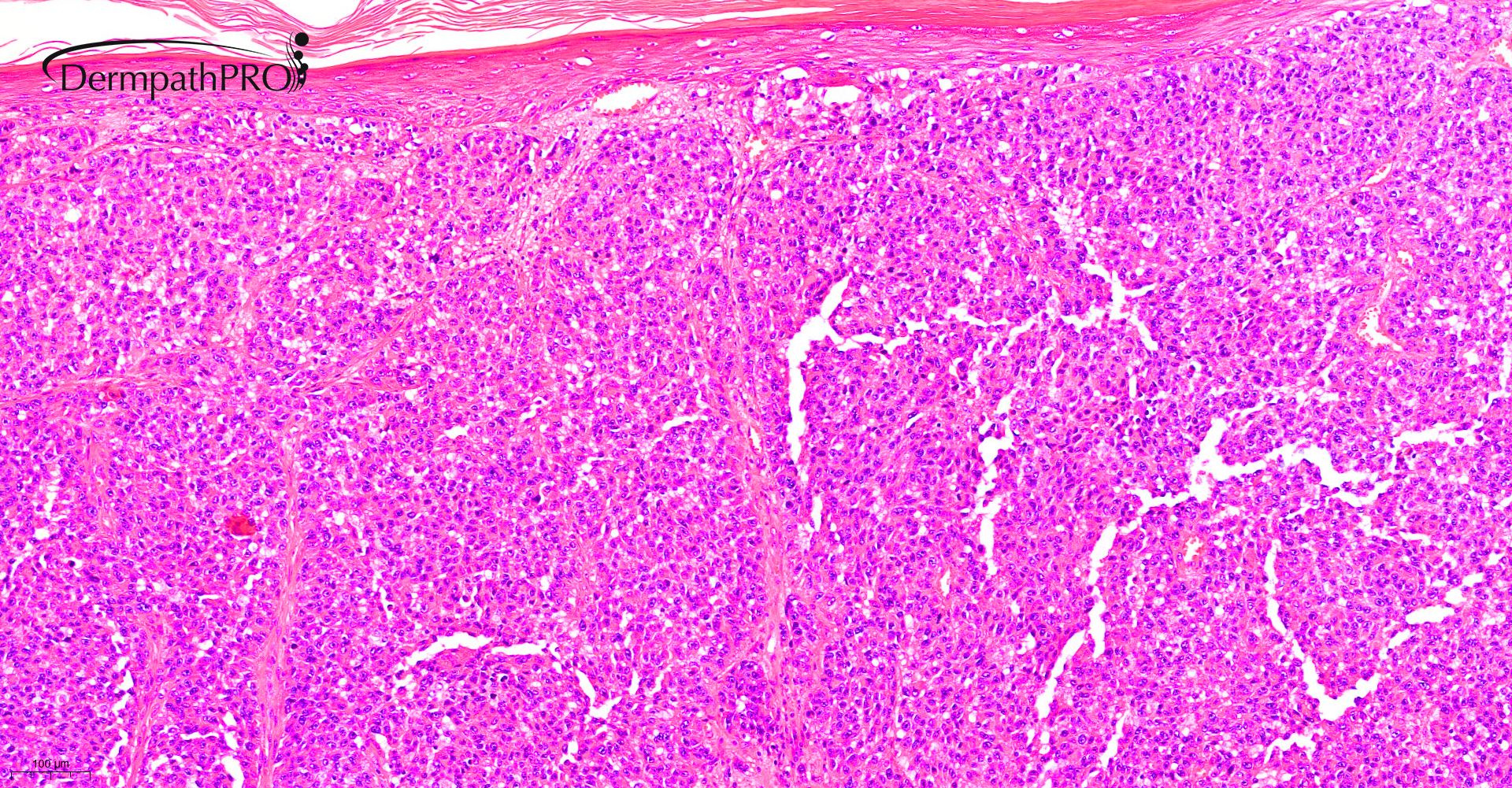
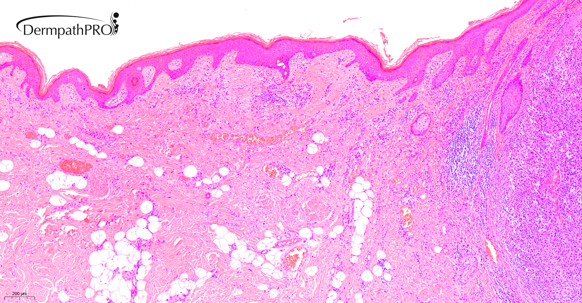
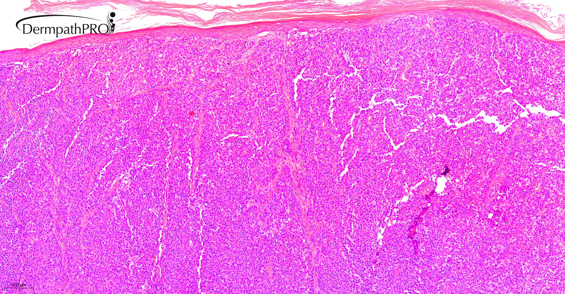
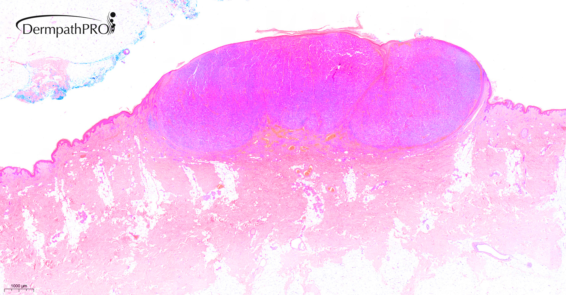
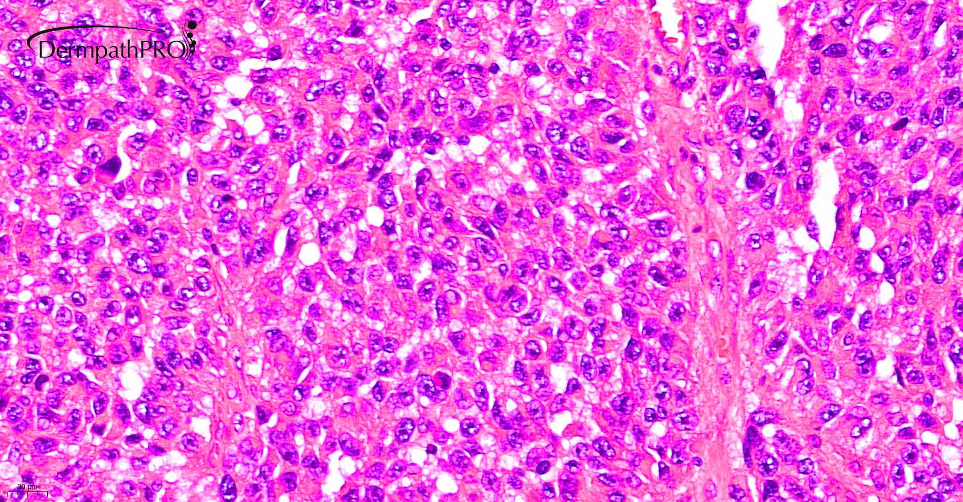
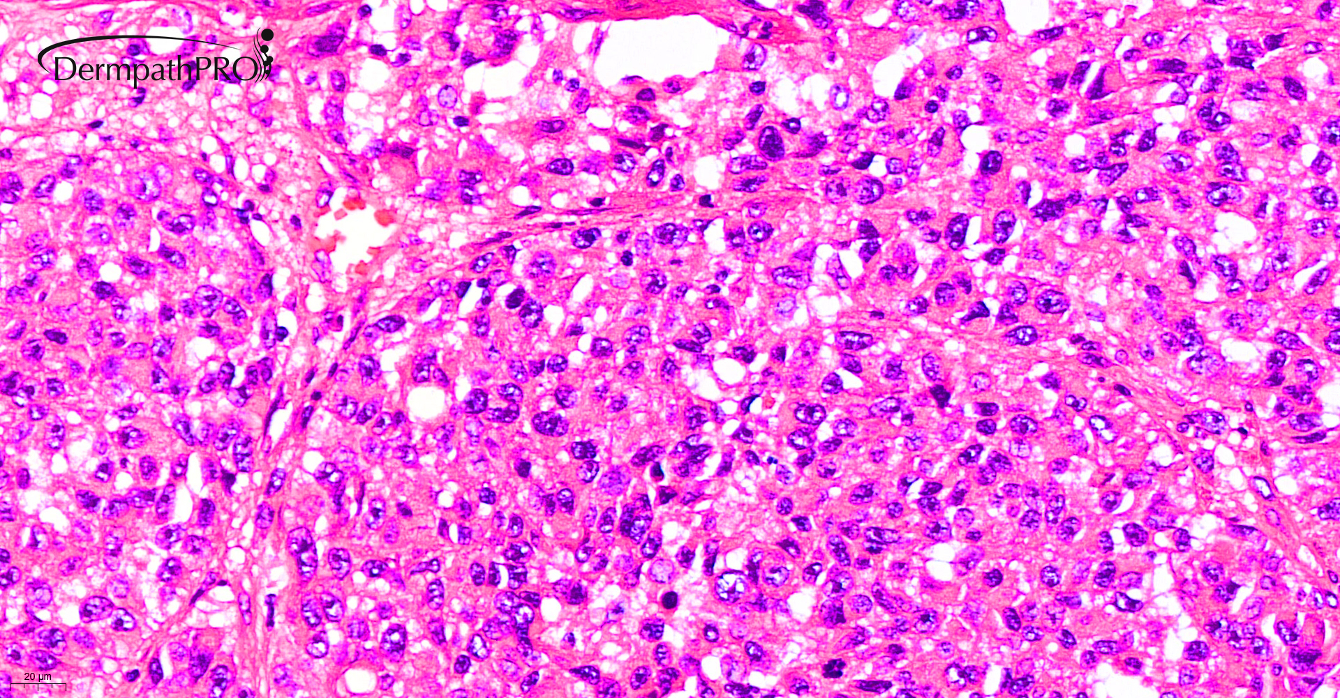
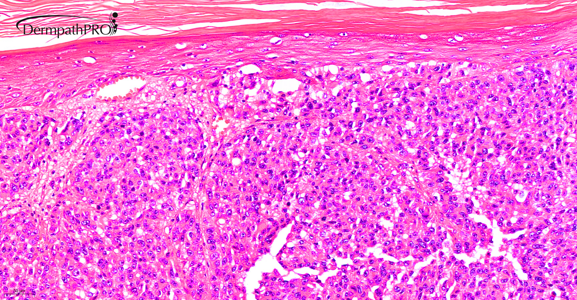
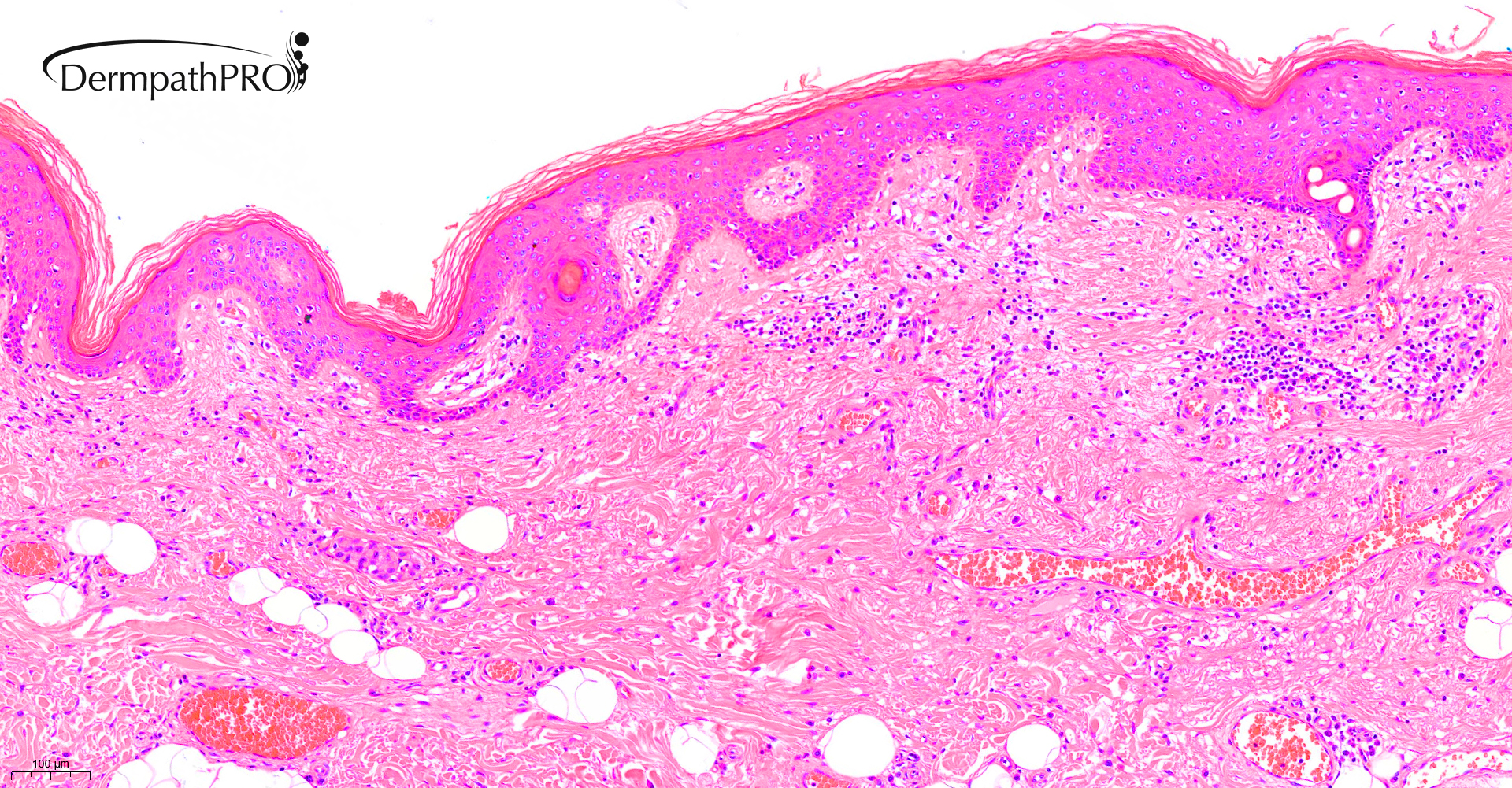
91M, 6 month history of scabby nodule on back


  Report Record

User Feedback


Richard Logan

Posted

For my last throw of the dice at the end of a year we'll all be glad to see the back of:

Neuroendocrine carcinoma. 

Some New Year immunohistochemistry awaited with bated breath!

Happy New Year everyone.

Share this comment


Link to comment
share_externally

Happy New Year !

agree with De Logan, it is consistent with Merkel Cell carcinoma, I can see some scattered similar cells in the epidermis. The chromatin is nice “ salt and peppers” neuroendocrine looking.  Question for the group discussion: how often do you do the Merkel cell polyomavirus test? Is it required nowadays for prognosis purpose?

Share this comment


Link to comment
share_externally

?Melanoma, with angiolymphatic infiltration ( second and last fig. 

Share this comment


Link to comment
share_externally

Saleem Taibjee

Posted

I'm not sure that the images uploaded that well initially. Hence see below.

S20-9169_1.0x.jpg

S20-9169_5.0x.jpg

S20-9169_40.0x.jpg

S20-9169_40.0xb.jpg

S20-9169_5.0xb.jpg

S20-9169_10.0xb.jpgS20-9169_20.0xb.jpg


Share this comment


Link to comment
share_externally

Richard Logan

Posted

Looking at these images I'm now less inclined towards a diagnosis of Merkel cell carcinoma.  The tumour cells have an eosinophilic cytoplasm and show vacuolation.  The last image shows intravascular spread.  Is this a metastatic SCC or suchlike?

Share this comment


Link to comment
share_externally

Saleem Taibjee

Posted

Happy New Year!

This is a recent case of amelanotic melanoma. The Melan-A shown below. The main reason for sharing it is that the intravascular micrometastasis was very subtle and could have been easily missed, especially during a busy sign out session. This is rather uncommon scenario in my experience, and of course, connotes a worse prognosis/upstaging.

Of course, the patient will undergo re-excision as per guidelines, but one is unsure whether this really affects the prognosis or outcome because in effect the disease is already 'metastatic'. For those that have not already read this excellent article by Wolfgang Weyers, I suggest reading Personalized Excision of Malignant Melanoma—Need for
a Paradigm Shift in the Beginning Era of Personalized Medicine in Am J Dermatopathol 2019;41:884–896.

BW, Saleem

S20-9169_18-12-2020_10-13-53_1.0x Melan-A.jpg

S20-9169_18-12-2020_10-13-53_2.0x pleomorphic dermal sarc.jpg

S20-9169_18-12-2020_10-13-53_20.0x Melan-A b.jpgS20-9169_18-12-2020_10-13-53_20.0x Melan-A.jpg


Share this comment


Link to comment
share_externally


Join the conversation

You can post now and register later. If you have an account, sign in now to post with your account.

Guest
Add a comment...

×   Pasted as rich text.   Paste as plain text instead

  Only 75 emoji are allowed.

×   Your link has been automatically embedded.   Display as a link instead

×   Your previous content has been restored.   Clear editor

×   You cannot paste images directly. Upload or insert images from URL.

Loading...

×
×
  • Create New...