Jump to content
In this section we have spot diagnoses posted on a daily basis since June 2010, now over 4000! You can review the archived cases and read the suggested diagnoses by users and the final comment by the contributors.
Case are uploaded each week day by 10 am UK time with the correct diagnosis will generally be posted at 8 pm UK time. Why not view the most recent spot diagnosis and proffer a diagnosis?

Case Number : Case 2511 - 20 February 2020 Posted By: Saleem Taibjee

Please read the clinical history and view the images by clicking on them before you proffer your diagnosis.
Submitted Date :
   (0 reviews)

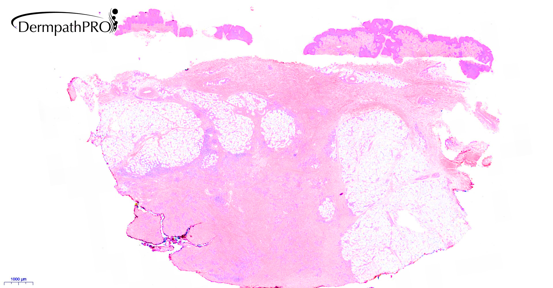
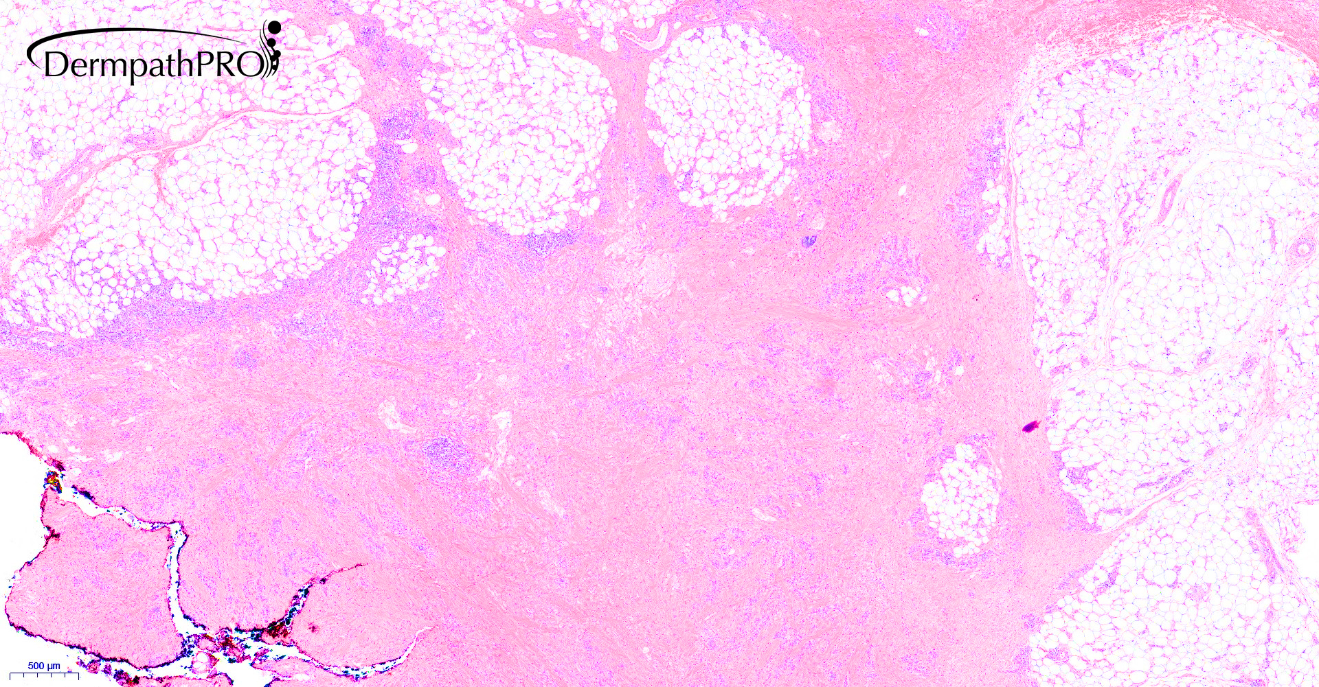
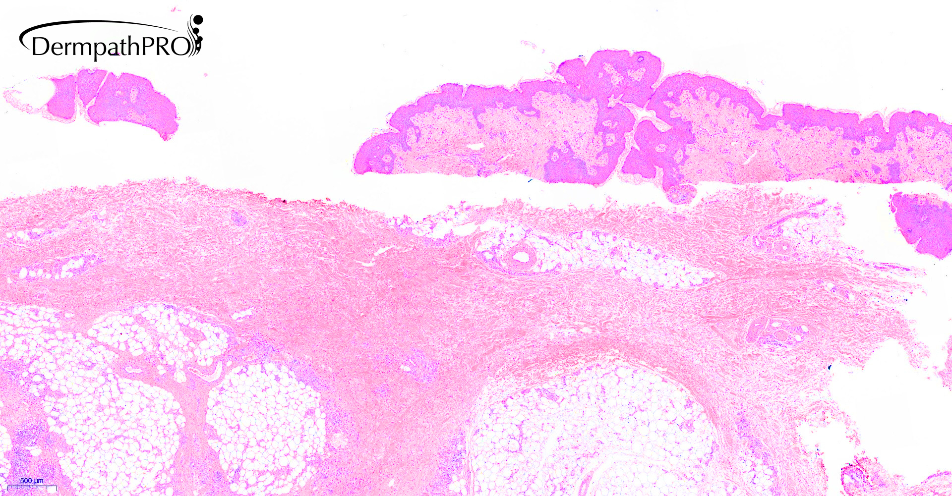
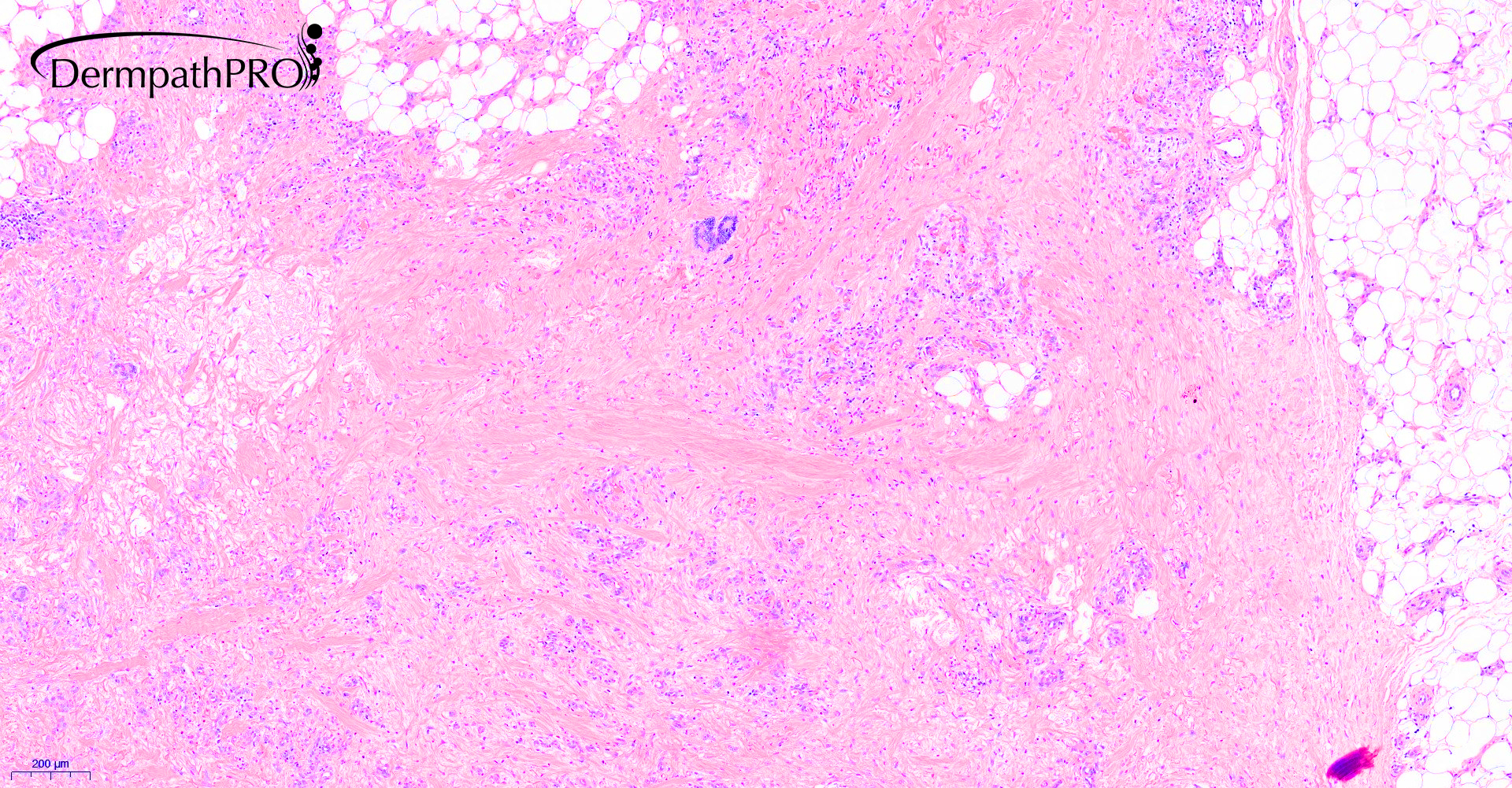
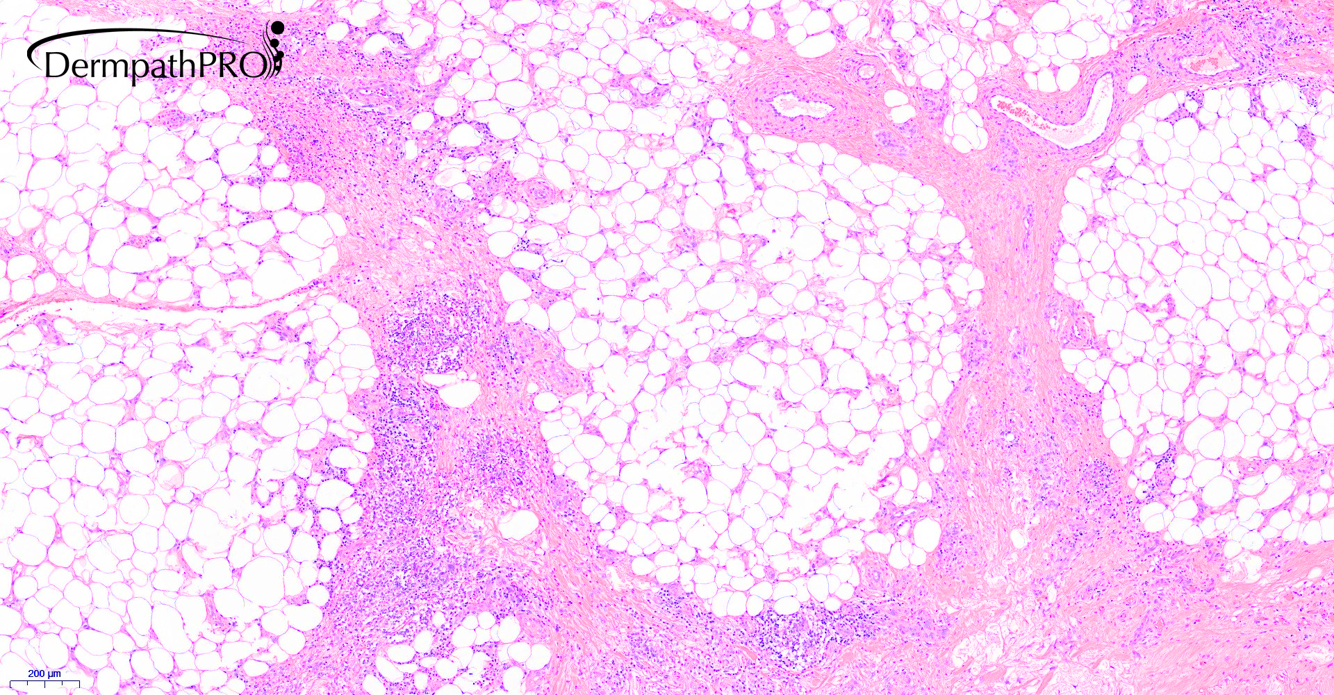
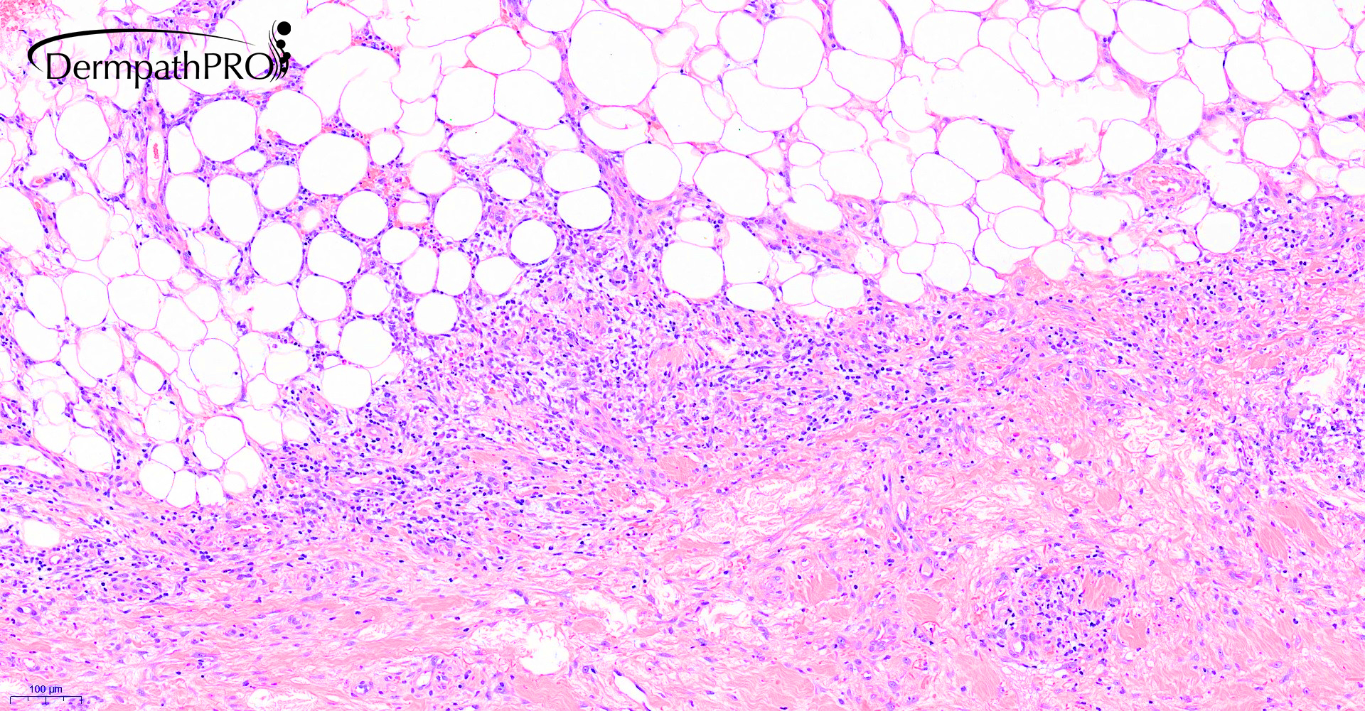
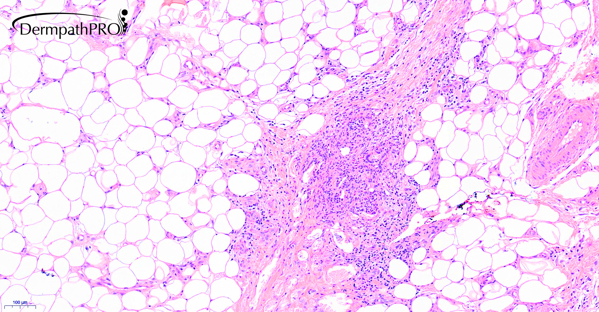
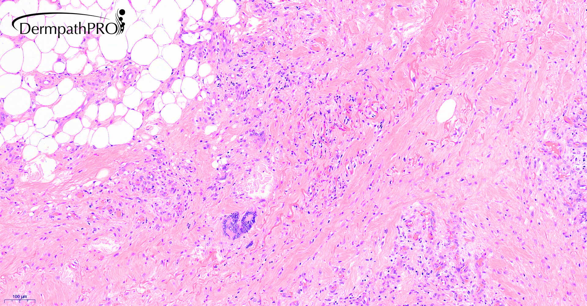
76M, an incisional biopsy from lumps behind the right knee.


  Report Record

User Feedback


Krishnakumar subramanian

Posted

granulomatous panniculitis

suggest PAS stain and examine under polarizer, something is there in the giant cells

Share this comment


Link to comment
share_externally

Dr. Mona Abdel-Halim

Posted

Granulomatous panniculitis with esinophils. Bug stains (especially PAS and silver methenamine), tissue culture and verification of foreign body

Share this comment


Link to comment
share_externally

Miescher radial granulomas...what about erythema nodosum?

Share this comment


Link to comment
share_externally

Saleem Taibjee

Posted

There were no organisms seen on special stains, and no polarising material on polarising microscopy.


The clinician was especially querying erythema induratum.


Here is the subsequent dialogue which I had with Professor Luis Requena (the 'god of panniculitis') about this case:


"Dear Professor Requena

I would be very grateful for your expert opinion on a panniculitis case sent to me for second opinion by one of our local hospitals/pathologists.

The patient is a 76 year old man with lumps behind the right knee. The clinician is querying erythema induratum. I do not have clinical images.
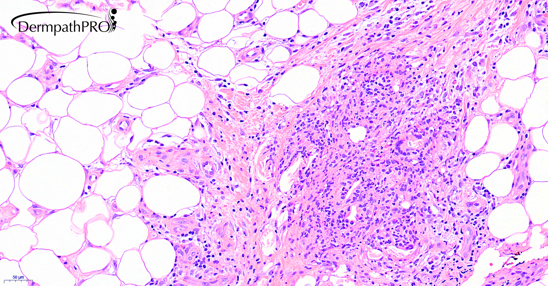
After attending your superb lecture on panniculitis previously some years ago, my reasoning is that this is more likely to be erythema nodosum (a chronic scarring variant) in view of the predominant septal (vs lobular) involvement, and absence of a clear-cut vasculitis. The histology is quite granulomatous (which as I noted can be a feature of late stages of erythema nodosum), but I could not appreciate definite Miescher’s radial granulomas or granulation type changes at the septal:lobule interface, but I must admit that I find these signs quite difficult to fully appreciate. From your lecture you had indicated that erythema induratum is very much predominantly lobular, which I think is not the case here.

Many thanks indeed for your advice and thoughts."


Professor Requena's reply:

"Dear Dr. Taibjee:

I entirely agree with your interpretation and a late stage erythema nodosum in a granulomatous phase is the best possibility.

Best regards. Luis Requena, MD"

Share this comment


Link to comment
share_externally

Krishnakumar subramanian

Posted

sir was it unilateral or bilateral

if bilateral  then EN if unilateral we need to think

Share this comment


Link to comment
share_externally

Saleem Taibjee

Posted

unilateral. Yes, agree unusual for EN, but still possible.

Share this comment


Link to comment
share_externally


Join the conversation

You can post now and register later. If you have an account, sign in now to post with your account.

Guest
Add a comment...

×   Pasted as rich text.   Paste as plain text instead

  Only 75 emoji are allowed.

×   Your link has been automatically embedded.   Display as a link instead

×   Your previous content has been restored.   Clear editor

×   You cannot paste images directly. Upload or insert images from URL.

Loading...

×
×
  • Create New...