Jump to content
In this section we have spot diagnoses posted on a daily basis since June 2010, now over 4000! You can review the archived cases and read the suggested diagnoses by users and the final comment by the contributors.
Case are uploaded each week day by 10 am UK time with the correct diagnosis will generally be posted at 8 pm UK time. Why not view the most recent spot diagnosis and proffer a diagnosis?

Case Number : Case 2486 - 15 January 2020 Posted By: Dr. Hafeez Diwan

Please read the clinical history and view the images by clicking on them before you proffer your diagnosis.
Submitted Date :
   (0 reviews)

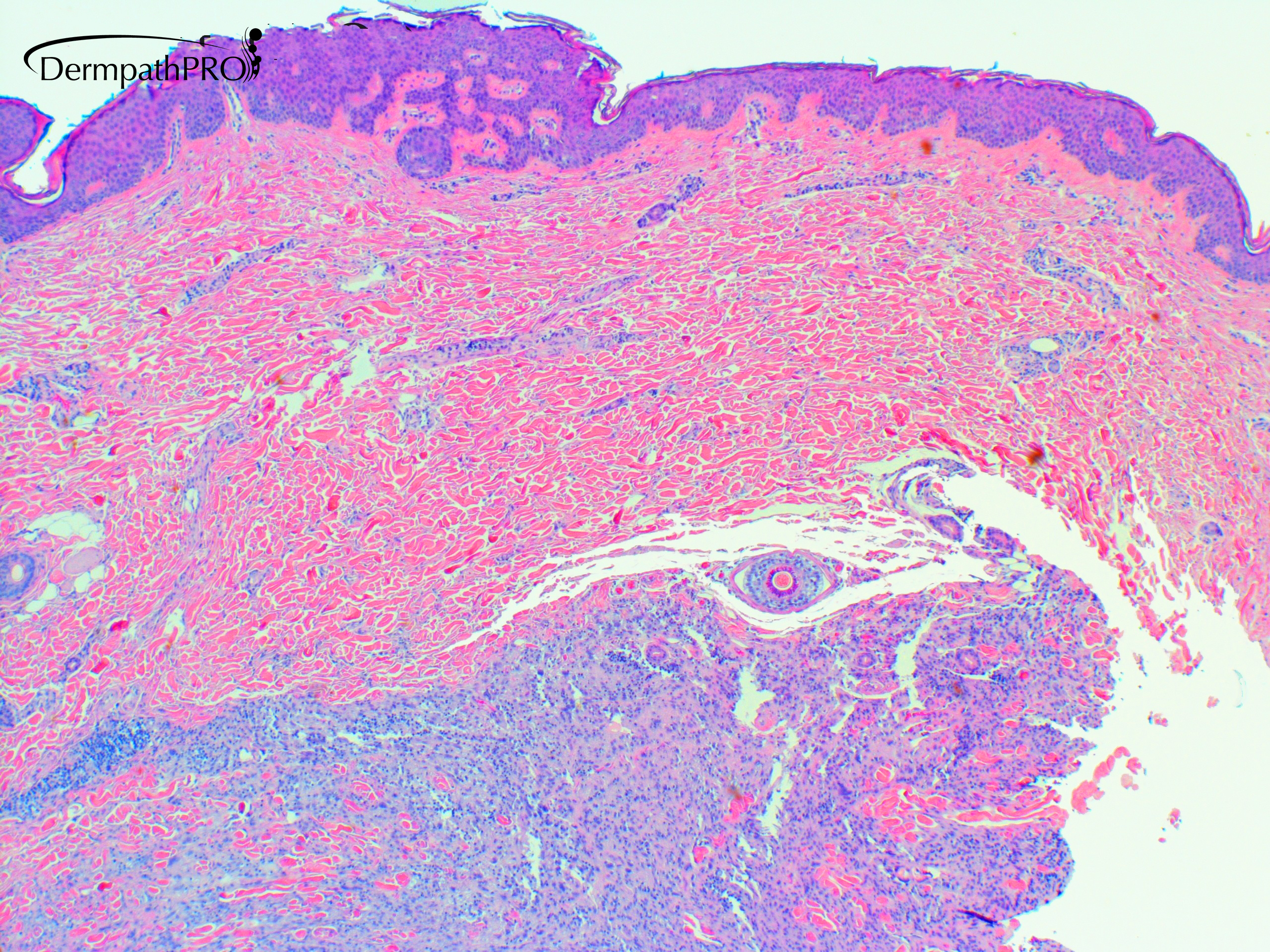
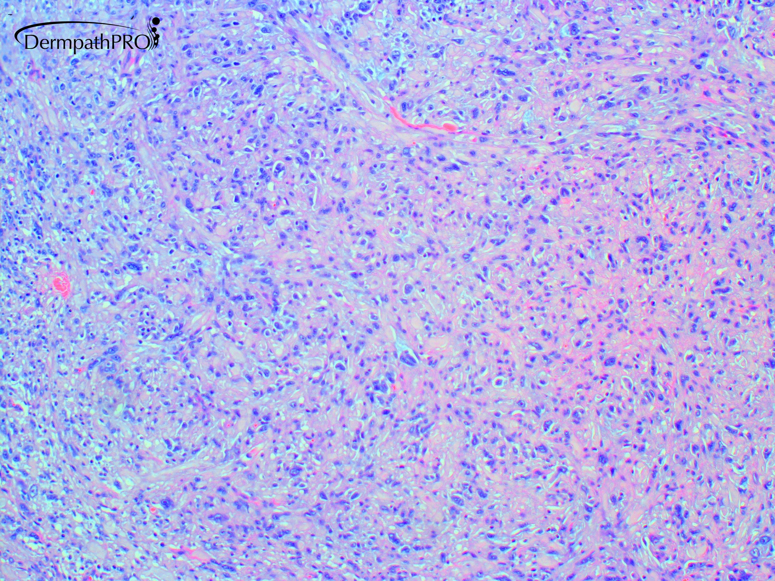
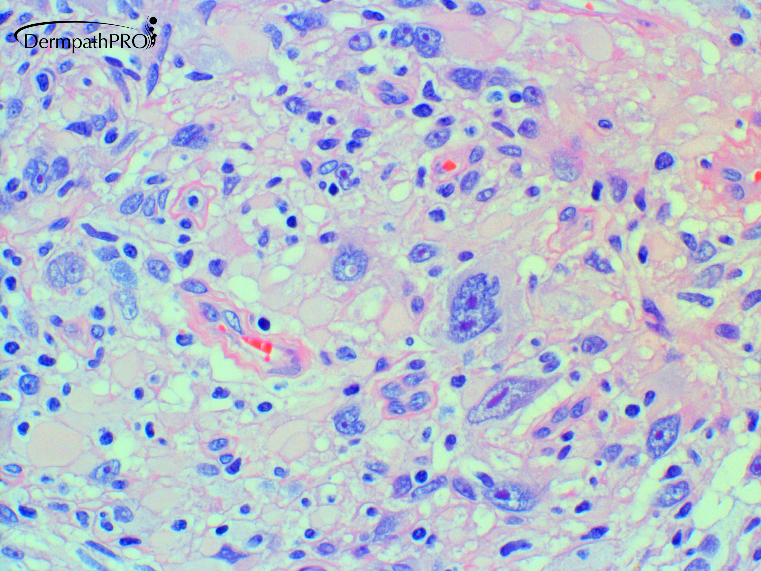
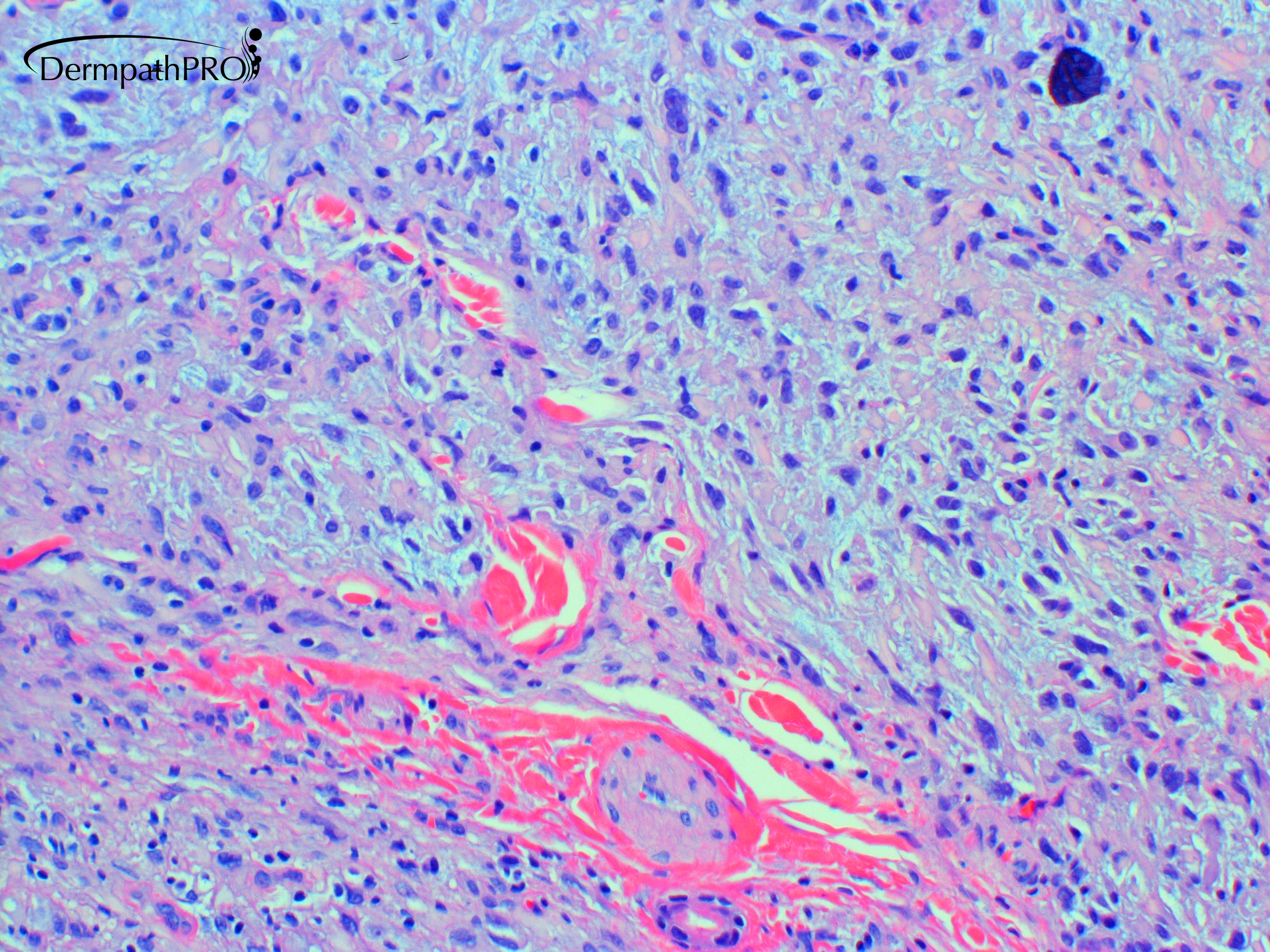
39 year-old female withleft lower limb mass


  • Like 1
  Report Record

User Feedback


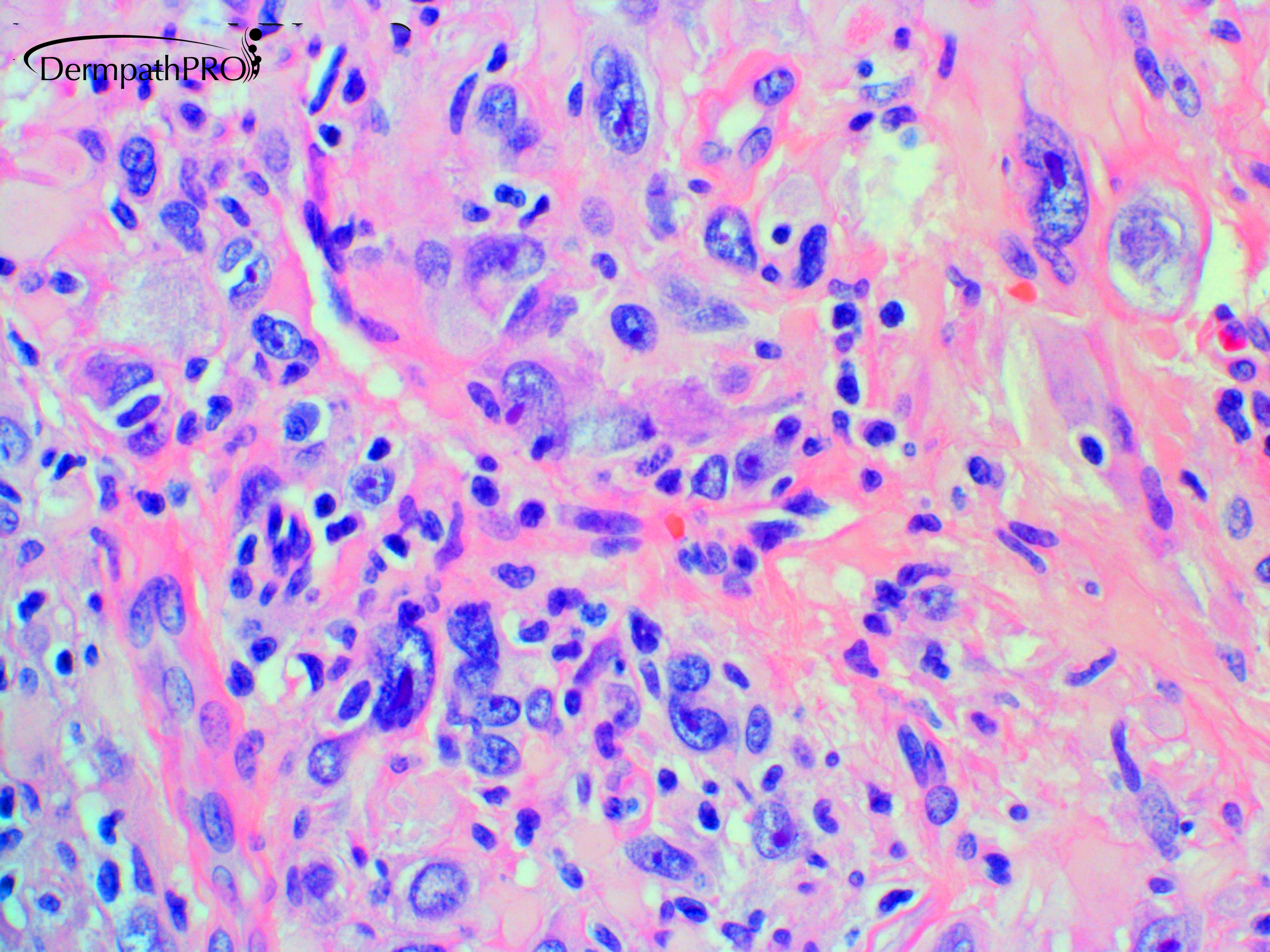
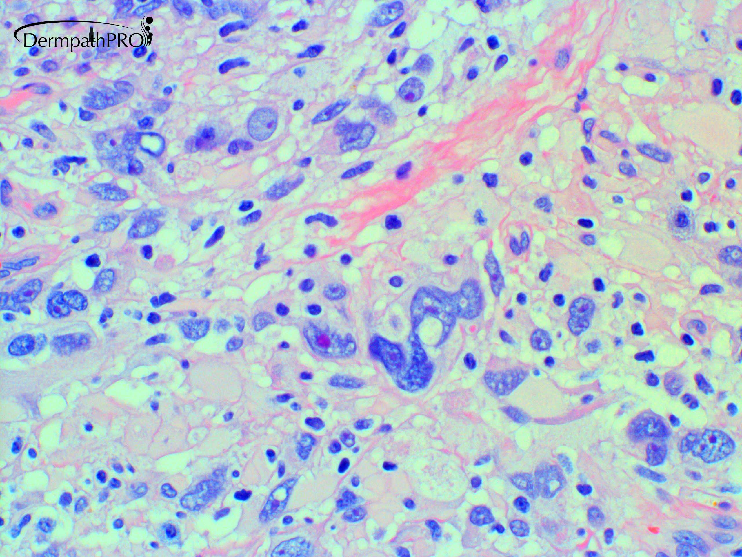
Large bizarre nuclei with cherry-red nucleoli .Options are broad and include melanoma / carcinoma (e.g sebaceous or metastatic )  / sarcoma (e.g angiosarcoma or leiomyosarcoma or  eptheloid or undifferentiated  ) / Chordoma / and Lymphoma (e.g Anaplastic large cell  or diffuse large B-cell leg type).

My gut feeling is for extra- axial chordoma.

For IHC tests.

Share this comment


Link to comment
share_externally

Deep cutaneous tumor (maybe soft parts extending to skin) with atypical virocyte-cells, inflammatory background plus focal myxoid stroma (last picture). My hypothesis is Myxoinflammatory fibroblastic sarcoma.

Share this comment


Link to comment
share_externally

First image is diagnostic for me: Fibrous Histiocytoma(DF), atypical variant. 

Share this comment


Link to comment
share_externally

Eman El-Nabarawy

Posted

1 hour ago, vincenzo said:

First image is diagnostic for me: Fibrous Histiocytoma(DF), atypical variant. 

Agree. Epidermal reaction, preserved adnexa and keloidal collagen.  

Share this comment


Link to comment
share_externally

Krishnakumar subramanian

Posted

dermatofibroma with pleomorphism and monster cells

Share this comment


Link to comment
share_externally

Dr. Hafeez Diwan

Posted

Myxoinflammatory fibroblastic sarcoma. CD34 was positive.

Share this comment


Link to comment
share_externally

Krishnakumar subramanian

Posted

Dear  respected madam, do we need fusion transcript studies to confirm the diagnosis or we relay on CD 34

as there is variable positivity for  CD68, CD34, c-kit,SMA,keratin, desmin, EGFR in these tumors

please let me know

since CD 34 is positive it has excluded fibrous histiocytoma with atypical cells


Share this comment


Link to comment
share_externally

Krishnakumar subramanian

Posted

also must we do S 100. Prof Fletcher discusses Cd34 and  s 100 positive tumors which have fusion genes

Genes Chromosomes Cancer. 2019 Nov;58(11):739-746. doi: 10.1002/gcc.22767. Epub 2019 Jun 4.

The histologic spectrum of soft tissue spindle cell tumors with NTRK3 gene rearrangements.

.

Abstract

NTRK3-rearranged tumors other than infantile fibrosarcomas (IFSs) harboring the canonical ETV6-NTRK3 fusions are uncommon, and include mainly inflammatory myofibroblastic tumors and gastrointestinal stromal tumors. Herein, we describe an additional subset of seven tumors sharing NTRK3 gene rearrangements. The cohort included five females and two males (age range 1-67 years). Tumors were located in extremities, trunk, retroperitoneum, or intra-abdominal. In all tumors, fluorescence in situ hybridization (FISH) revealed rearrangements in NTRK3 accompanied by NTRK3 amplification in two cases. In three cases, RNA sequencing identified a fusion transcript composed of NTRK3 exon 14 fused to ETV6, TFG, and TPM4, respectively, retaining the NTRK3 kinase domain. All tumors were positive for pan-TRK by immunohistochemistry (IHC). Two cases showed low- to intermediate-grade histology composed of monomorphic spindle cells arranged in a patternless architecture, stromal bands, and perivascular rings of hyalinized collagen and coexpression of S100 and CD34. The remaining five cases were high-grade fascicular monomorphic spindle cell sarcomas, morphologically somewhat reminiscent of either malignant peripheral nerve sheath tumors (MPNSTs) or fibrosarcomas (FSs). Two high-grade NTRK3 sarcomas showed aggressive clinical behavior with development of lung metastases. Identification of high-grade NTRK3-rearranged sarcomas is clinically important, since the development of selective NTRK inhibitors has opened new avenues for targeted therapy. Although IHC for pan-TRK can be applied as a screening tool, molecular studies are recommended for a conclusive diagnosis of NTRK-rearranged neoplasms.

Share this comment


Link to comment
share_externally

Nabil Mansouri2

Posted

Dear friends and brothers. I Do agree with atypical FH since there is an epidermal reaction. May be with hyperpigmentation. Green zone , lateral entrapement of collagen. CD34 Is not enough . Can be positf and week in FH

Share this comment


Link to comment
share_externally

daniellindsay

Posted

I would also consider Pleomorphic Hyalinising Angiectatic Tumour of soft parts here

Share this comment


Link to comment
share_externally


Join the conversation

You can post now and register later. If you have an account, sign in now to post with your account.

Guest
Add a comment...

×   Pasted as rich text.   Paste as plain text instead

  Only 75 emoji are allowed.

×   Your link has been automatically embedded.   Display as a link instead

×   Your previous content has been restored.   Clear editor

×   You cannot paste images directly. Upload or insert images from URL.

Loading...

×
×
  • Create New...