Jump to content
In this section we have spot diagnoses posted on a daily basis since June 2010, now over 4000! You can review the archived cases and read the suggested diagnoses by users and the final comment by the contributors.
Case are uploaded each week day by 10 am UK time with the correct diagnosis will generally be posted at 8 pm UK time. Why not view the most recent spot diagnosis and proffer a diagnosis?

Case Number : Case 2492 - 23 January 2020 Posted By: Saleem Taibjee

Please read the clinical history and view the images by clicking on them before you proffer your diagnosis.
Submitted Date :
   (0 reviews)

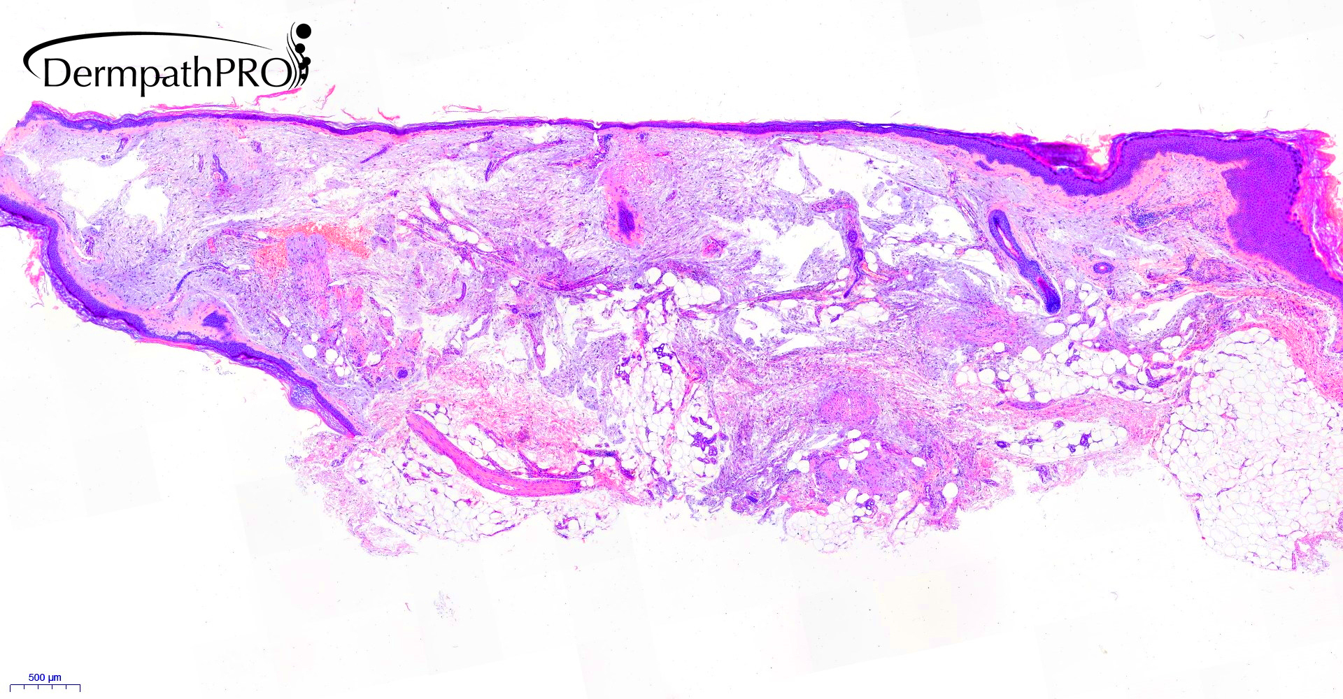
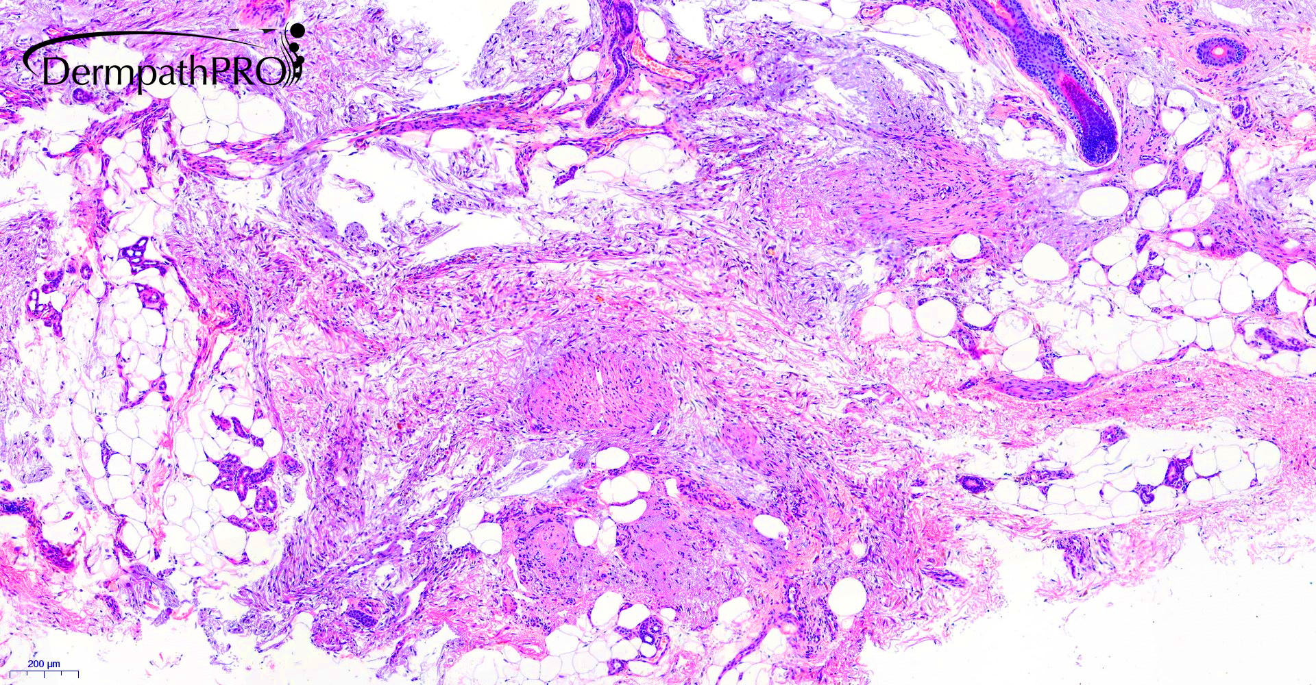
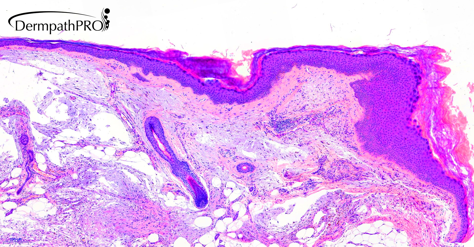
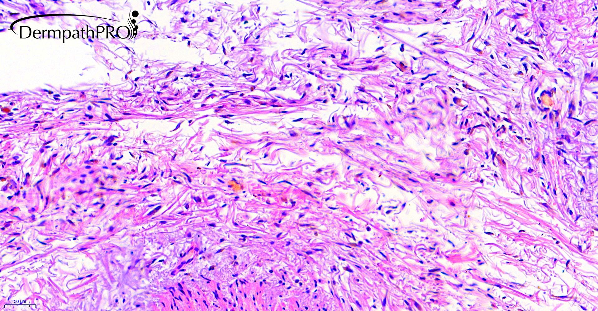
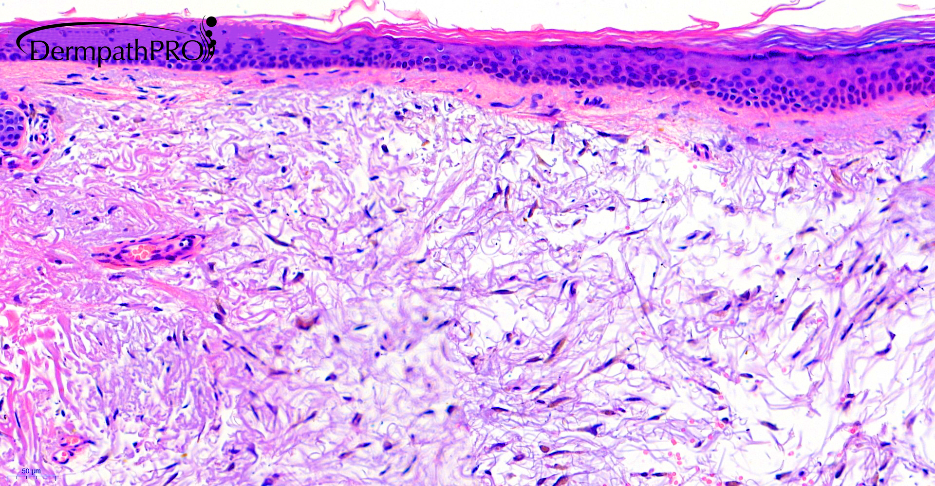
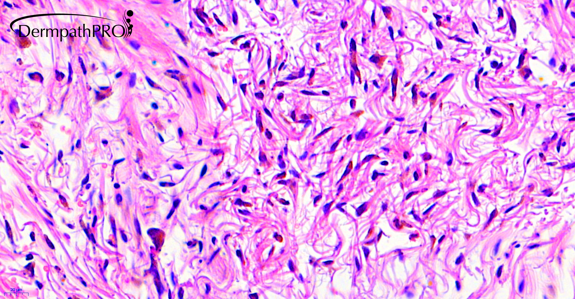
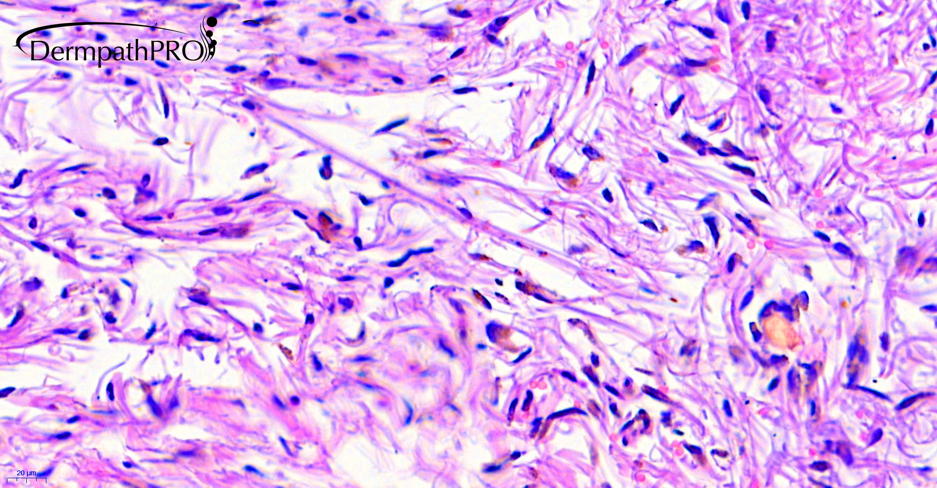
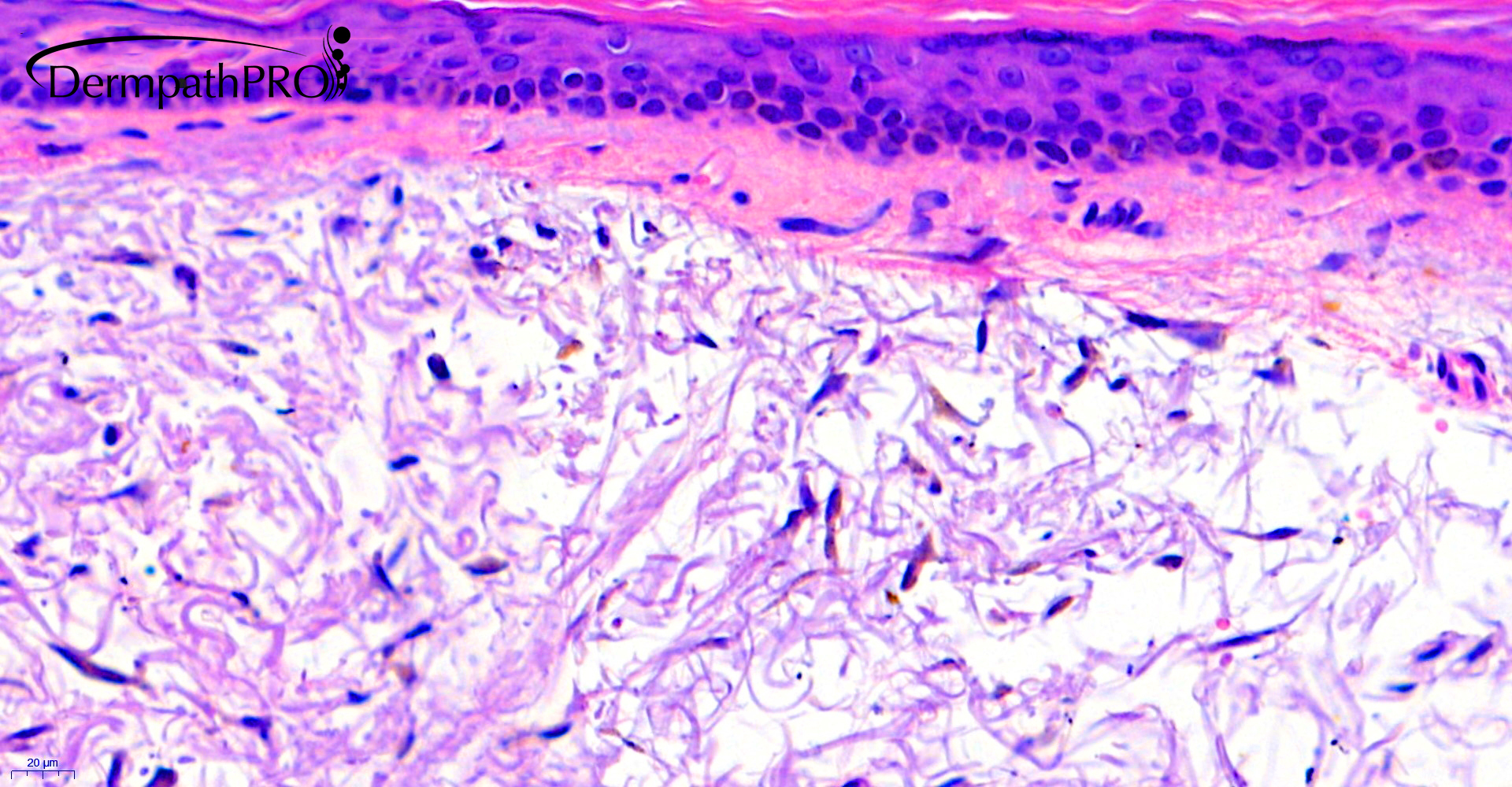
84F, incisional biopsy lower leg ?lentigo


  Report Record

User Feedback


Difficult case. It seems that tissue inclusion is not so correct, so it looks an "hamartoma" at the beginning. I see some dermal cells with brown pigment, but it doesn´t look dermal melanocytes, I think it could have some relation with medication. Maybe minocycline-induced hyperpigmentation, Type-II. I would do Perls and Fontana Masson stains.

Share this comment


Link to comment
share_externally

I don't know what this is!

Histologically it seems a scleromixedematous/nephrogenic systemic fibrosis-like, with an strange brown pigment. Clinically it seems a melanocytic-like lesion ( lentigo ). 

?Blue nevus with mucinous and cystic changes...

Share this comment


Link to comment
share_externally

Eman El-Nabarawy

Posted

Difficult. Ideas: Sclerosing mucinous blue nevus, angiomatoid cellular blue nevus, melanotic plexiform mucinous schwannoma.


Share this comment


Link to comment
share_externally

30 minutes ago, Eman El-Nabarawy said:

Difficult. Ideas: Sclerosing mucinous blue nevus, angiomatoid cellular blue nevus, melanotic plexiform mucinous schwannoma.


Like the last : melanotic mucinous schwannoma. 

Share this comment


Link to comment
share_externally

like to add superficial angiomyxoma , may be co-existing with post-inflammatory hyperpigmentation.

Share this comment


Link to comment
share_externally

Dr. Mona Abdel-Halim

Posted

Or melanocytic myxoid spindle cell tumor with ALK rearrangement?? Difficult and interesting lesion!

Share this comment


Link to comment
share_externally

Saleem Taibjee

Posted

OK, so no-one has got it yet, difficult case. Clue: The pigment is Perl's positive, Masson-Fontana negative.

Share this comment


Link to comment
share_externally

Dr. Mona Abdel-Halim

Posted

So with the clue given: hemosiderotic fibrohistiocytic lipomatous lesion???

Share this comment


Link to comment
share_externally

9 minutes ago, Dr. Mona Abdel-Halim said:

So with the clue given: hemosiderotic fibrohistiocytic lipomatous lesion???

Yes you are right, Mona. But it's very difficult for me. Superficial angiomyxoma is a my spot hypotheses because of the silhouette and the vascular and myxoid mix in a superficial site. However this is a predominant hemosiderotic lesion. I don't know. Waiting for the diagnosis.

Share this comment


Link to comment
share_externally

Eman El-Nabarawy

Posted

20 hours ago, Dr. Mona Abdel-Halim said:

So with the clue given: hemosiderotic fibrohistiocytic lipomatous lesion???

Fits well!

Share this comment


Link to comment
share_externally

Saleem Taibjee

Posted

Hi Guys.

Yes you are quire right, the working diagnosis at the moment (suggested by Richard Carr and Raul Perrett) is haemosiderotic fibrolipomatous tumour. It does seem to have the various components including prominent haemosiderin, spindle cell and fatty components. I have indicated this possibility in my original report, and suggested consideration of complete excision given the uncertain biological potential. I have since heard back from the local histopathologist (patient is from another hospital) after MDT discussion that the lesion is clinically a flat pigmented lesion, with no obvious lump. The clinical team plan to arrange an ultrasound scan in the first instance. Will keep you posted if I hear more.

BW
Saleem


Share this comment


Link to comment
share_externally


Join the conversation

You can post now and register later. If you have an account, sign in now to post with your account.

Guest
Add a comment...

×   Pasted as rich text.   Paste as plain text instead

  Only 75 emoji are allowed.

×   Your link has been automatically embedded.   Display as a link instead

×   Your previous content has been restored.   Clear editor

×   You cannot paste images directly. Upload or insert images from URL.

Loading...

×
×
  • Create New...