Jump to content
In this section we have spot diagnoses posted on a daily basis since June 2010, now over 4000! You can review the archived cases and read the suggested diagnoses by users and the final comment by the contributors.
Case are uploaded each week day by 10 am UK time with the correct diagnosis will generally be posted at 8 pm UK time. Why not view the most recent spot diagnosis and proffer a diagnosis?

Case Number : Case 2493 - 24 January 2020 Posted By: Dr. Richard Carr

Please read the clinical history and view the images by clicking on them before you proffer your diagnosis.
Submitted Date :
   (0 reviews)

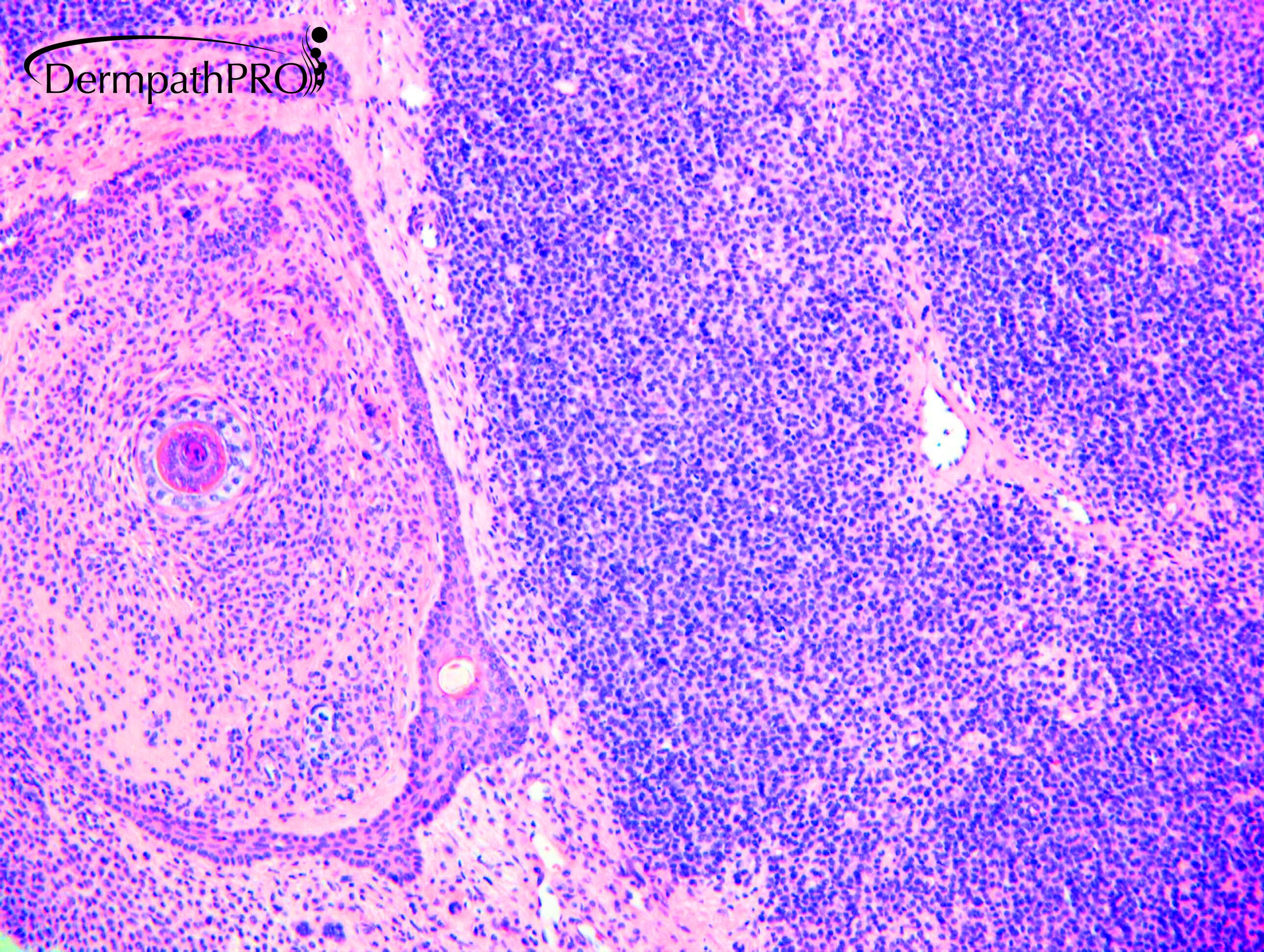
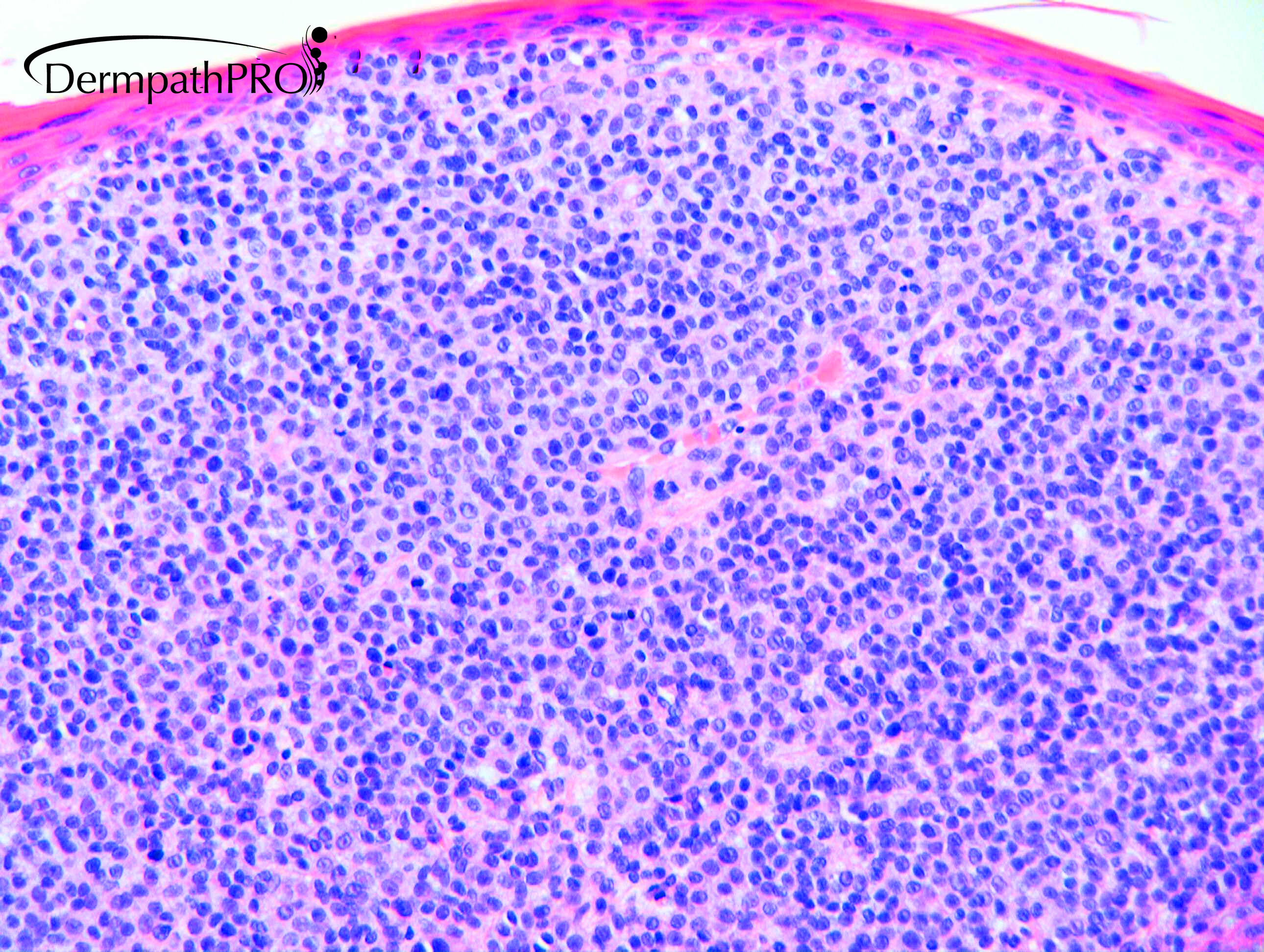
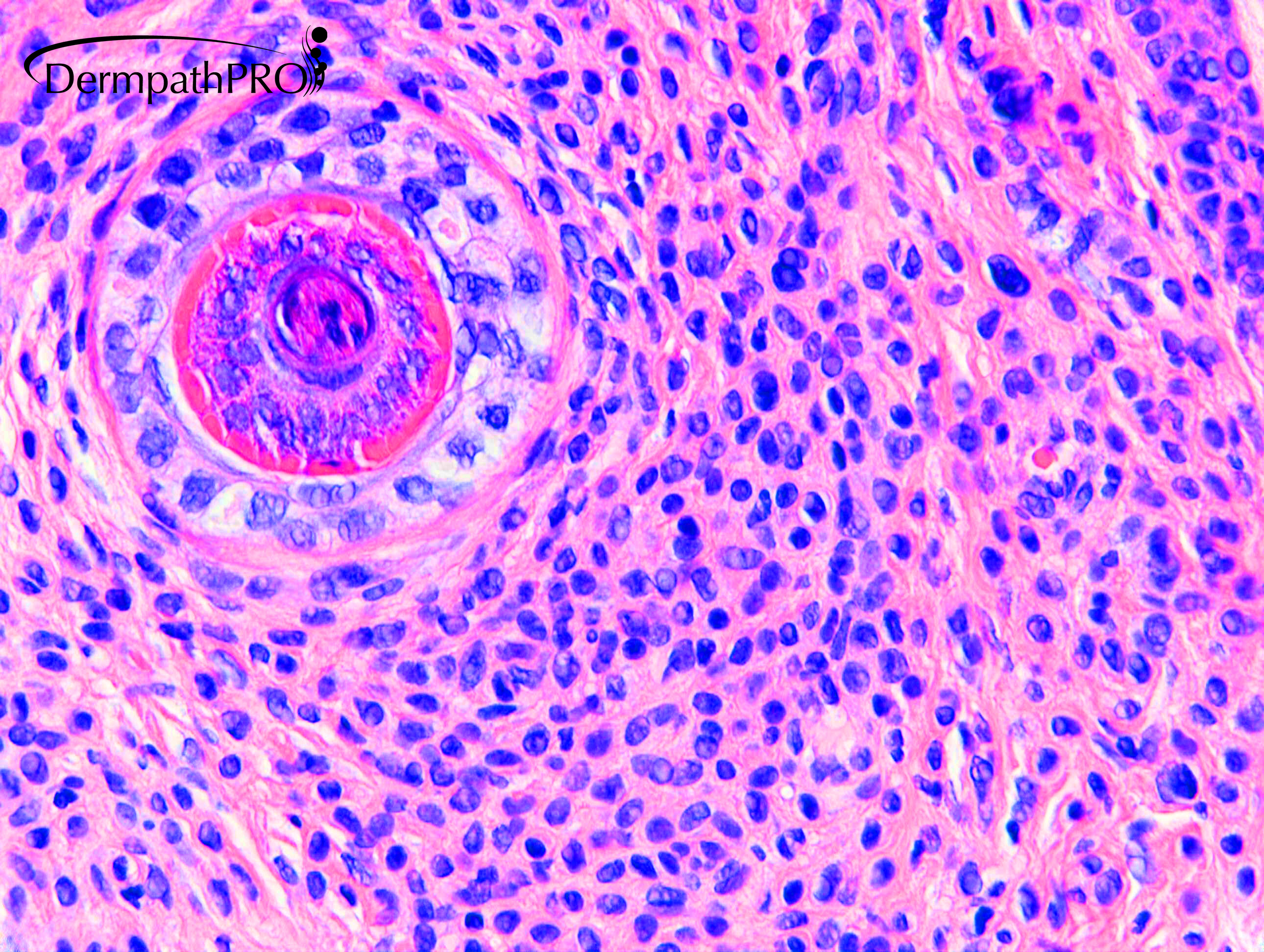
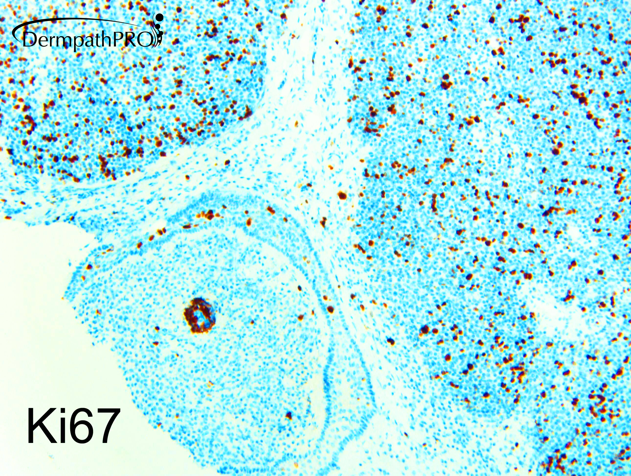
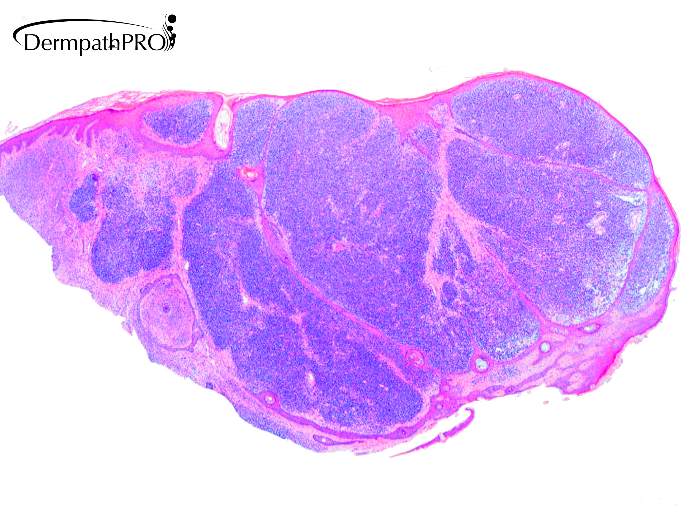
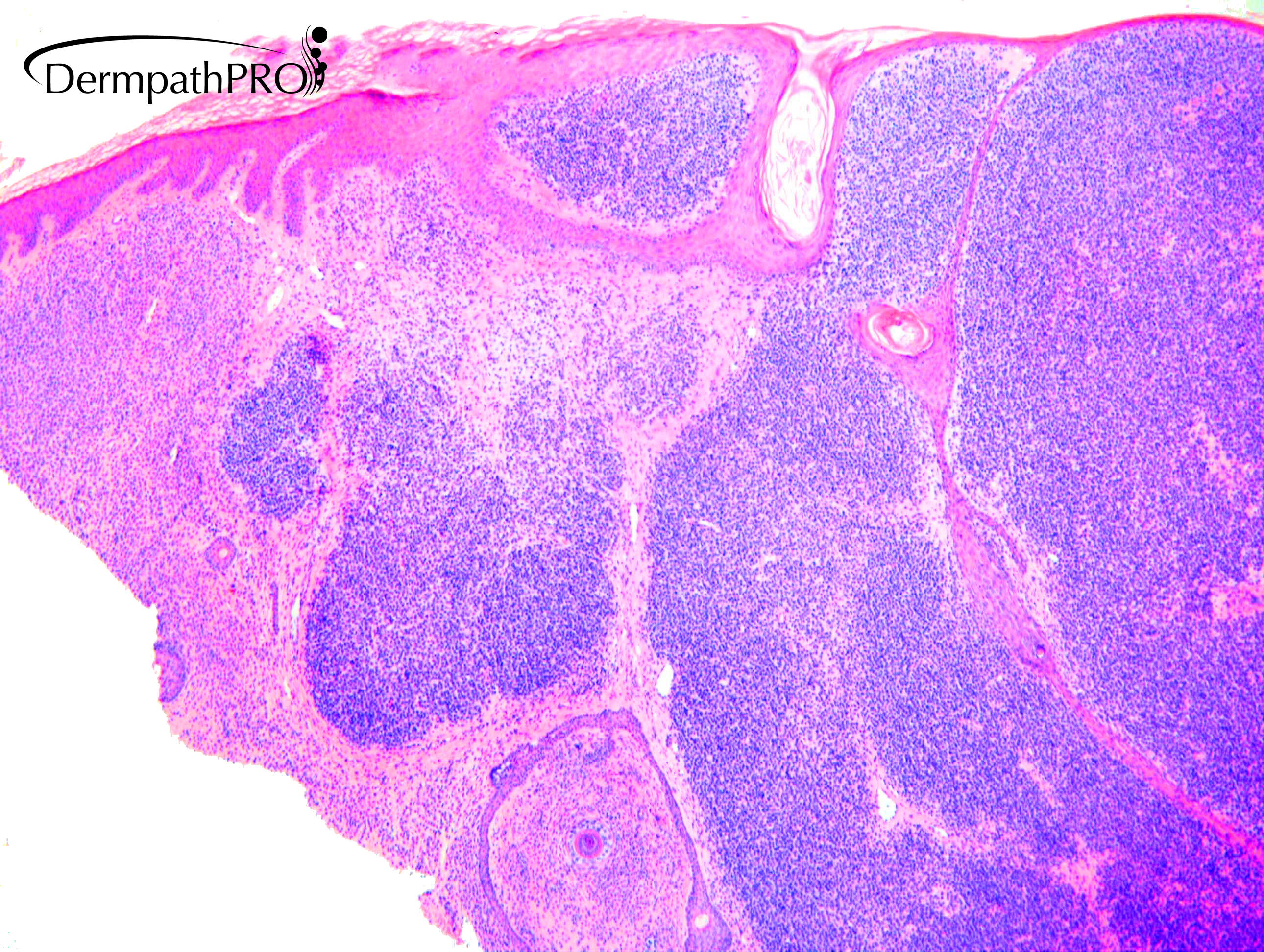
F71 Cheek. Longstanding globular congenital naevus cheek. Recurrently traumatised & bleeds. No atypical features.

  • Thanks 1
  Report Record

User Feedback


Dr. Mona Abdel-Halim

Posted

Congenital nevus with proliferation nodule? if it is a longstanding lesion with no clinical atypical features, then the mitotic activity is mostly just a result of repeated trauma. 

Share this comment


Link to comment
share_externally

Eman El-Nabarawy

Posted

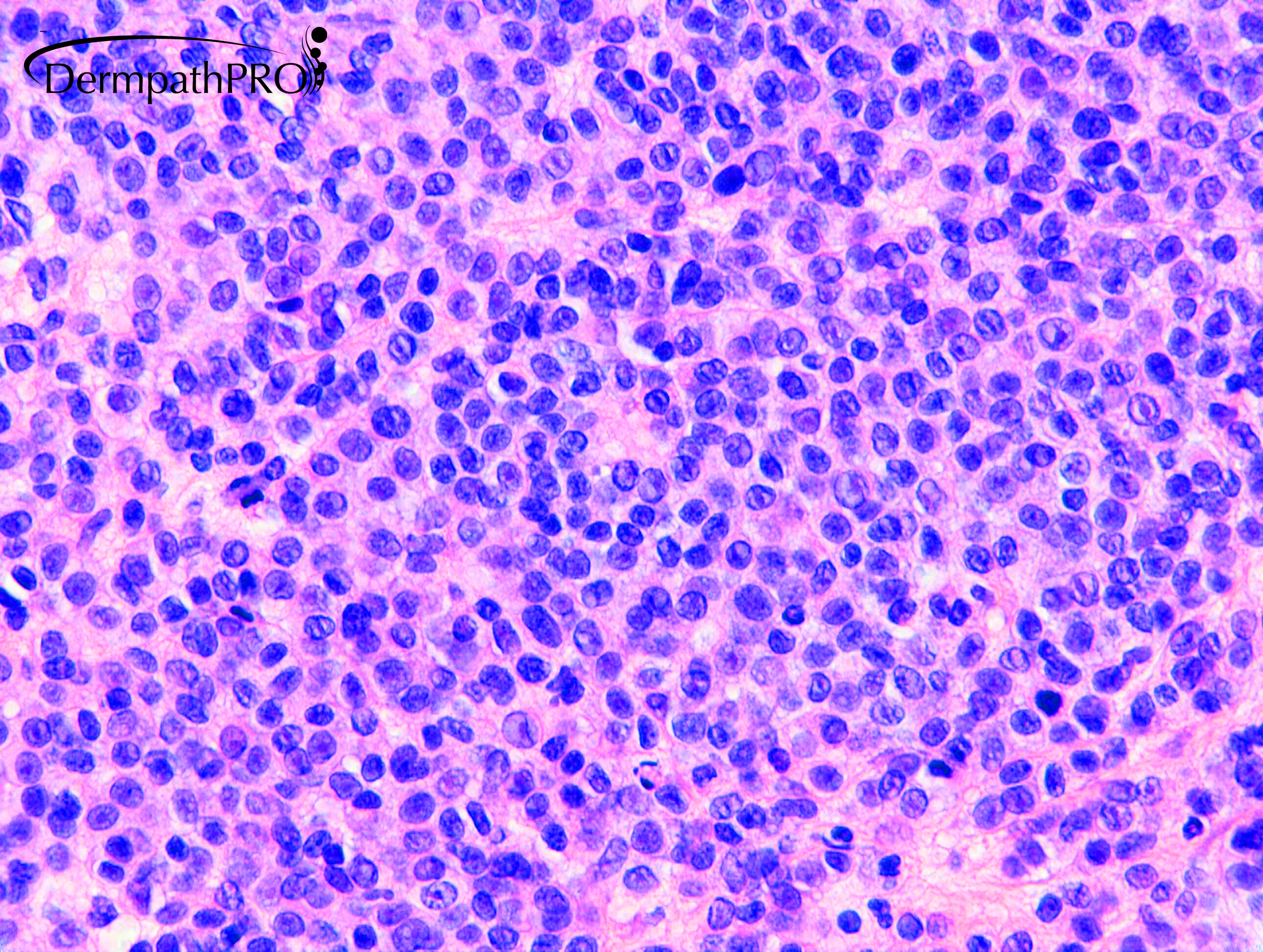
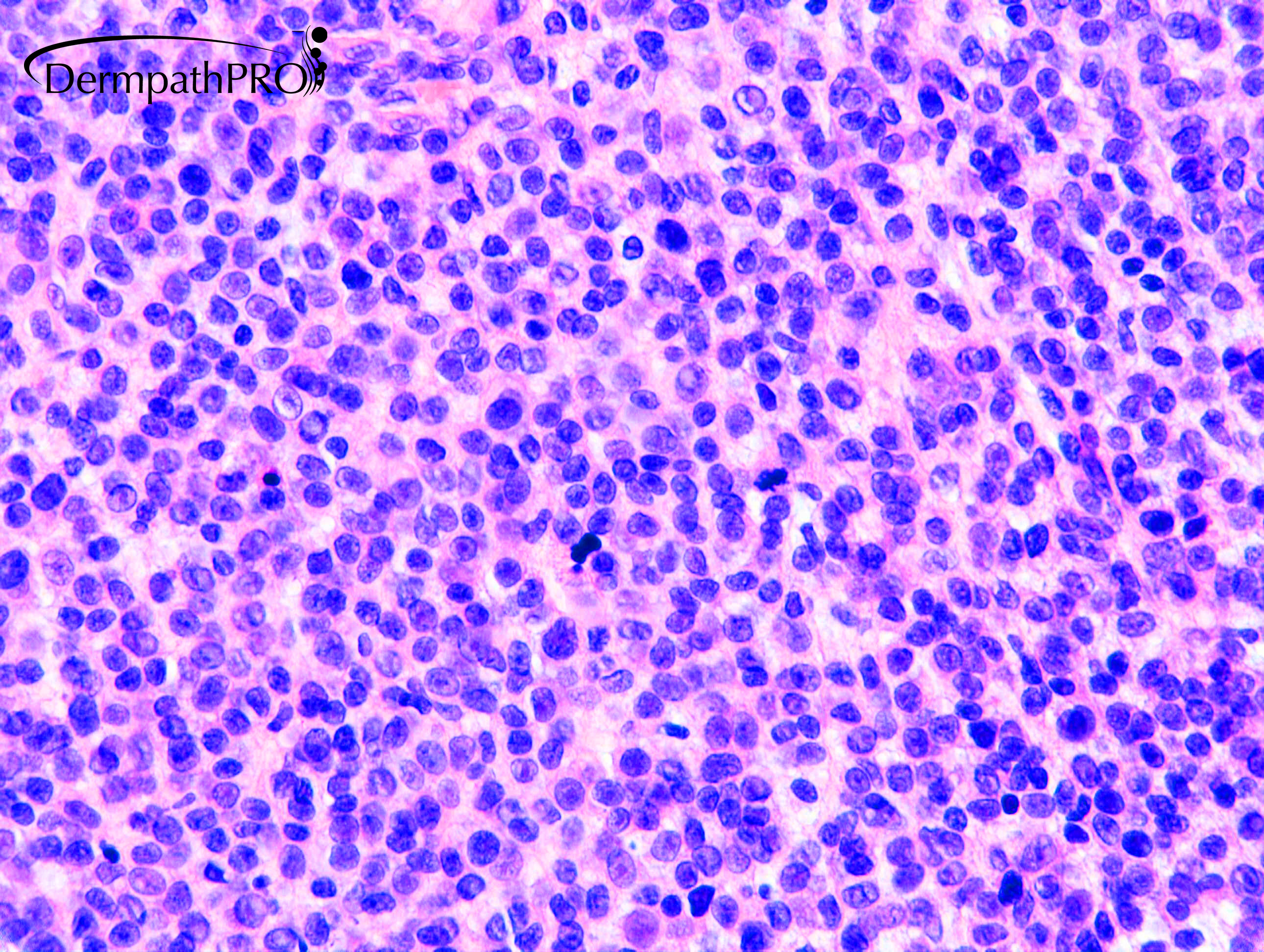
Looks innocent! Hope so. But old age, epidermal consumption (? trauma), deep mitoses, ? maturation, can't see enough stroma inbetween the nevus cells. Before I go to Congenital nevus with proliferation nodule, I want to see P16, ki67, HMB45, MelanA? To exclude nevoid melanoma. 

Share this comment


Link to comment
share_externally

Krishnakumar subramanian

Posted

I agree

it could be nevoid melanoma

also it is difficult to differentiate between atypical proliferation nodule or melanoma

IHC could be helpful

Share this comment


Link to comment
share_externally

Dr. Mona Abdel-Halim

Posted

Ki67 is high, so it could be nevoid melanoma but still how to differentiate it from nevoid melanoma like pattern of proliferation nodule? Especially that the clinical is describing a long standing congenital lesion with no atypical features? Will molecular be of need/help here? Interesting case! 

Share this comment


Link to comment
share_externally

Very interesting case. The blue population is atypical if compared with the residual congenital nevus in fig 7...but I hope never have to go trough this lesion during my routine work!!! 

Share this comment


Link to comment
share_externally

Dr. Richard Carr

Posted

Good discussion. Agree this is a challenge and therefore somewhat subjective. I reported it quite a while back (it was a referral). For me the density of the infiltrate, fairly obvious mitotic activity and high Ki67 proliferation, when comparing with the benign dermal naevus was enough to favour a diagnosis of dermal melanoma arising in a pre-existing benign naevus. Another feature which I don't associate with benign naevus is the presence of apoptotic bodies. I think this is also a clue to naevoid melanoma and not discussed in the textbooks.

Share this comment


Link to comment
share_externally

Nabil Mansouri2

Posted

. Dr Richard. I favor nevoid melanoma too . No maturation ki67 and paralel layout of cells. Thanks for sharing.

Share this comment


Link to comment
share_externally


Join the conversation

You can post now and register later. If you have an account, sign in now to post with your account.

Guest
Add a comment...

×   Pasted as rich text.   Paste as plain text instead

  Only 75 emoji are allowed.

×   Your link has been automatically embedded.   Display as a link instead

×   Your previous content has been restored.   Clear editor

×   You cannot paste images directly. Upload or insert images from URL.

Loading...

×
×
  • Create New...