Jump to content
In this section we have spot diagnoses posted on a daily basis since June 2010, now over 4000! You can review the archived cases and read the suggested diagnoses by users and the final comment by the contributors.
Case are uploaded each week day by 10 am UK time with the correct diagnosis will generally be posted at 8 pm UK time. Why not view the most recent spot diagnosis and proffer a diagnosis?

Case Number : Case 2497 - 31 January 2020 Posted By: Dr. Richard Carr

Please read the clinical history and view the images by clicking on them before you proffer your diagnosis.
Submitted Date :
   (0 reviews)

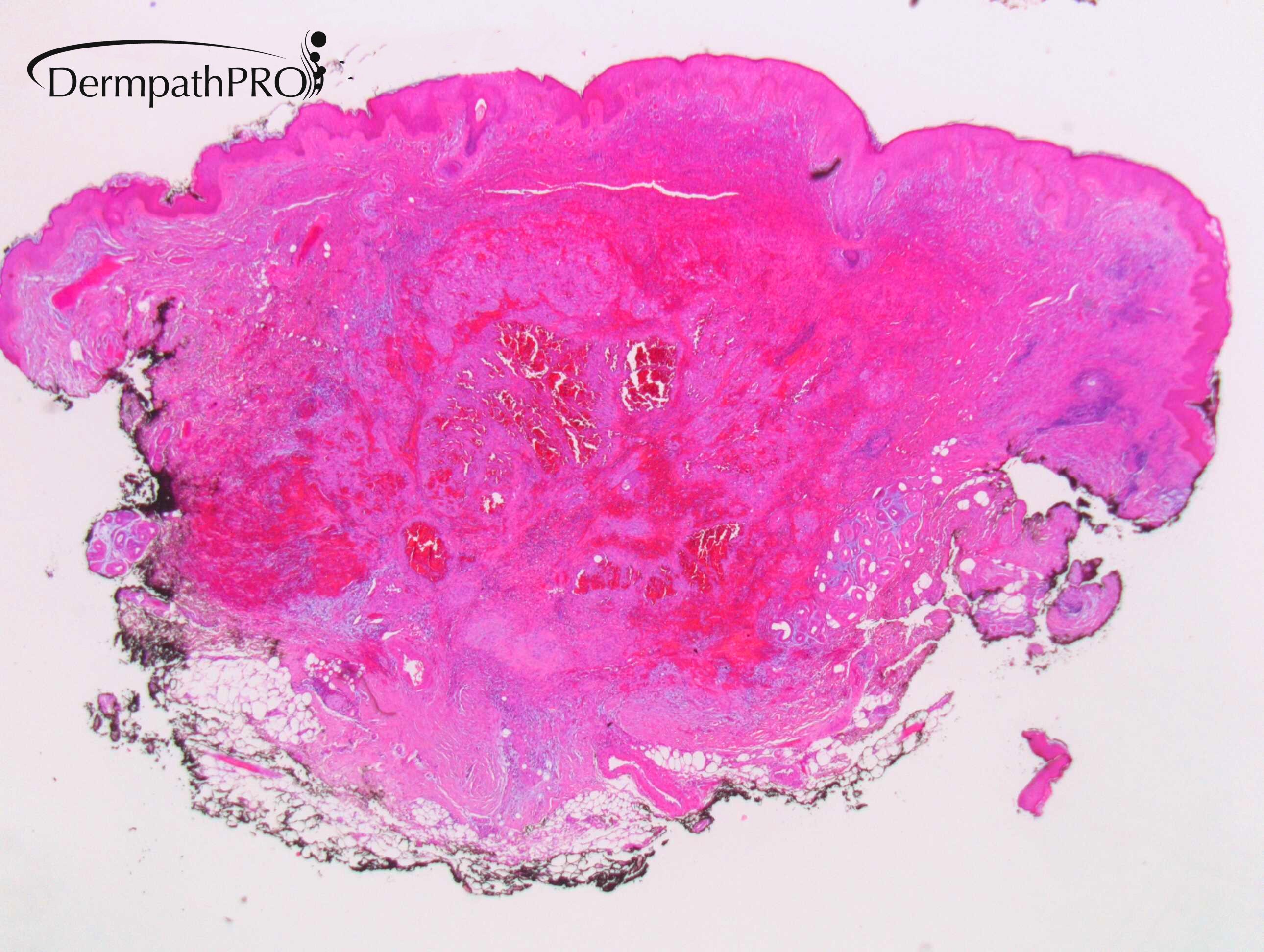
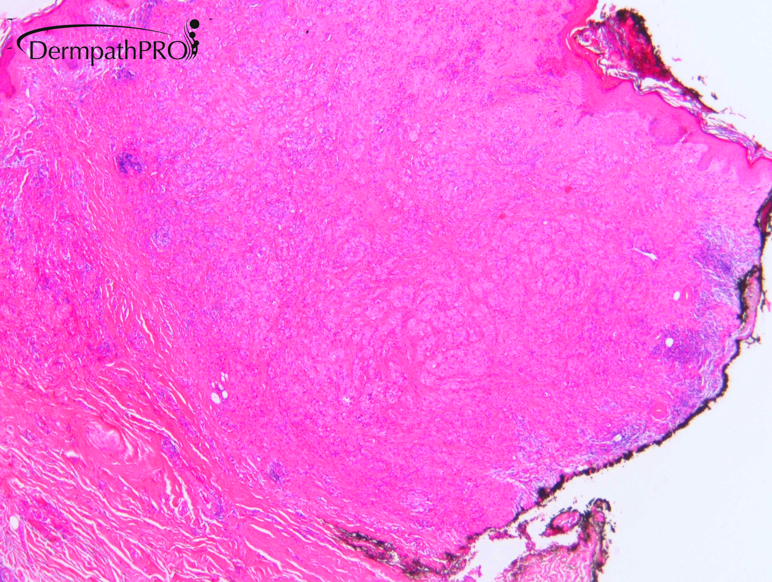
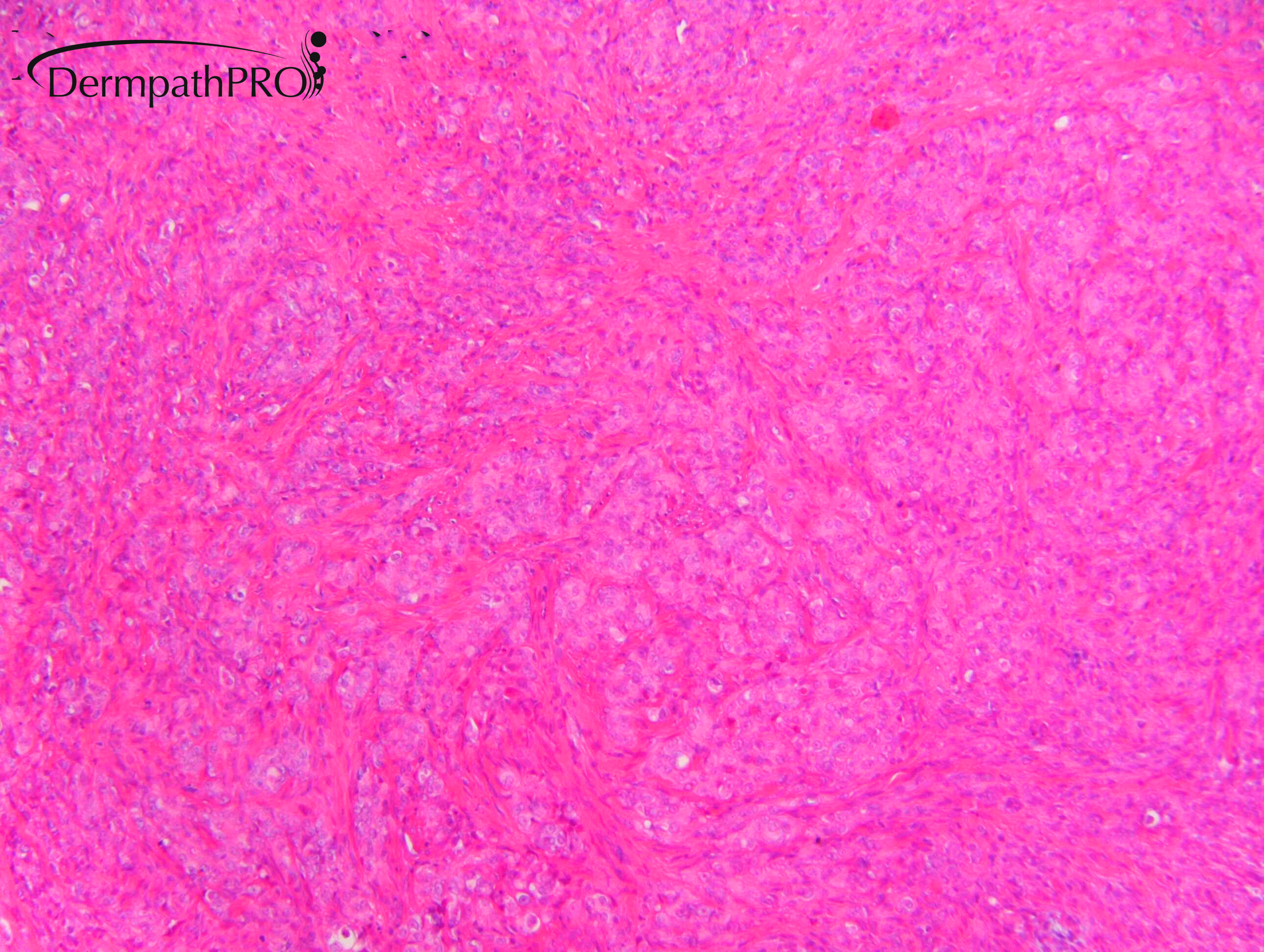
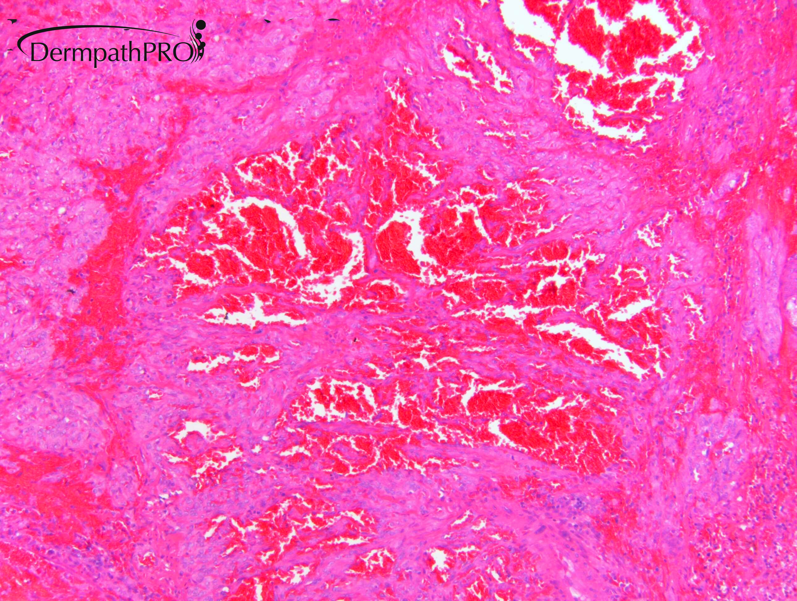
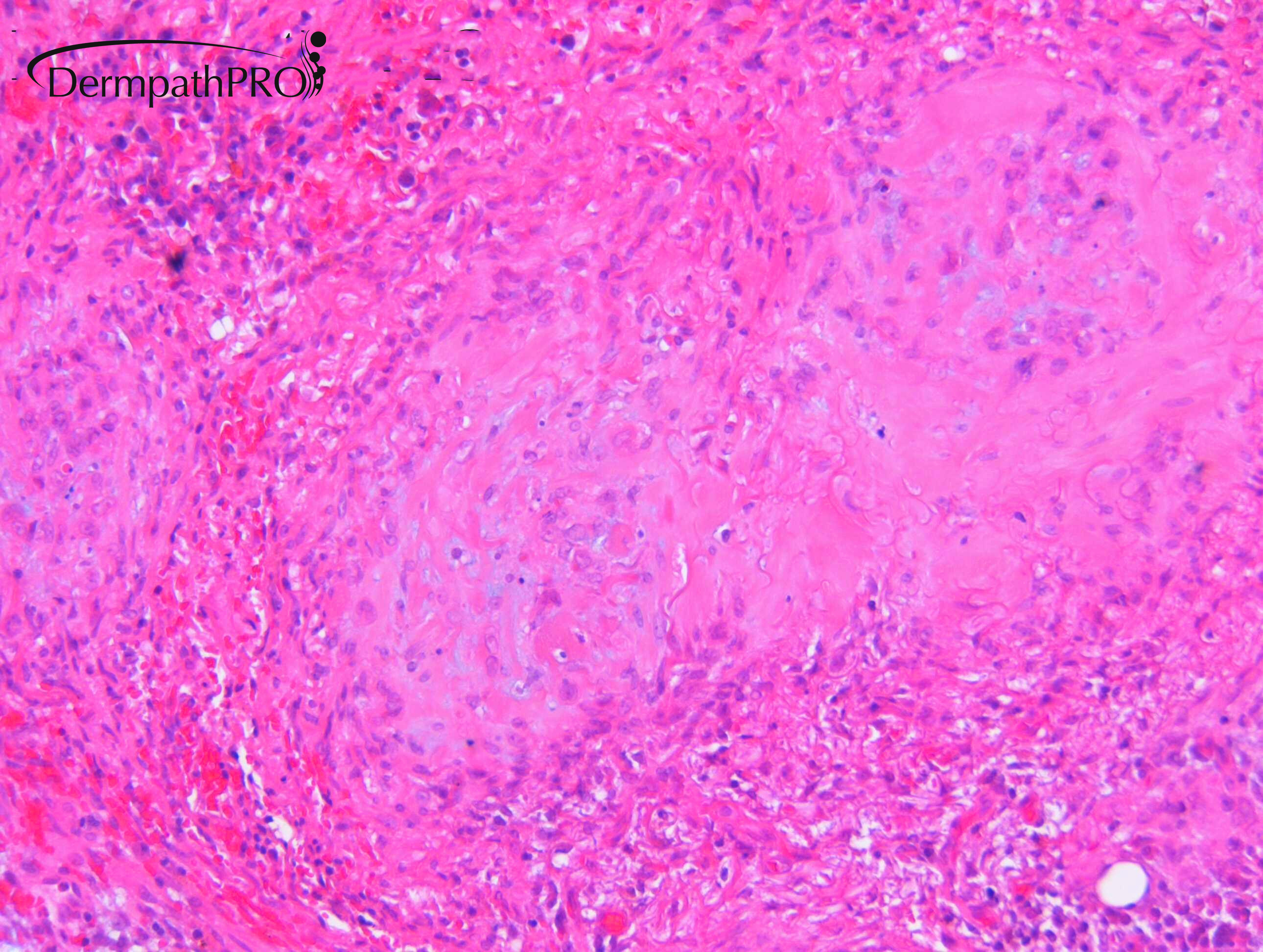
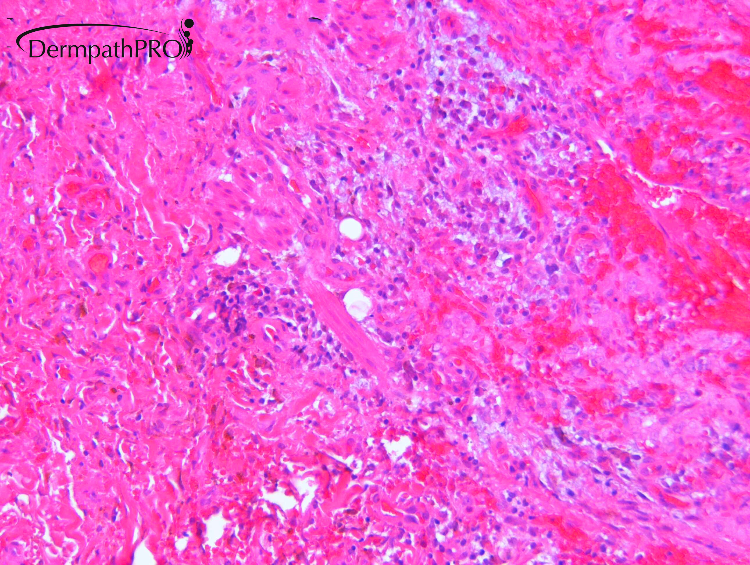
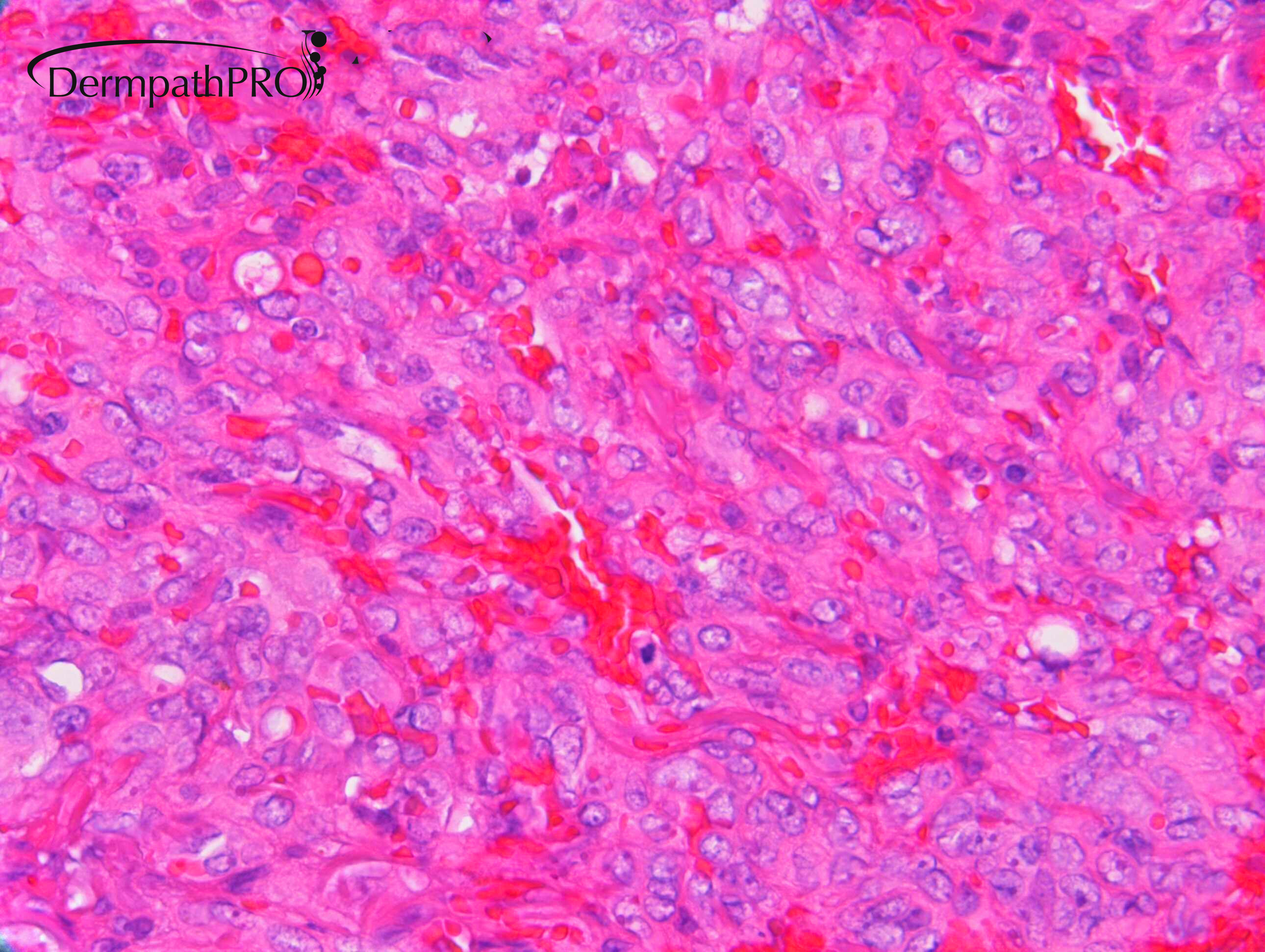
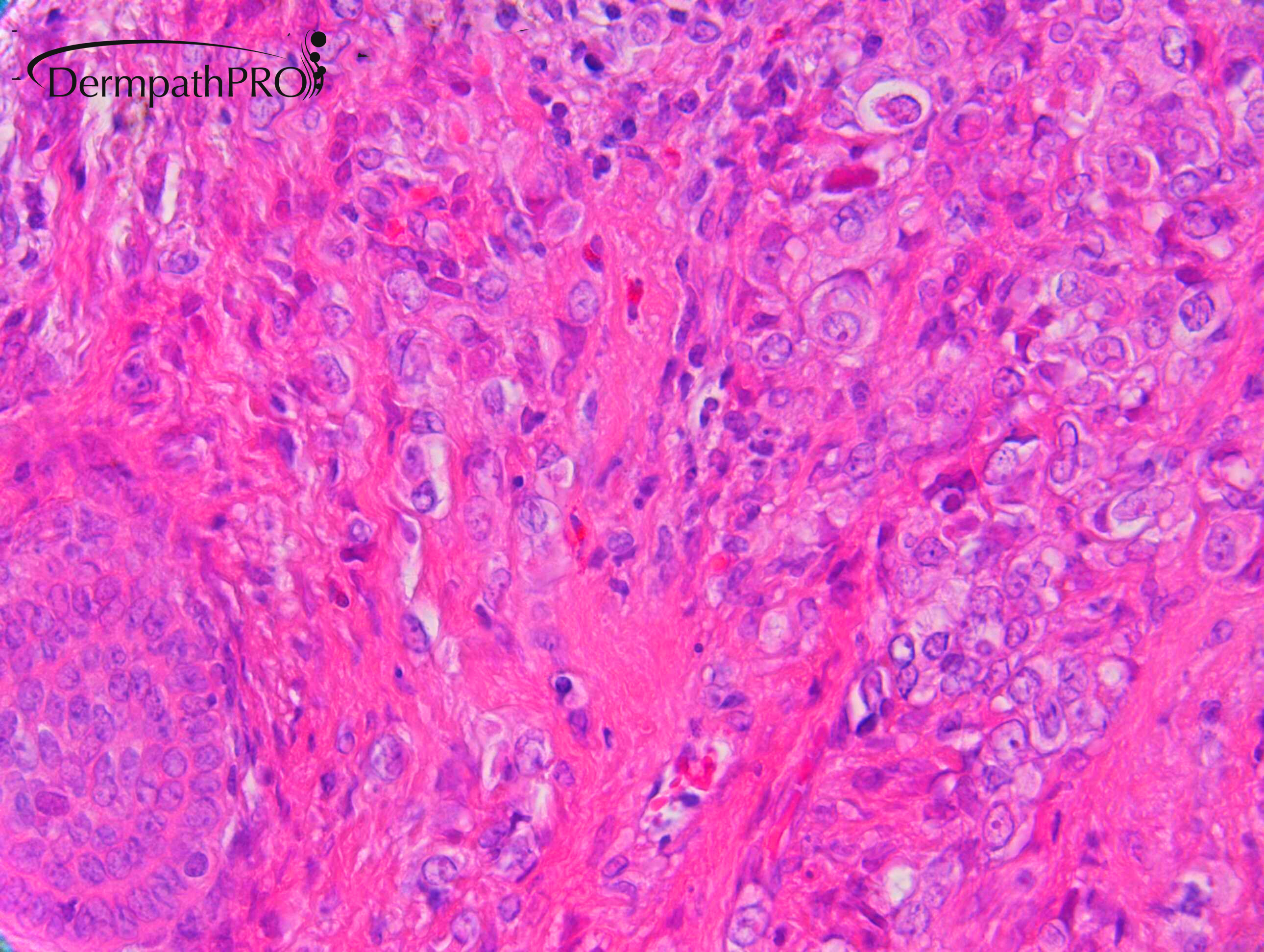
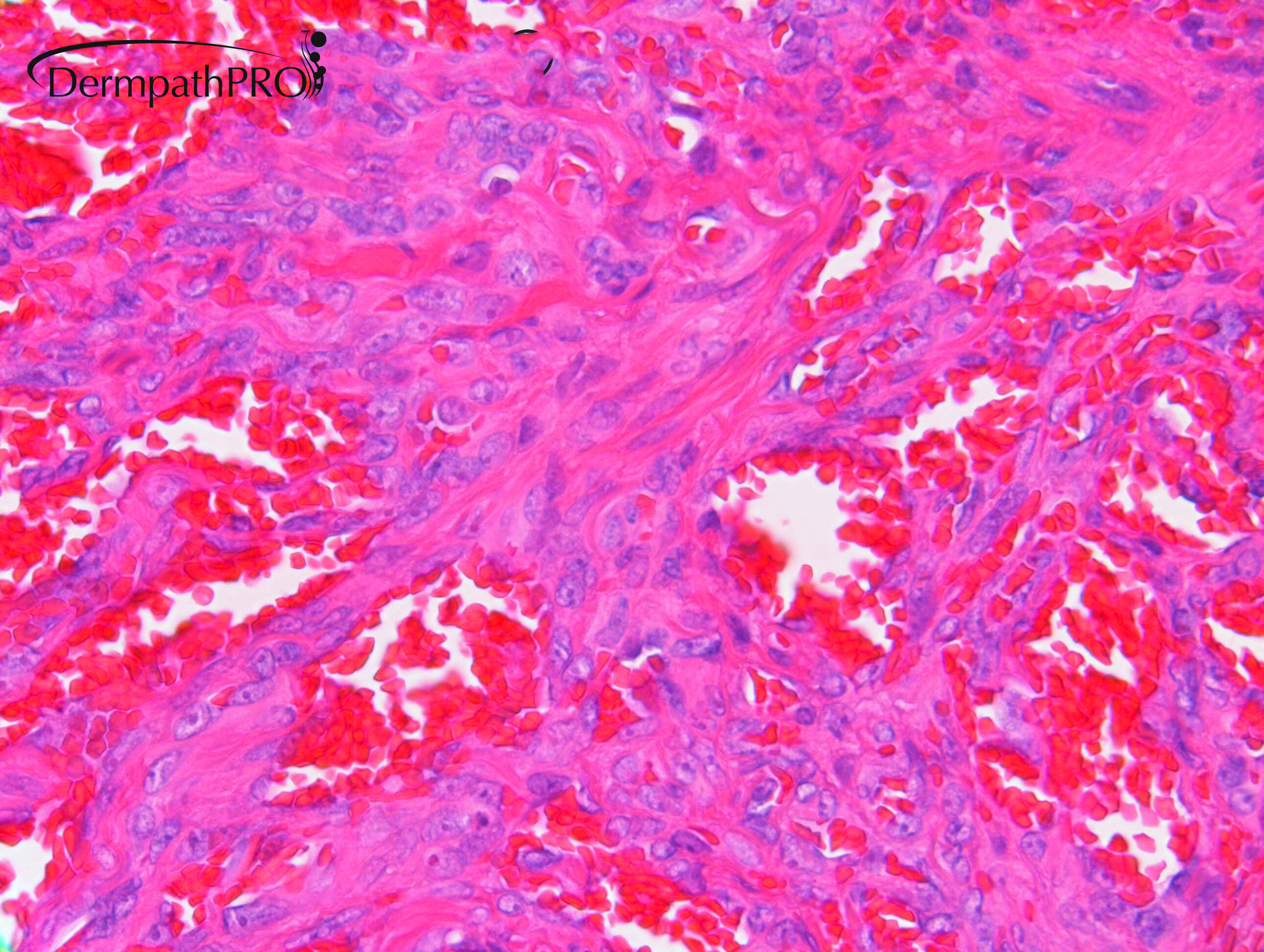
F70. Below right clavicle. Had chest radiotherapy 1973 for Hodgkin’s disease.


  Report Record

User Feedback


Dr. Mona Abdel-Halim

Posted

 Of course epithelioid angiosarcoma is a possibility but some images make me also consider recurrent Hodgkin (cutaneous Hodgkin?). Awaiting IHC.

Share this comment


Link to comment
share_externally

Eman El-Nabarawy

Posted

Looks epitheioid angiosarcoma. But waiting for the trick and immunos! 

Share this comment


Link to comment
share_externally

Dr. Richard Carr

Posted

This is a challenging and controversial case.

Reported initially as a fibrohistiocytic lesion in keeping with an aneurysmal fibrous histiocytoma (CD34 negative, also negative for PanK, SMA, S100, MelanA with scattered macrophages. No other vascular markers performed.

I was asked to review a small dermal local recurrence at two years  which showed widespread CD31 positive staining, CD34 neg.

My differential of this relatively small dermal based lesion including: epithelioid angiomatous nodule or epithelioid haemangioendothelioama (favoured).

Colleagues in Friedricshafen kindly reviewed the case (Thomas Mentzel) and opined this was epithelioid angiosarcoma (CAMTA1 and HHV8 negative, CD31 & ERG +ve). They were not happy with the presence of mitotic activity, some apoptosis.

I confess I was uncomfortable with the implication of a high grade angiosarcoma for such a small dermal tumour.

Raul Perret commented for me (on selected images) as follows: Dear Rick, Based on these images, I am personally in favour of an intermediate malignancy vascular tumor (rarely metastasizing) and would have suggested (as TM) complete excision and clinical follow-up. I thought of pseudomyogenic hemangioendothelioma and composite hemangioendothelioma but I confess morphology is not completely clear cut. Personally, I would have added FOSb immunohistochemistry (typically positive in pseudomyogenic hemangioendothelioma). It is important to remark that vascular tumors are one of the most difficult areas in Pathology so we can all commit mistakes. Hope I helped a little.  Big hug, Raul

Interestingly the case was subsequently reviewed by one of our National experts in the UK and they favoured epithelioid haemangioendothelioma.

I guess we learn from this that such cases are challenging and there is not necessarily agreement between experts.

Share this comment


Link to comment
share_externally


Join the conversation

You can post now and register later. If you have an account, sign in now to post with your account.

Guest
Add a comment...

×   Pasted as rich text.   Paste as plain text instead

  Only 75 emoji are allowed.

×   Your link has been automatically embedded.   Display as a link instead

×   Your previous content has been restored.   Clear editor

×   You cannot paste images directly. Upload or insert images from URL.

Loading...

×
×
  • Create New...