Jump to content
In this section we have spot diagnoses posted on a daily basis since June 2010, now over 4000! You can review the archived cases and read the suggested diagnoses by users and the final comment by the contributors.
Case are uploaded each week day by 10 am UK time with the correct diagnosis will generally be posted at 8 pm UK time. Why not view the most recent spot diagnosis and proffer a diagnosis?

Case Number : Case 2612 - 10 July 2020 Posted By: Dr. Richard Carr

Please read the clinical history and view the images by clicking on them before you proffer your diagnosis.
Submitted Date :
   (0 reviews)

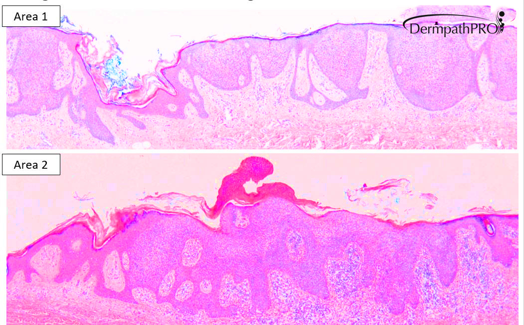
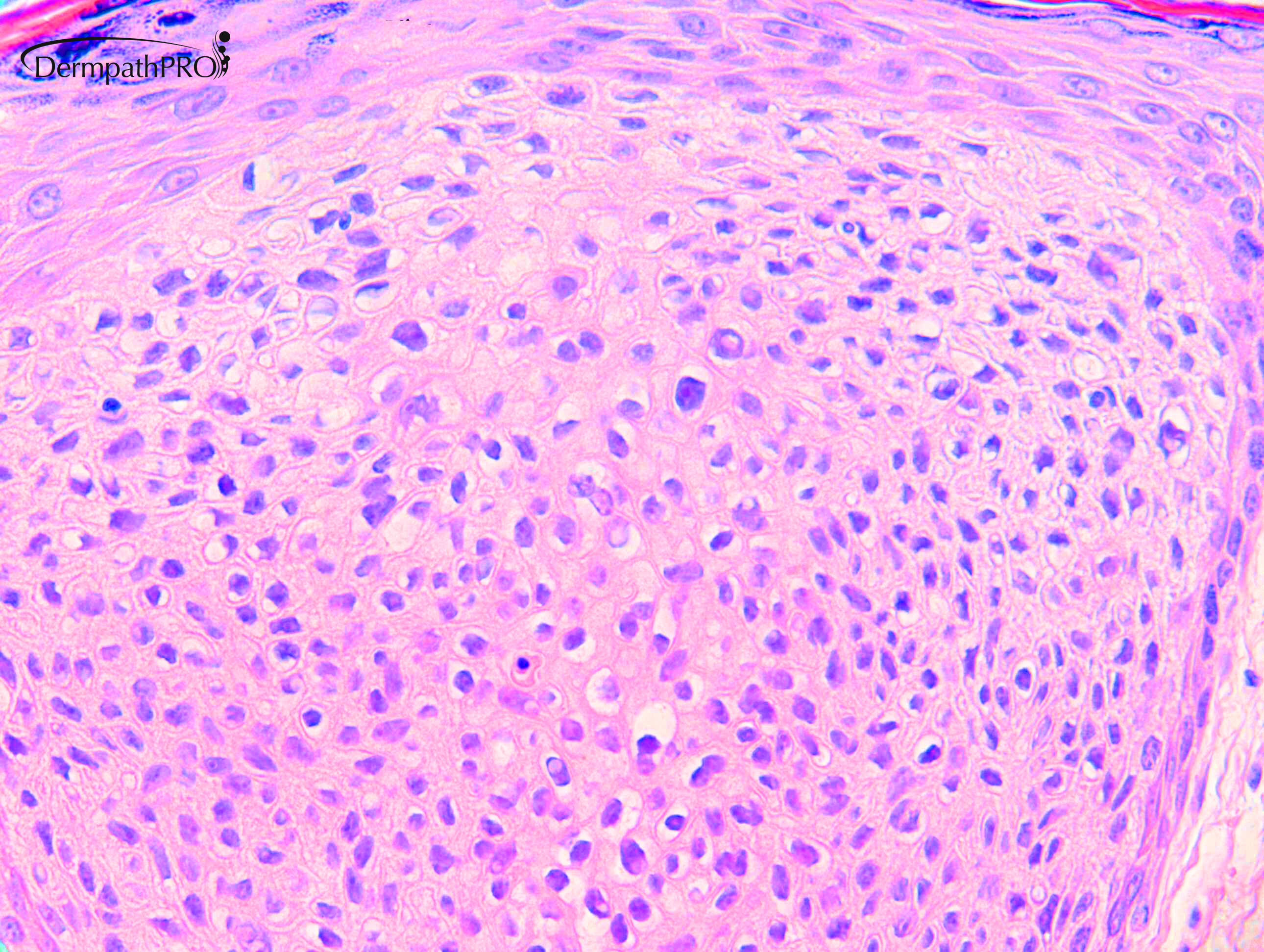
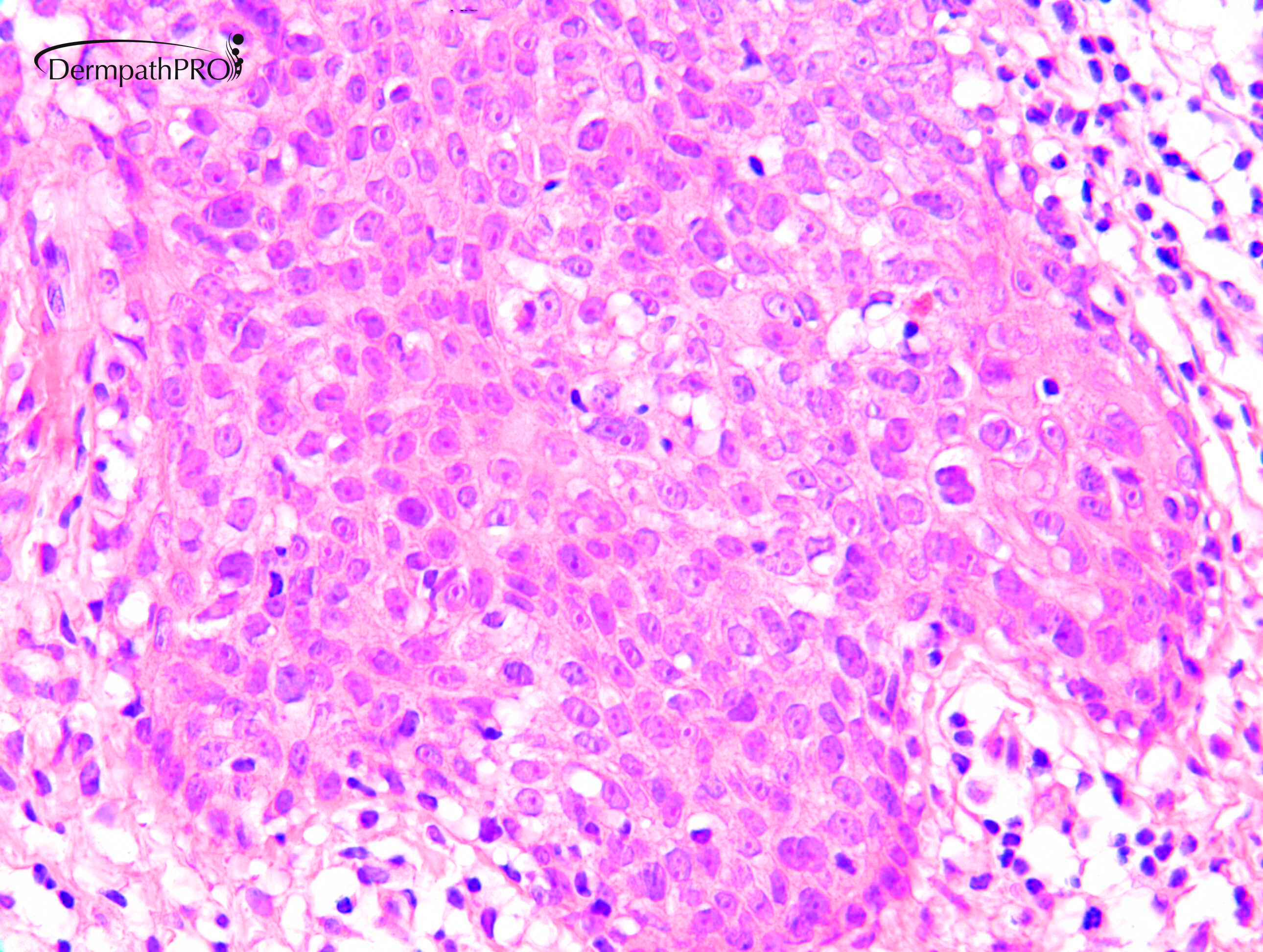
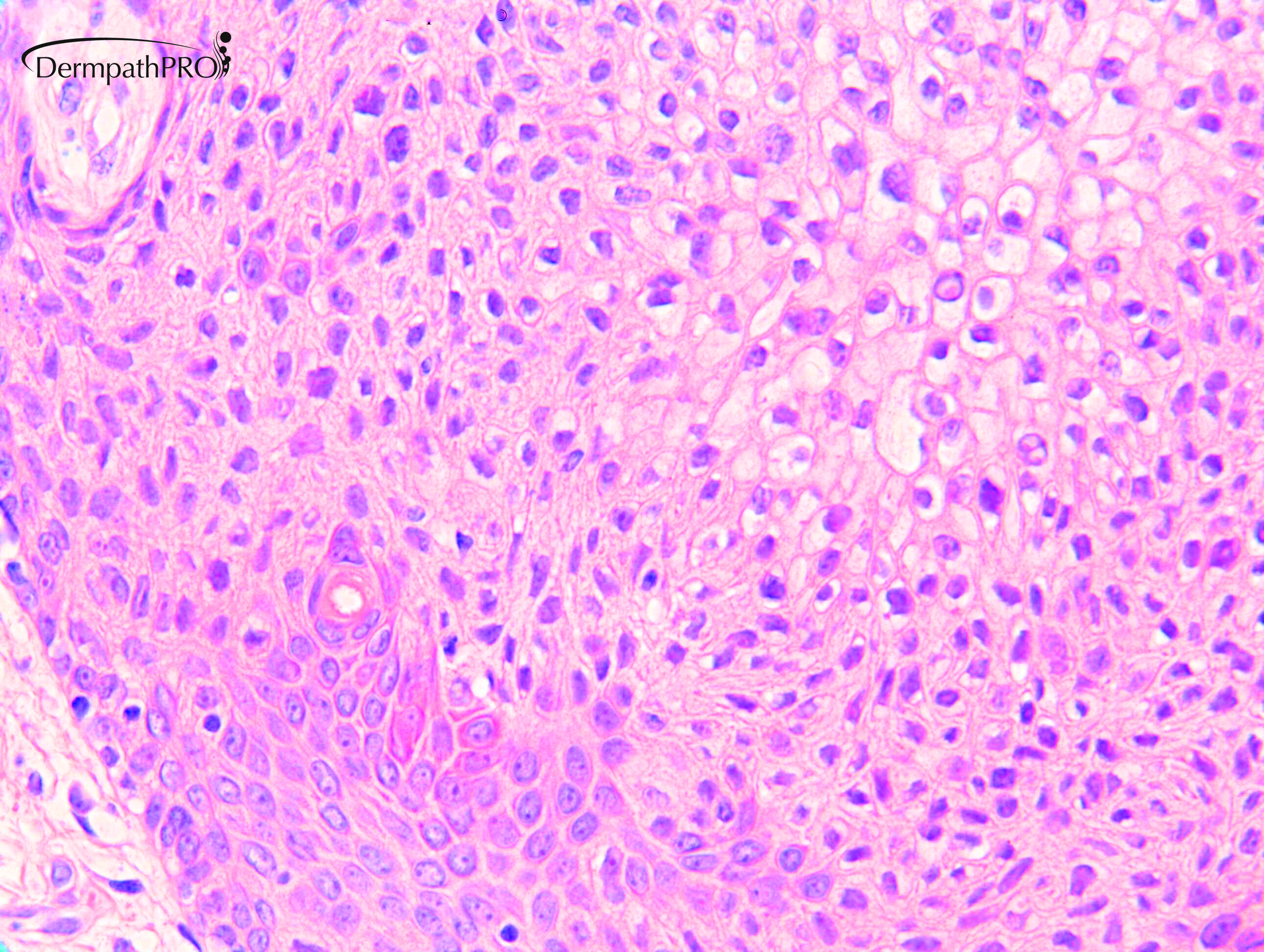
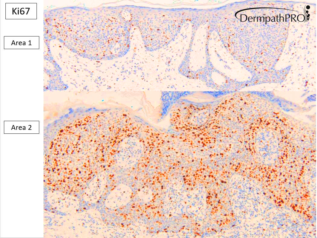
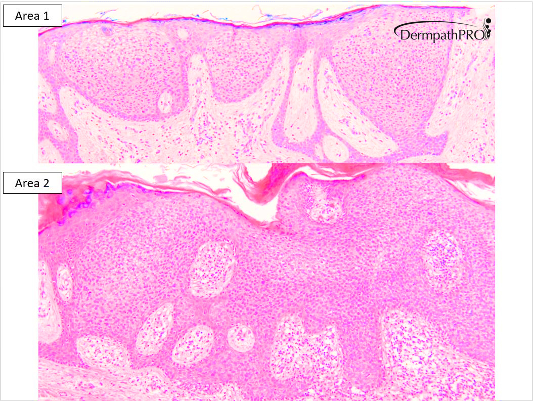
F90. 20 year history of lesion on left buttock, now 60 x 55mm, enlarging, intermittent bleeding.


  • Like 1
  • Thanks 2
  Report Record

User Feedback


Richard Logan

Posted

Hidroacanthoma simplex preferred to clonal seborrhoeic wart.  IH may help.

Share this comment


Link to comment
share_externally

Krishnakumar subramanian

Posted

hidracanthoma simplex

Share this comment


Link to comment
share_externally

Porocarcinoma in situ, ex hidroacanthoma simplex. 

Share this comment


Link to comment
share_externally

On 10/07/2020 at 11:16, vincenzo said:

Porocarcinoma in situ, ex hidroacanthoma simplex. 

I agree.

Did you see in Area 2 the figure of keratine? Seems a lover couple.

Share this comment


Link to comment
share_externally

Yeahh! Brilliant point, Liluga. Very nice. 

Share this comment


Link to comment
share_externally

Krishnakumar subramanian

Posted

Thanks for a good learning case sir

Share this comment


Link to comment
share_externally

Dr. Richard Carr

Posted

Sorry for delay - on hols this week. I'd like to thank Dr Natalie Brearley for sharing this case. Thanks Liluga for pointing out the lovers - wonderful!

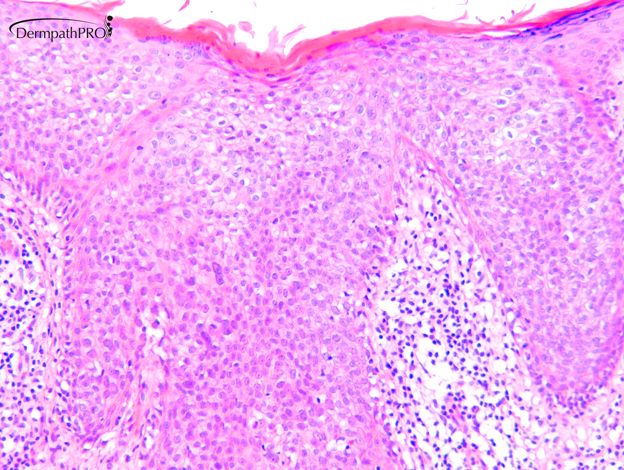
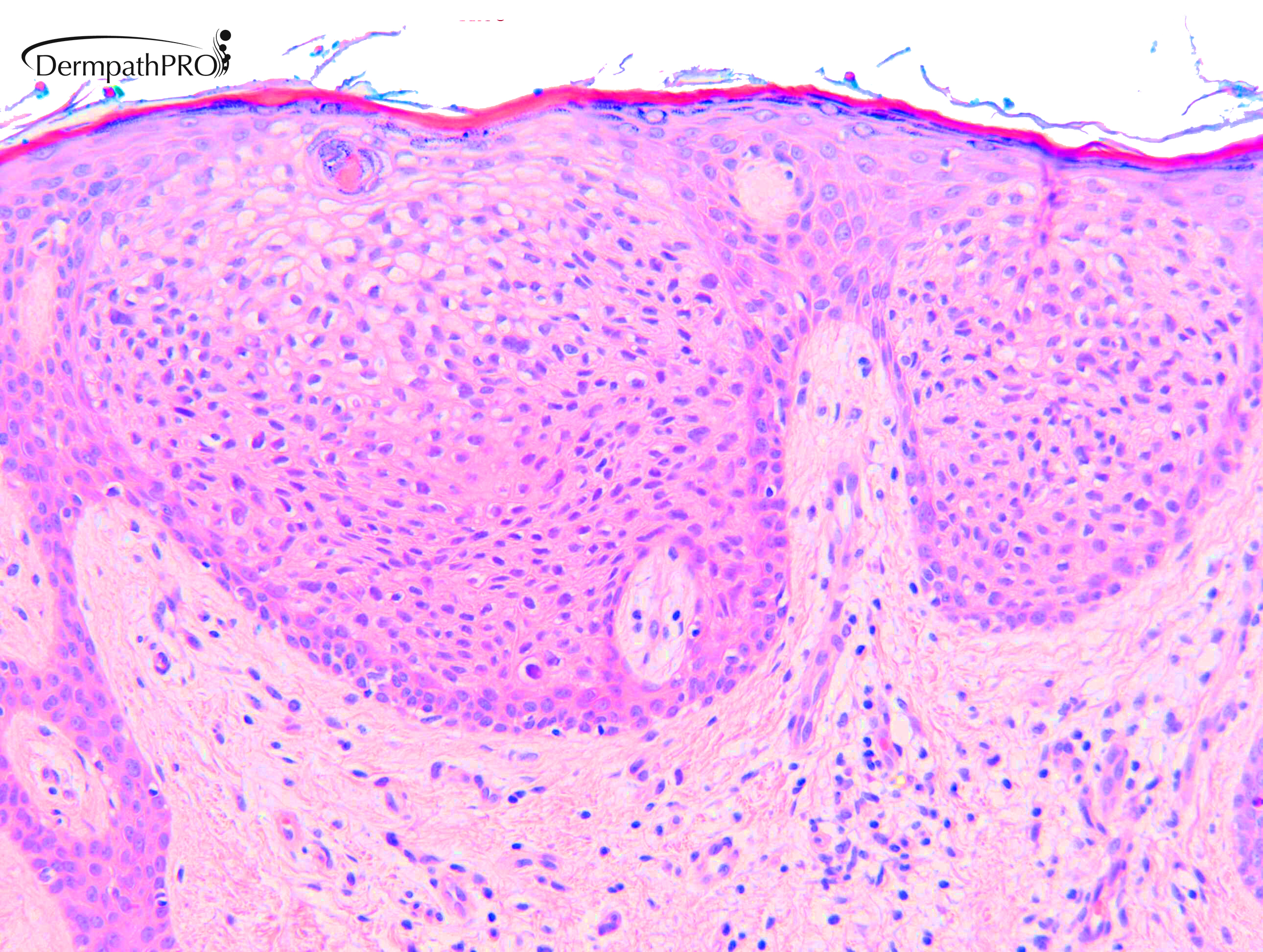
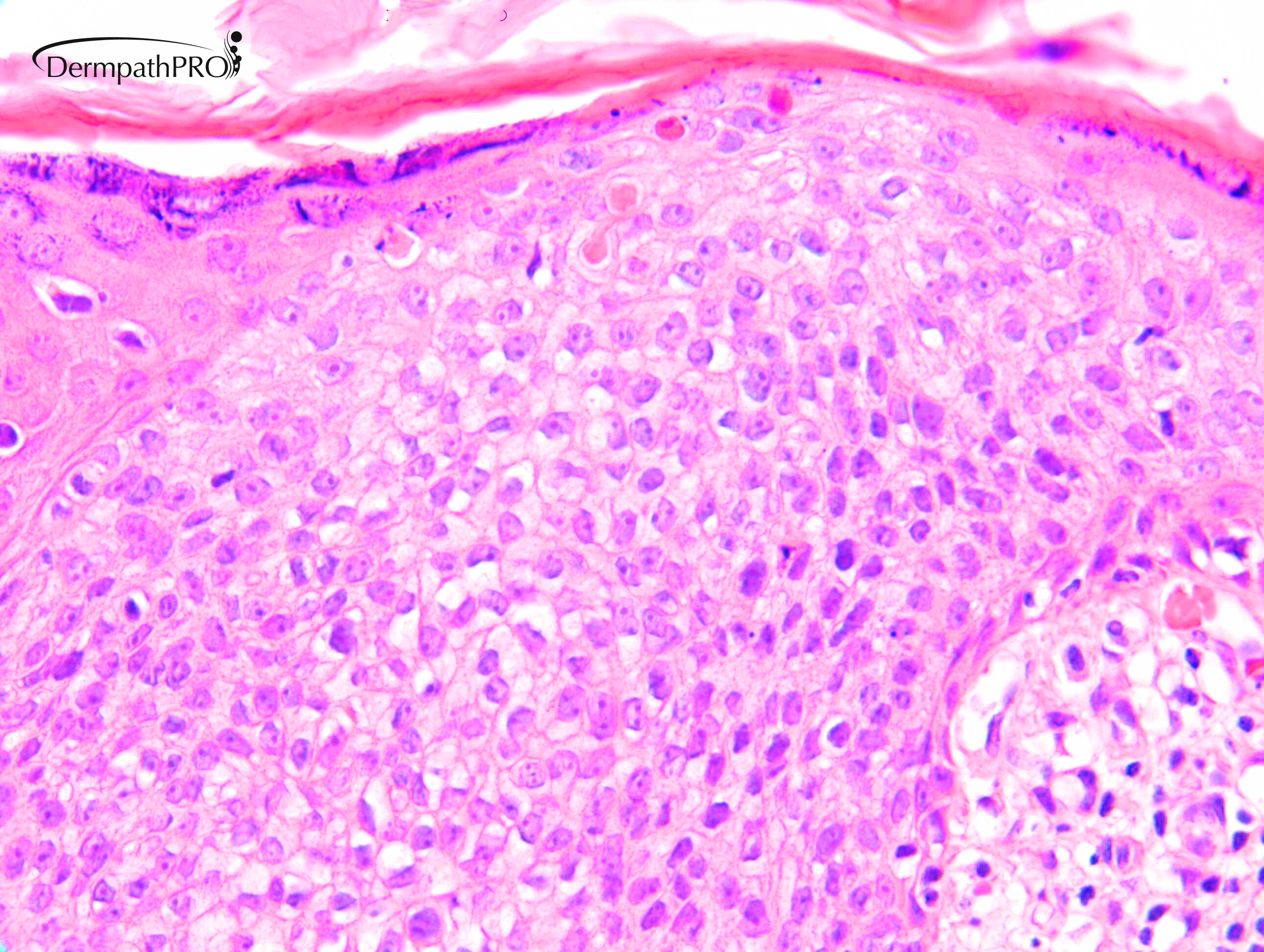
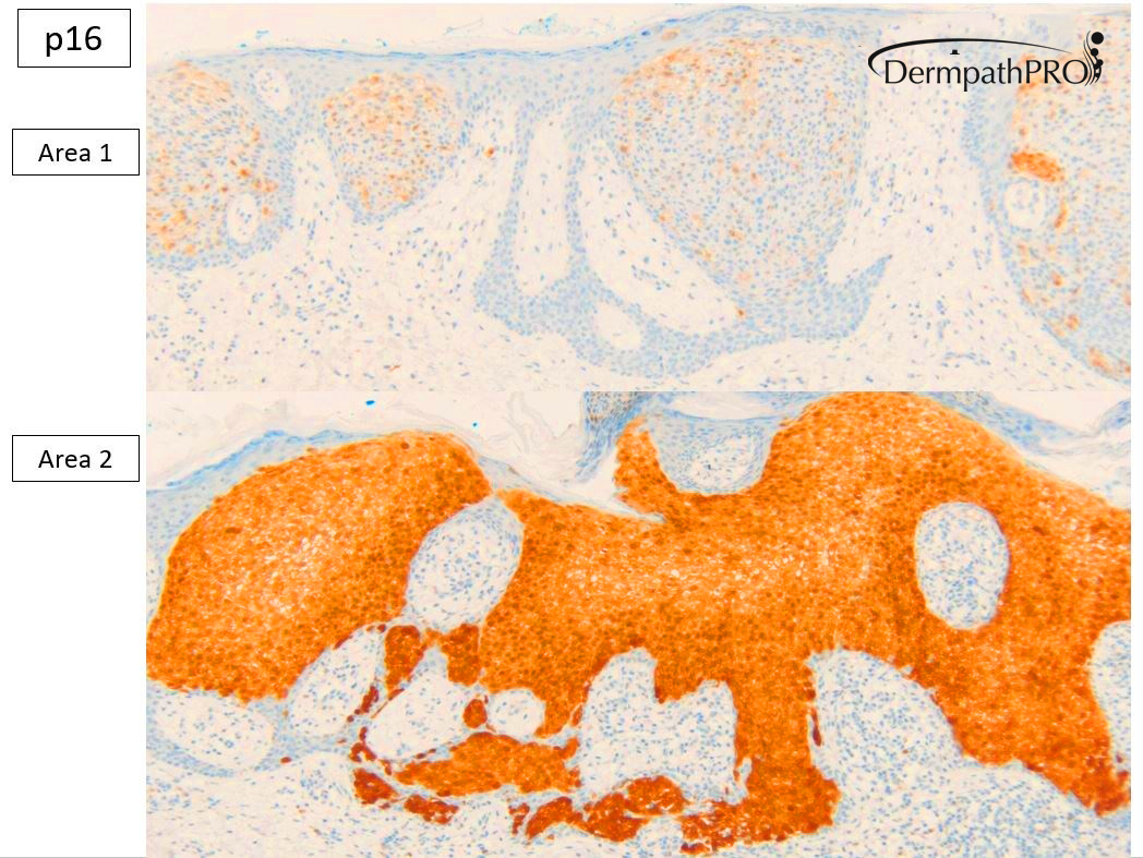
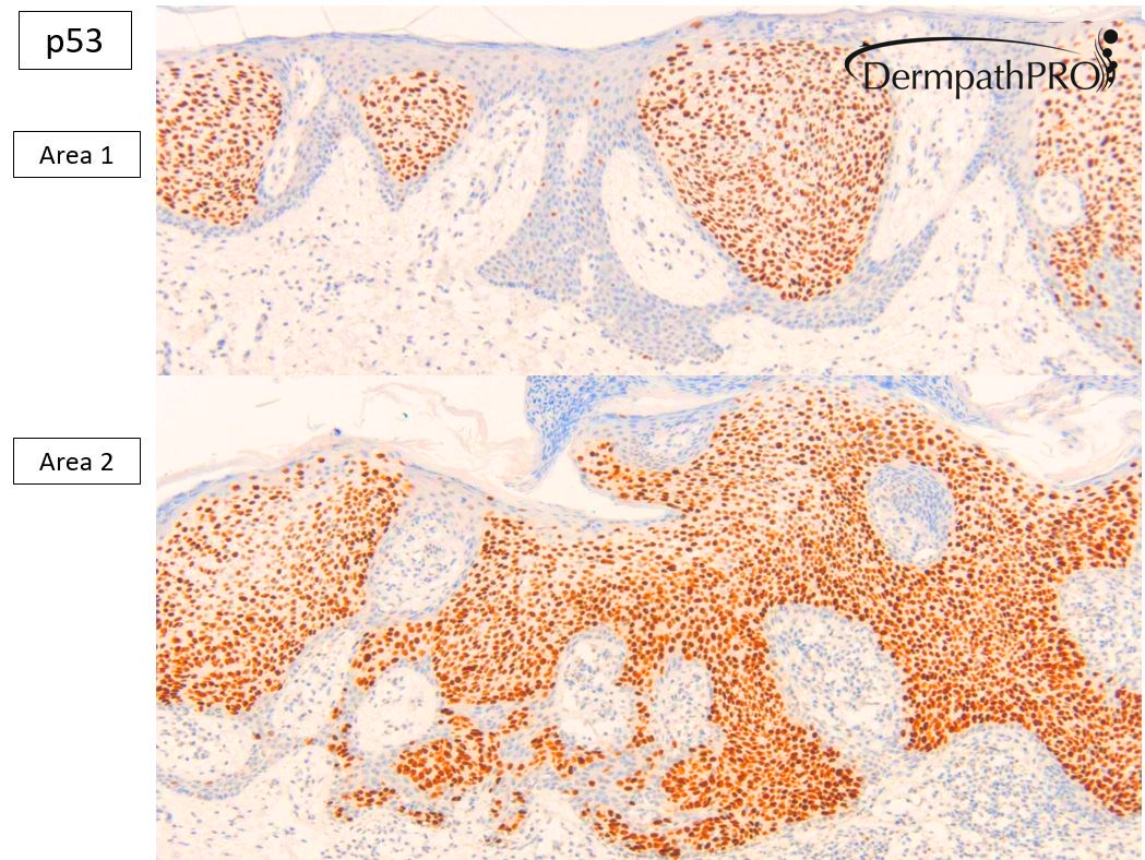
My opinion as follows: Many thanks for sending this extensively sampled lesion. We have a clonal proliferation in the epidermis with rather banal appearing areas resembling clonal seborrhoeic keratosis or hidroacanthoma simplex (clonal intraepidermal poroma). The distinction between the two is rather academic. In areas we have increased nuclear cytoplasmic ratio highly suggestive of Bowenoid change within the lesion. I was somewhat surprised to find that p53 was moderately strong in greater than 50% of nuclei throughout all areas of the specimen. To me this is incompatible with a fully benign lesion, so despite the areas being rather banal at the peripheries, I think these are potentially a precursor to frank Bowenoid dysplasia. Interestingly the p16 showed a different pattern with chequer board/intermittent staining in the more banal areas, but block diffuse positivity in the obviously more Bowenoid areas. Importantly the lesion is non-invasive. I can well remember two previous highly similar cases in which patient had either clonal seborrhoeic keratosis or intraepidermal poroma who subsequently developed Bowenoid transformation with similar immunohistochemical findings. Importantly, there is no invasive disease. The options for management would include those for Bowen’s disease. 

This case did not have demonstrable ductal differentiation but as I say the distinction of porocarcinoma in situ from bowenoid dysplasia, athough fun for us morphologists, is largely academic.

Later in the year I will be sharing many cases of highly challenging squamoproliferative lesions with interesting and some novel findings for p16 and p53.

Share this comment


Link to comment
share_externally

Yousra

Posted (edited)

I can't see any ducts, why hidracanthoma simplex or porocarcinoma

On 10/07/2020 at 16:12, Krishnakumar subramanian said:

hidracanthoma simplex


On 10/07/2020 at 16:12, Krishnakumar subramanian said:

hidracanthoma simplex


On 10/07/2020 at 18:16, vincenzo said:

Porocarcinoma in situ, ex hidroacanthoma simplex. 


On 11/07/2020 at 09:21, Meenakshi Batrani said:

Hidroacanthoma simplex


Edited by Yousra
Add an inquiry

Share this comment


Link to comment
share_externally


Join the conversation

You can post now and register later. If you have an account, sign in now to post with your account.

Guest
Add a comment...

×   Pasted as rich text.   Paste as plain text instead

  Only 75 emoji are allowed.

×   Your link has been automatically embedded.   Display as a link instead

×   Your previous content has been restored.   Clear editor

×   You cannot paste images directly. Upload or insert images from URL.

Loading...

×
×
  • Create New...