Jump to content
In this section we have spot diagnoses posted on a daily basis since June 2010, now over 4000! You can review the archived cases and read the suggested diagnoses by users and the final comment by the contributors.
Case are uploaded each week day by 10 am UK time with the correct diagnosis will generally be posted at 8 pm UK time. Why not view the most recent spot diagnosis and proffer a diagnosis?

Case Number : Case 2616 - 16 July 2020 Posted By: Saleem Taibjee

Please read the clinical history and view the images by clicking on them before you proffer your diagnosis.
Submitted Date :
   (0 reviews)

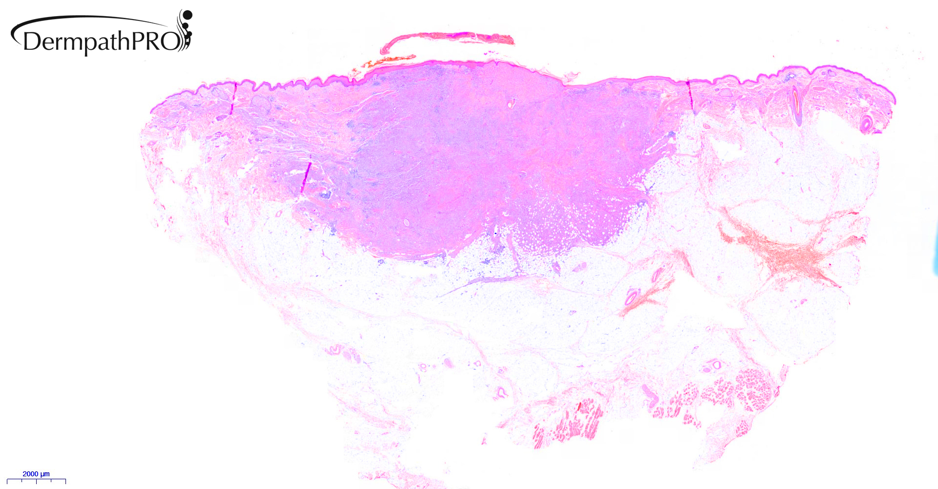
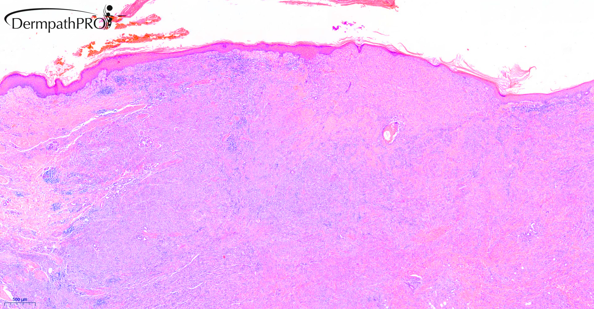
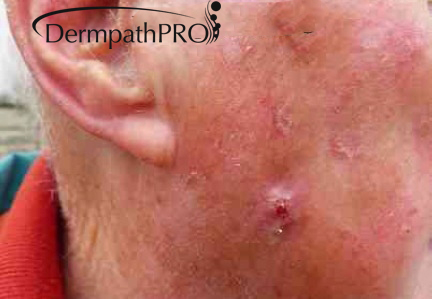
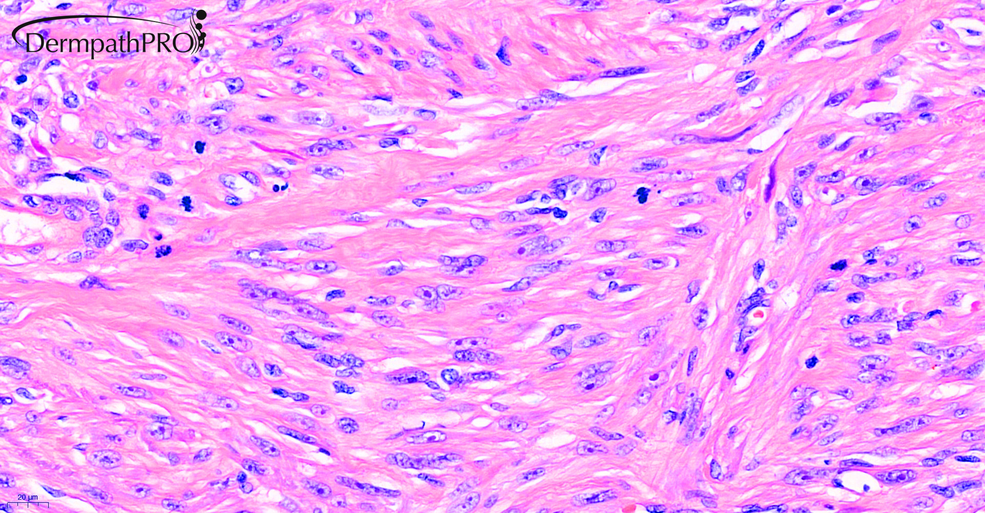
85M excision R cheek ?BCC ?SCC

  Report Record

User Feedback


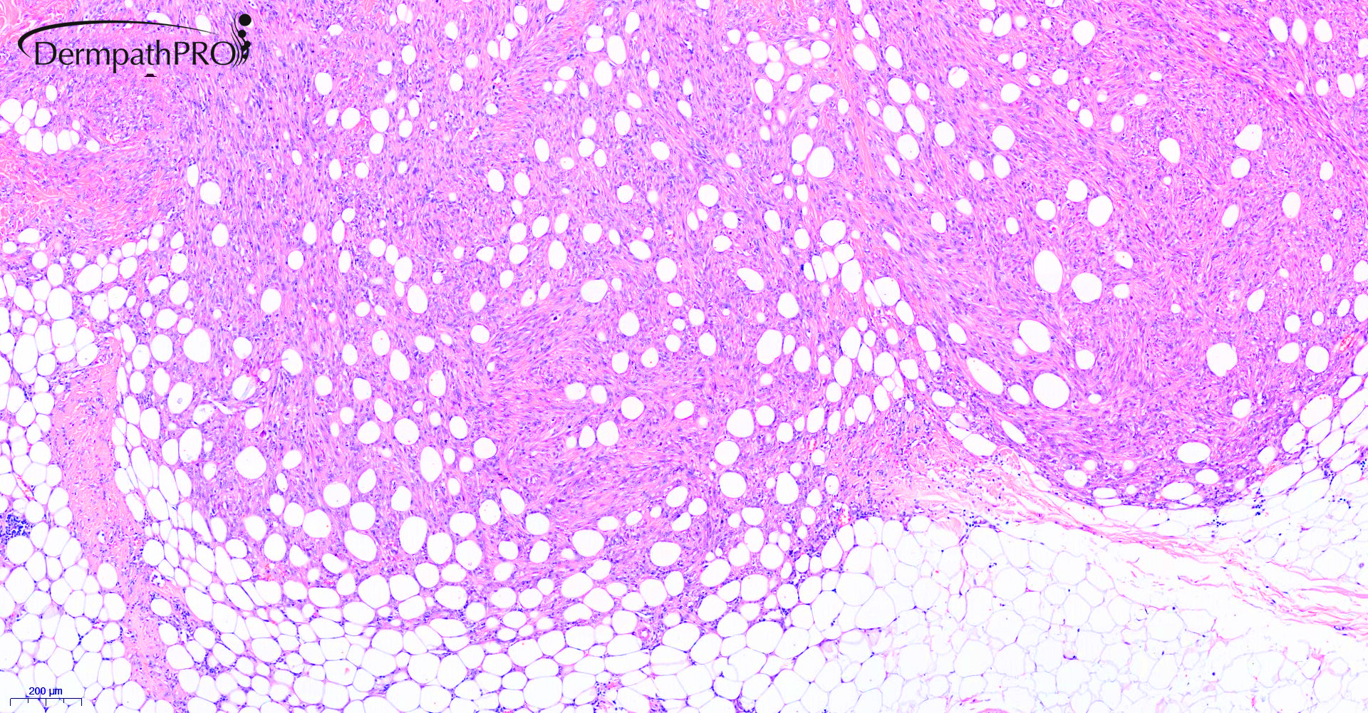
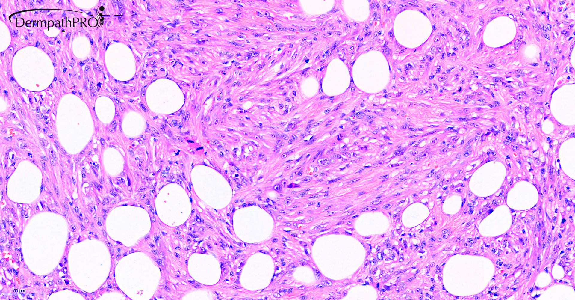
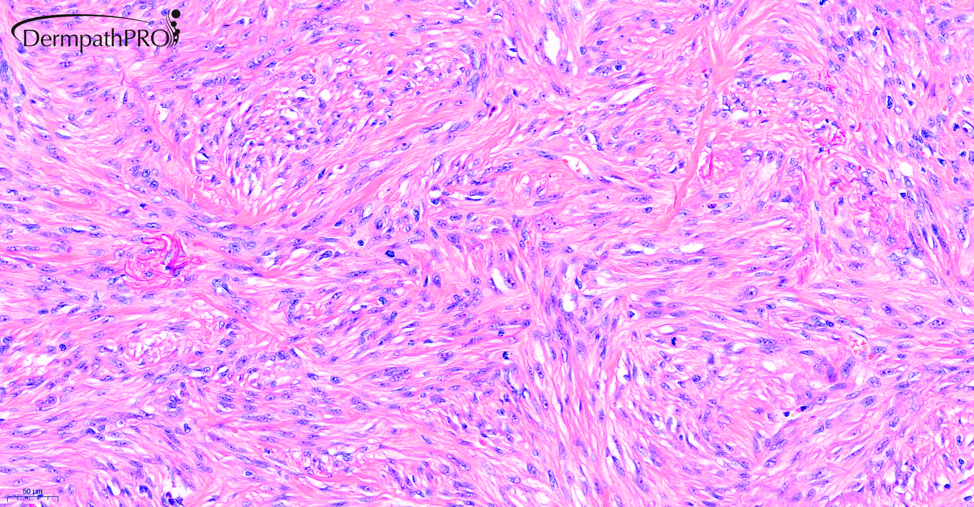
I think this is a lightly difficult case of DermatoFibroSarcoma Protuberans, or, as we pathologists say, a DFSP who hasn’t studied the pathology books. Instead there are too mitosis, as in fibrosarcomatous transformation ( but I think this is not the case ), and the largest part of the tumor is sclerotic/hyalinized and intradermal, as a sclerotic DF ( but this isn’t a DF ).

The cartwheel growth pattern, the focal but striking honeycombing subcutaneous infiltration and the typical sparing of skin adnexa definitely win. 

Share this comment


Link to comment
share_externally

Richard Logan

Posted

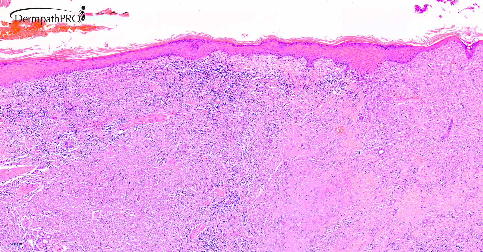
Histologically this looks like a typical DFSP to me although as Vincenzo says there are rather more mitoses than usual.  The other thing is I don't ever recall seeing a DFSP on the face.  I think some precautionary immunohistochemistry is in order.

Share this comment


Link to comment
share_externally

If this is the whole lesion, it looks somewhat circumscribed compared to the usual DFSP. I would consider the possibility of a cellular dermatofibroma / fibrous histiocytoma which would explain the unusual high mitotic activity. Since both DF/ fibrous histiocytoma and DFSP are rare in the head, I would definitely need IHC to rule out other more common pathologic processes on the face of an 85 year old patient. 

Share this comment


Link to comment
share_externally

Saleem Taibjee

Posted

This was a case sent to me for second opinion. I am including some of the immunohistochemistry from the original laboratory below. A number of melanocytic and epithelial/keratin stains were all negative. Does this alter your opinions?

S2042_20.0x SMA labelled.jpg

S2042_2.0x Caldesmon labelled.jpg

S2042_2.0x Desmin labelled.jpg

S2042_1.0x CD10 labelled.jpg


Share this comment


Link to comment
share_externally

Dr. Mona Abdel-Halim

Posted

Spindle cell AFX (pleomorphic dermal sarcoma)

Share this comment


Link to comment
share_externally

Richard Logan

Posted

Yes, the clinical and immunohistochemistry features fit better with PDS, although it is not as "pleomorphic" as might be expected.

Share this comment


Link to comment
share_externally

Yes! I was wrong! Not thought about this, because of absence of pleomorphism. But I should thing so, because of clinician features. 
Nevertheless I think morphology and IHC are more in keeping with DF, cellular variant. 

Share this comment


Link to comment
share_externally

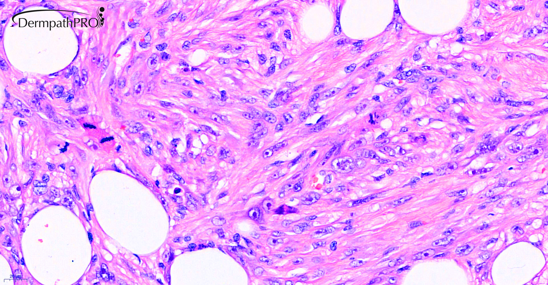
I’m struggling with this case. A red cell in a cellular vacuole. Very atypical cells, different than in DFSP ( macrocleola and vescicolar chromatin ). 
?CD31

?ERG. 

Share this comment


Link to comment
share_externally

Saleem Taibjee

Posted

In my opinion this is a case of pleomorphic dermal sarcoma. I have observed this honeycomb (DFSP-like) pattern of infiltration of the subcutaneous fat in a previous case also. However, as you have pointed out, the cytology is too pleomorphic, nuclei rather vesicular, for this to be DFSP. And the clinical context is much more in keeping with PDS, arising on the sun-damaged skin (adjacent solar elastosis) in an elderly man. As Vincenzo suggests, I will try to request a vascular marker (as well as CD34 for interest), and let you know if anything interesting transpires,

The clinician had requested my second opinion when the case was signed out by the original pathologist (from another lab) as leiomyosarcoma. As we know, SMA staining is not discriminatory, and I would tend to request Desmin (negative in this case) if genuinely considering leiomyosarcoma.

However, the positive Caldesmon staining was certainly thought-provoking, and on reviewing the literature, some of the papers seem to indicate that AFX/PDS is usually negative for caldesmon compared to leiomyosarcoma. I did come across this paper though: Beck EM, et al. A tale of two clones: Caldesmon staining in the differentiation of cutaneous spindle cell neoplasms J Cutan Pathol 2018, in which the authors highlight that the choice of Caldesmon clone/platform (Ventana vs Dako) can affect the results in AFX/PDS. As a result I requested a repeat of caldesmon (this is outsourced from our lab, and I am not sure which platform was used), and this is now negative (see below). I think this further supports PDS rather than leiomyosarcoma in this case.10560_5.0x Repeat Caldesmon labelled.jpg

BW

Saleem

Share this comment


Link to comment
share_externally

Krishnakumar subramanian

Posted

Oh god sir then which IHC to believe we usually do ventana, what about the controls for caldesmon both the times . we cannot be repeating like this suggest

Share this comment


Link to comment
share_externally

Saleem Taibjee

Posted

The arrector pili muscles serve as good internal positive control (see image above). I think bottom line is probably not to request caldesmon in the first place! Desmin should suffice to rule out leiomyosarcoma if that is a genuine consideration. However, the distinction between PDS and leiomyosarcoma is somewhat academic anyway, it will have little bearing on clinical management. In both instances clinical follow up will be advised.

Share this comment


Link to comment
share_externally

Saleem Taibjee

Posted

Just to let you know that subsequent CD31 and CD34 are negative in tumour cells (background vessels staining only).

Share this comment


Link to comment
share_externally

Dr. Richard Carr

Posted

BTW the treatment is the same whether its AFX, AFX-type sarcoma (PDS), leiomyosarcoma, spindle cell carcinoma, DFSP or spindle cell melanoma. i.e. complete excision with clear margins. Lesson don't bother with SMA or Desmin or CD34 (unless angiosarcoma is a possibility morphologically). Keep it simple: S100, p63, CD10, Pa-keratin. I add p53 for uncertain margins as AFX (and many of these diagnoses) often highly expresses the marker and it's easier sometimes to interpret c/w CD10 which is capricious.

Share this comment


Link to comment
share_externally


Join the conversation

You can post now and register later. If you have an account, sign in now to post with your account.

Guest
Add a comment...

×   Pasted as rich text.   Paste as plain text instead

  Only 75 emoji are allowed.

×   Your link has been automatically embedded.   Display as a link instead

×   Your previous content has been restored.   Clear editor

×   You cannot paste images directly. Upload or insert images from URL.

Loading...

×
×
  • Create New...