Jump to content
In this section we have spot diagnoses posted on a daily basis since June 2010, now over 4000! You can review the archived cases and read the suggested diagnoses by users and the final comment by the contributors.
Case are uploaded each week day by 10 am UK time with the correct diagnosis will generally be posted at 8 pm UK time. Why not view the most recent spot diagnosis and proffer a diagnosis?

Case Number : Case 2620 - 22 July 2020 Posted By: Dr. Hafeez Diwan

Please read the clinical history and view the images by clicking on them before you proffer your diagnosis.
Submitted Date :
   (0 reviews)

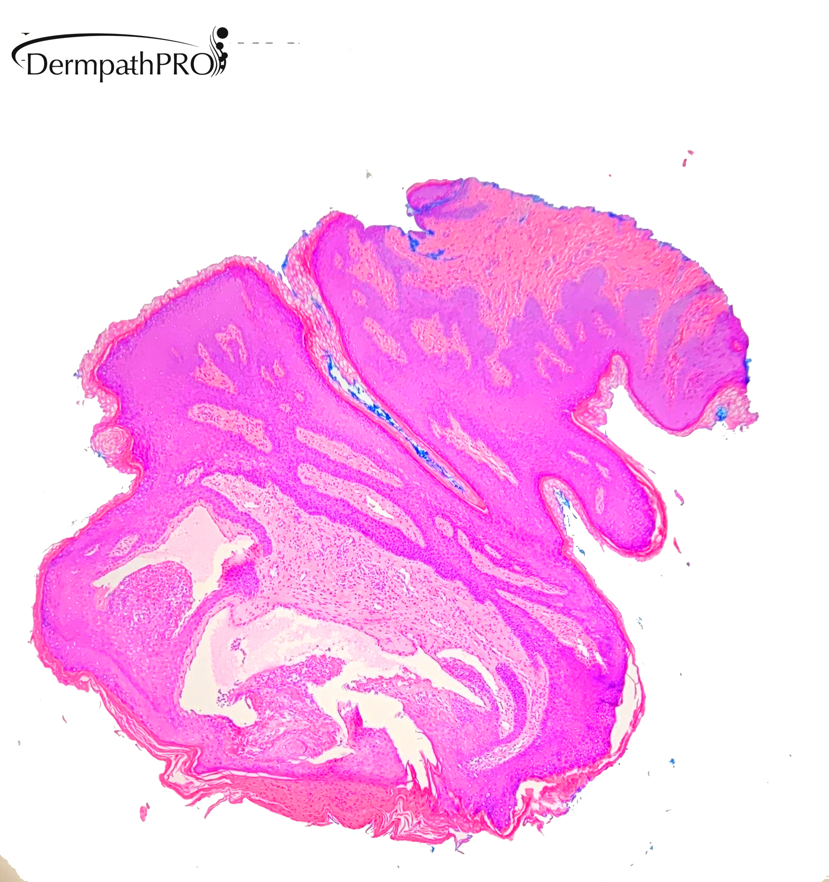
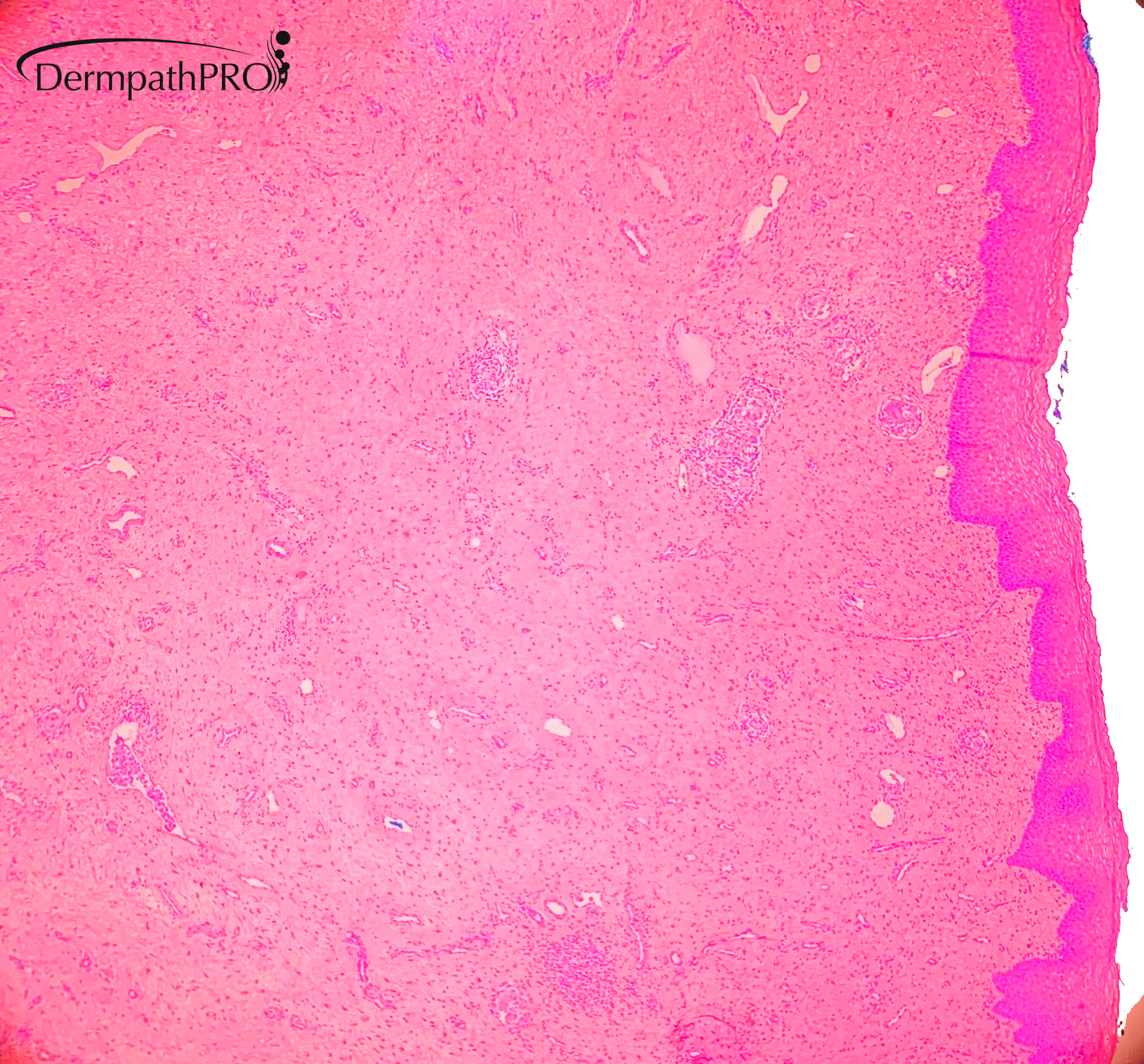
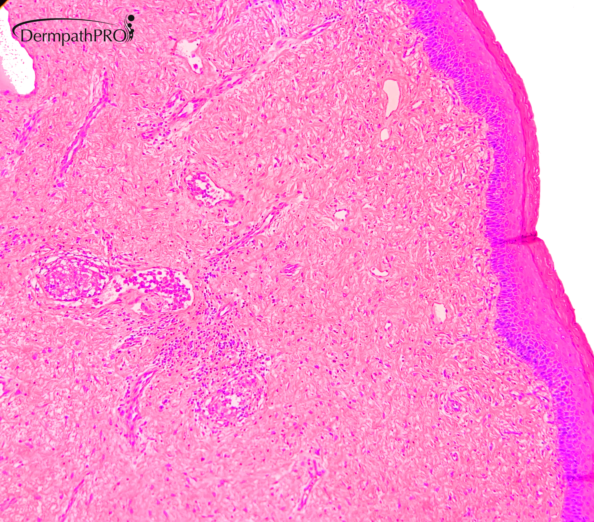
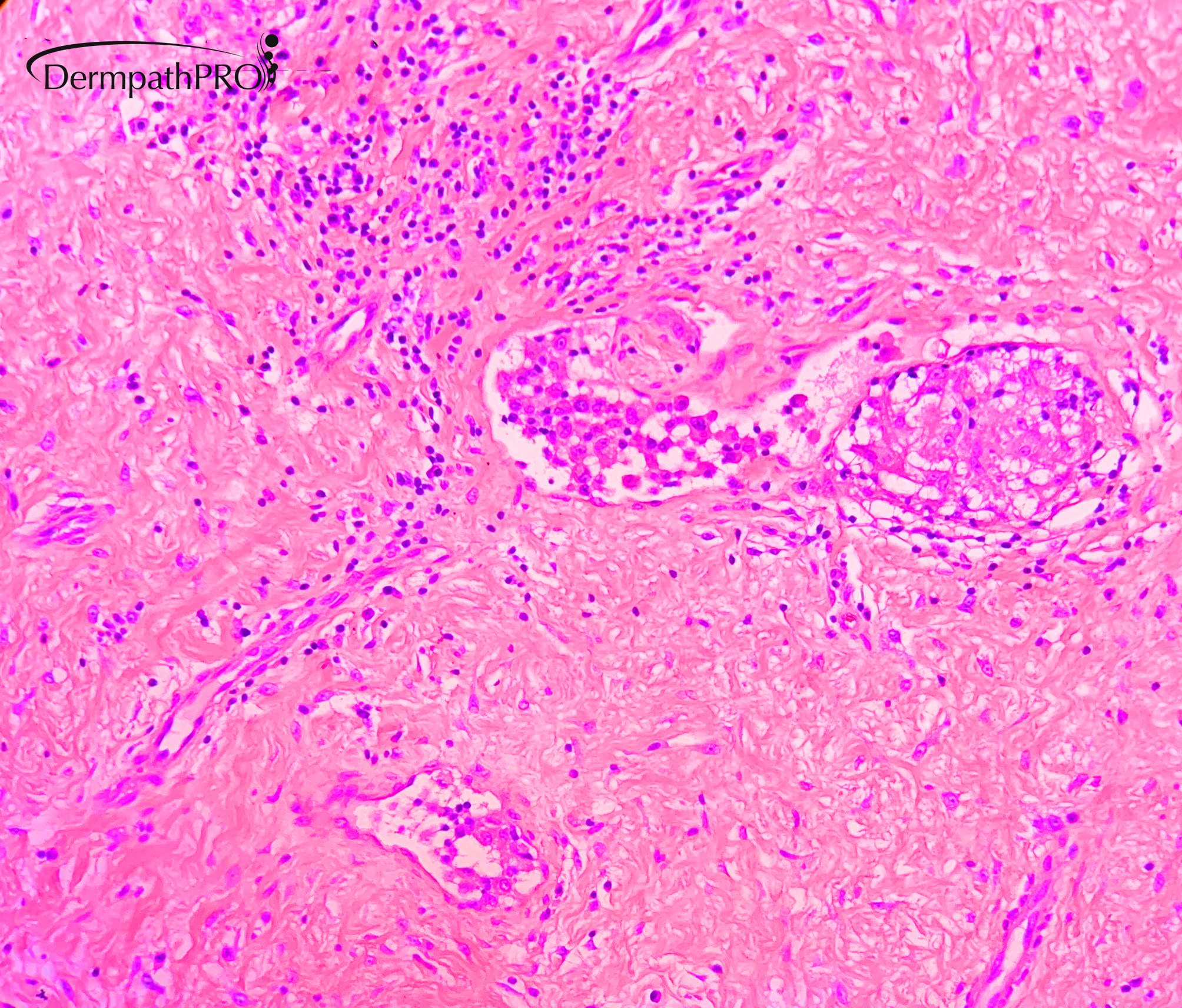
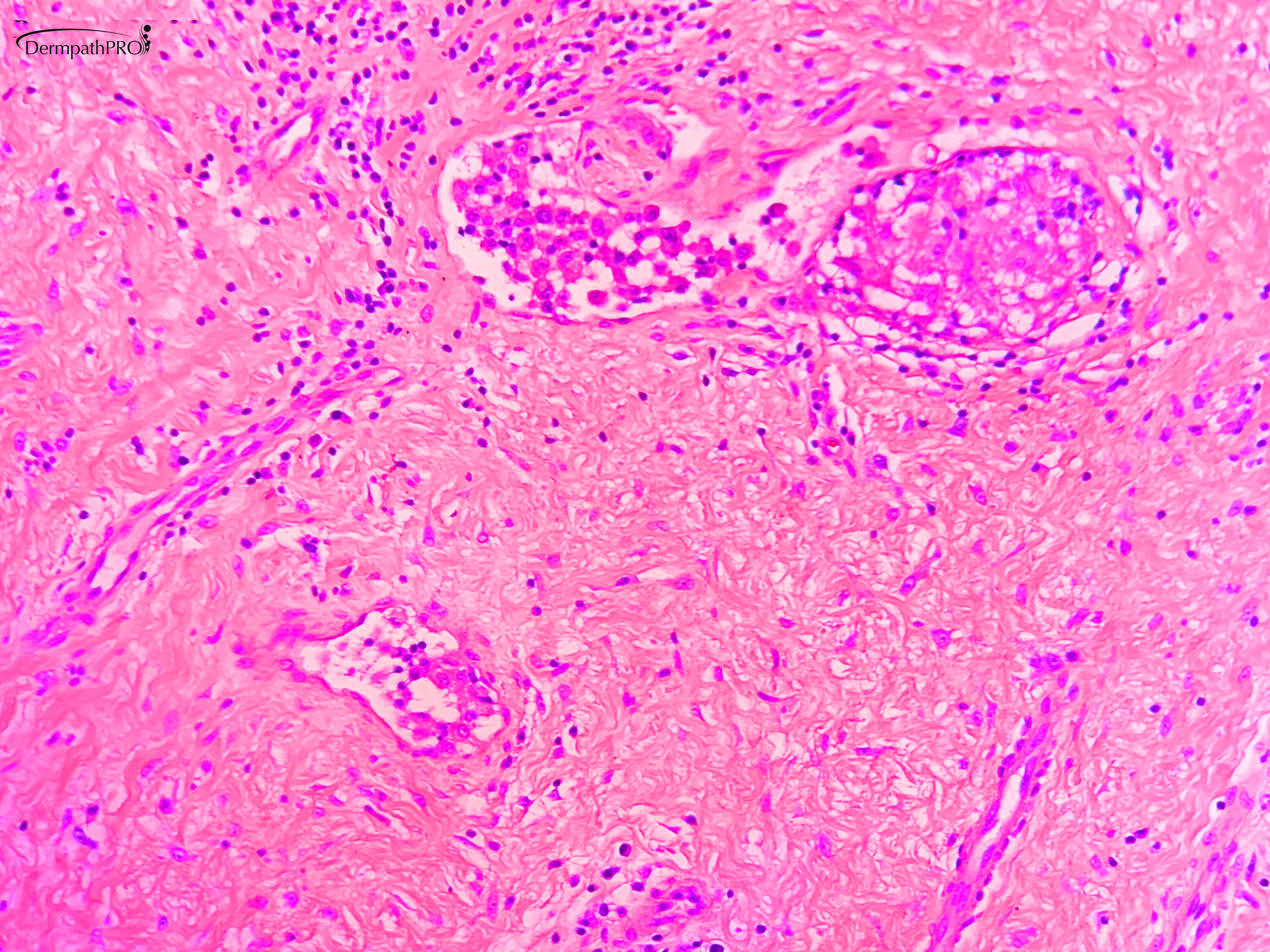
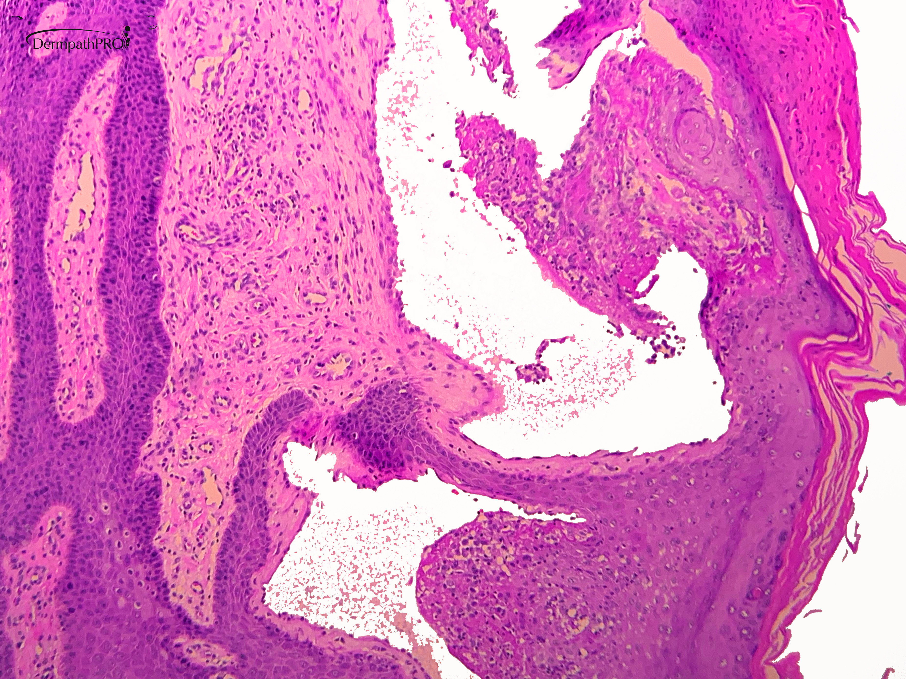
14 year old female with PMH of asthma, eczema, and intermittent/relapsing predominantly unilateral vulvar swelling. There was a prior biopsy in 2008. It showed “lymphatic proliferation compatible with lymphangioma circumscriptum”. No significant history of GI symptoms, or sarcoidosis.
Physical exam: multiple pearly vesicular appearing lesions in a cluster predominantly on the right side of the vulva (biopsy part B), also at the same side, a pedunculated flesh colored lesion with a firm consistency (part A). Clinically it has been attributed to CALME syndrome.


  Report Record

User Feedback


Richard Logan

Posted

I had never heard of CALME syndrome before, so I guess it is extremely rare.  The reported associated histology is really of normal constituents of vulvar tissue.  Here we have lymphangiectiasiae, fibrosis and rather sarcoidal granolomata.  I think this might be cutaneous Crohn's disease.

Share this comment


Link to comment
share_externally

John Zhang

Posted

Interesting case. Those small aggregates of epithelioid cells can be intravascular Langerhan cells (to be confirmed by IHC). Is there a consideration for Langerhan cell histiocytosis?

Share this comment


Link to comment
share_externally

Dr. Hafeez Diwan

Posted

There were two lesions biopsied.  The top left and bottom right panels are from the same lesion.  The other images are of the other lesion, which was pedunculated and fleshy.  There is no history of Crohn's disease.  These were interpreted as lymphangioma.  The fleshy lesion in addition shows intravascular histiocytosis, which is probably reactive in this setting.

CALME stands for "Childhood asymmetry labium majus enlargement."

I wanted to thank Dr. Sarah Al-Awami (who provided the photomicrographs and the history).

Share this comment


Link to comment
share_externally

Dr. Richard Carr

Posted

Interesting case. Certainly I've seen some very lymphangioma-like lesions in patient's with Crohn's or other reasons for lymphatic obstruction e.g. radiotherapy, obesity, prior surgery and pressure related etc.

Share this comment


Link to comment
share_externally


Join the conversation

You can post now and register later. If you have an account, sign in now to post with your account.

Guest
Add a comment...

×   Pasted as rich text.   Paste as plain text instead

  Only 75 emoji are allowed.

×   Your link has been automatically embedded.   Display as a link instead

×   Your previous content has been restored.   Clear editor

×   You cannot paste images directly. Upload or insert images from URL.

Loading...

×
×
  • Create New...